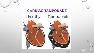
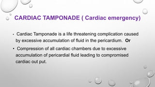
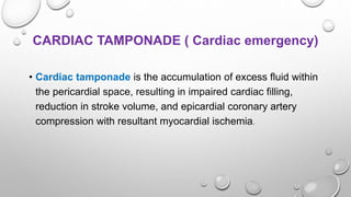
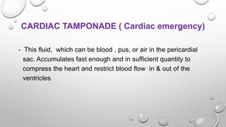
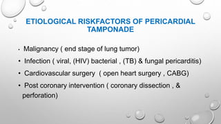
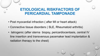
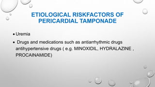
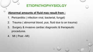
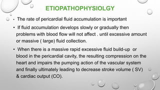
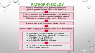
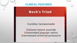
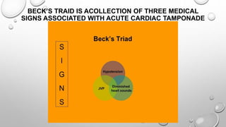
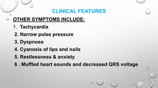
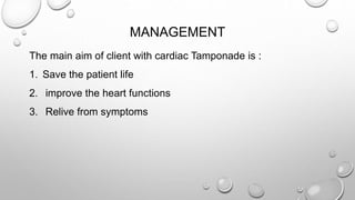
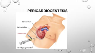
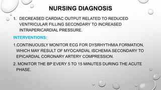
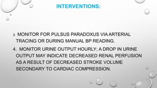
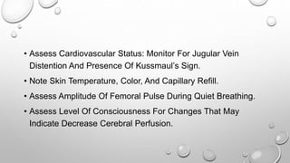
CARDIAC TAMPONADE ( Cardiac emergency) • Cardiac Tamponade is a life threatening complication caused by excessive accumulation of fluid in the pericardium. Or • Compression of all cardiac chambers due to excessive accumulation of pericardial fluid leading to compromised cardiac out put.
Kindly leave your comment if you found this helpful ;)
Some of the slides, i hide it from my real presentations for my own reference. Download to see all of them.
Coronary artery bypass grafting (CABG) is a type of surgery that improves blood flow to the heart. It's used for people who have severe coronary heart disease (CHD), also called coronary artery disease.
Cardiogenic shock is a condition of diminished cardiac output that severely impairs cardiac perfusion. In this condition in which the heart suddenly can't pump enough blood to meet the body's needs.
commonly used for medical students, and helpful to use this ppt to study for them, and also a common man can understand very easily what is coarctation of aorta.
Myocardial infraction or Heart attack are terms used anonymously, but the preferred term is MI.
In an MI an area of the myocardium is permanently destroyed.
MI is usually caused by reduced or decreased blood flow in a coronary artery due to rupture of an atherosclerotic plaque and subsequent occlusion of the artery by a thrombus.
Myocardial infarction (MI) death of the cells of an area of the heart muscle (myocardium) as a result of oxygen deprivation, which in turn is caused by obstruction of the blood supply; commonly referred to as a “heart attack.”
MI refers to the processes by which myocardial tissue is destroyed in regions of the heart that are deprived of an adequate blood supply because of reduced coronary artery blood flow.
Eighty percent to 90% of all acute MI are secondary to thrombus formation. When thrombus develops , perfusion to the myocardium distal to the occlusion is halted, resulting in necrosis.The myocardium receives its blood supply from the two large coronary arteries and their branches.
Occlusion of one or more of these blood vessels (coronary occlusion) is one of the major causes of myocardial infarction.
The occlusion may result from the formation of a clot that develops suddenly when an athermanous plaque ruptures through the sub layers of a blood vessel, or when the narrow, roughened inner lining of a scleroses artery leads to complete thrombosis.
The acute MI process takes time. Cardiac cells can withstand in ischemic conditions for approximately 20 minutes before cellular death begins.
The earliest tissue to become ischemic is the sub endocardium (the innermost layer of tissue in the cardiac muscle)
If ischemia persists, it takes approximately 4 to 6 hours for the entire thickness if the heart muscle to become necrosis.
Cardiac tamponade
Synonyms Pericardial tamponade
Hemorragic effusion.jpg
A very large pericardial effusion resulting in tamponade as a result of bleeding from cancer as seen on ultrasound. Closed arrow - the heart; open arrow - the effusion
Specialty Cardiac surgery
Symptoms Shortness of breath, weakness, lightheadedness, cough[1]
Usual onset Rapid or more gradual[2]
Causes Cancer, kidney failure, chest trauma, pericarditis, tuberculosis[2][1]
Diagnostic method Symptoms and ultrasound of the heart[2]
Treatment Drainage (pericardiocentesis, pericardial window, pericardiectomy)[2]
Frequency 2 per 10,000 per year (US)[3]
Cardiac tamponade, also known as pericardial tamponade, is when fluid in the pericardium (the sac around the heart) builds up, resulting in compression of the heart.
Bell’s palsy
Trigeminal Neuralgia ( Tic Douloreux)
Cranial & spinal neuropathies
Bell’s palsy (facial paralysis) is due to unilateral inflammation of the ( CN VII Facial nerve) seventh cranial nerve, which results in weakness or paralysis of the facial muscles on the affected side.
Polices for intensive care units / critical care units ANILKUMAR BR
What is a Policy?
A Policy is a statement, verbal, written or implied, of those principles and rules that are set by Board of Directors as guidelines on organizations actions.
There should be written polices for the intensive care units or critical care units which will guide the personnel working there.
The polices making body, there should be representation from administrative team, medical team and the nursing team.
ADMISSION POLICES: This should specify whether the patients can be admitted directly to CCU /ICU or through the casualty department.
There should be polices regarding the admission of medico-legal cases.
RESUSCIATION EQUIPMENTS IN INTENSISIVE CARE UNITSANILKUMAR BR
Intensive care unit (ICU) equipment includes patient monitoring, respiratory and cardiac support, pain management, emergency resuscitation devices, and other life support equipment .
They are designed to care for patients who are seriously injured, have a critical or life-threatening illness, or have undergone a major surgical procedure thereby requiring 24-hour care and monitoring.
Intensive care unit equipment includes
Patient monitoring devices
Life support and emergency resuscitation devices, and
Diagnostic devices.
Nursing management of critically ill patient in intensive care unitsANILKUMAR BR
Critical care nursing: it is the field of nursing with a focus on the utmost care of the critically ill (or) unstable patients.
Critically ill patients : critically ill patients are those who are at risk for actual (or) potential life threatening health problems.
Admission QGeneral appearance (consciousness)
Airway: Patency Position of artificial airway (if present)
Breathing: Quantity and quality of respirations (rate, depth, pattern, symmetry, effort, use of accessory muscles) Breath sounds Presence of spontaneous breathing.
Circulation and Cerebral Perfusion: ECG (rate, rhythm, and presence of ectopy) Blood pressure Peripheral pulses and capillary refill Skin, color, temperature, moisture Presence of bleeding Level of consciousness, responsiveness.
quick Check Assessment in CCU.
Infection control protocols in intensive care unitsANILKUMAR BR
Hospital acquired infections (HAIs) are common in intensive care unit (ICU) patient and are associated with increased morbidity and mortality.
The main reason being severity of illness, interruption of normal defense mechanism (e.g. mechanical ventilation), malnutrition & inability to ambulate make it more susceptible to multi drug resistant organism (MDRO).
The most frequent mode of transmission is Contact transmission, this may be direct or indirect other modes include droplet transmission, airborne transmission, common vehicle such as ventilator etc.
Abnormal development or deformities of the ear anatomy can cause a range of complications, from cosmetic issues to hearing and development problems.
An estimated 6 to 45 percent of children are born with some sort of congenital ear deformity.
Normally, the pleural space contains a small amount of fluid (5 to 15 mL), which acts as a lubricant that allows the pleural surfaces to move without friction.
But if fluid builds up from either increased production or inadequate removal pleural effusion results.
Pleural effusion B/L or unilateral (parapneumonic process)
Refers to any significant collection of fluid within pleural space.
Any imbalance in formation, absorption lead accumulation of pleural fluid. Common condition:
CHF
Bacterial pneumonia
Malignancy(chest tumor)
Pulmonary embolism
Pleura effusion is a condition refers to a collection of fluid in the pleural space. It is almost secondary to other conditions.
Hearing is one of our primary modes of communication. 360 million people worldwide have disabling hearing loss.
Hearing loss may result from genetic causes, complications at birth, certain infectious diseases, chronic ear infections, the use of particular drugs, exposure to excessive noise and ageing.
Hearing loss may be mild, moderate, severe or profound.
It can affect one ear or both ears, and leads to difficulty in hearing conversational speech or loud sounds. it may occur in one or both ear.
CONDUCTIVE HEARING LOSS (CHL)
SENSORINEURAL HEARING LOSS (SHL)
MIXED HEARING LOSS (MHL)
There are many types of cancer treatment. The types of treatment that patient receive will depend on the type of cancer, stage of cancer and how advanced it is.
Some people with cancer will have only one treatment. But most people have a combination of treatments, such as surgery with chemotherapy and/or radiation therapy.
Prostate cancer or tumor is the most common cancer in men other than non-melanoma skin cancer.
The majority (more than 75%) of cases occur in men over age 65.
Prostate cancer is a malignant tumor of the prostate gland.
Nursing management of patients with oncological conditionsANILKUMAR BR
Cancer is a group of diseases characterized by uncontrolled growth and spread of abnormal cells.
Cancer is caused by external factors and internal factors which may act together to initiate or promote carcinogenesis.
External Factors - chemicals, radiation, viruses, and lifestyle.
Internal Factors – hormones, immune condition, and inherited mutations.
Oncology branch of medicine deals with etiology, diagnosis, treatment and prevention of cancer.
Onco - is a Greek word meaning tumor .
A group of eye disorders, glaucoma is characterized by high intraocular pressure (IOP) that damages the optic nerve.
Glaucoma is one of the leading causes of irreversible blindness in the world and is the leading cause of blindness among adults in the United States.
Glaucoma may occur as primary or congenital disease or secondary to other causes, such as injury, infection, surgery, or prolonged use of topical corticosteroids.
Primary glaucoma has mainly two forms :
1. Open angle glaucoma ( chronic, simple, or wide angle glaucoma)
2. Angle –closure glaucoma( Acute or narrow angle glaucoma)
Angle –closure glaucoma occurs suddenly and may cause permanent or irreversible vision loss in 48 to 72 hours.
An inflammation of the conjunctiva commonly known as pink eye. Conjunctivitis is usually acute condition and self- limiting.
Conjunctivitis may be unilateral or bilateral.
It may also be chronic, possibly indicating degenerative changes or damage from repeated attacks.
It transmitted by contaminated towels, wash cloths, or the client own hands and it usually spreads very rapidly from one eye to otCommon causes are
Bacterial
Viral and chlamydial infection
Less common causes
Allergy
Parasitic disease and fungal infection
Occupational irritants
her eye.
Nursing assessment and management of patients with hepatic disordersANILKUMAR BR
Liver or Hepatic disorders are common and may result from a virus or exposure to toxic substances such as alcohol.
Another liver disorder is cancer: hepatocellular carcinoma is a highly malignant tumor that is difficult to treat and often fatal.
Liver function is complex, and liver dysfunction affects all body systems.
For this reason, the nurse must understand how the liver functions and must have expert assessment and clinical management skills to care for patients undergoing complex diagnostic and treatment procedures.
The liver plays additional roles in detoxification of chemicals and synthesis and storage of important nutrients and The liver is especially important in the regulation of glucose and protein metabolism .
Ototoxicity is, quite simply, ear poisoning (oto = ear, toxicity = poisoning), which results from exposure to drugs or chemicals that damage the inner ear or the vestibulo-cochlear nerve (the nerve sending balance and hearing information from the inner ear to the brain).
Nursing assessment and Management clients with Pancreatic disordersANILKUMAR BR
The pancreas, located in the upper abdomen, has endocrine as well as exocrine functions .
The secretion of pancreatic enzymes into the gastrointestinal tract through the pancreatic duct represents its exocrine function.
The secretion of insulin, glucagon, and somatostatin directly into the bloodstream represents its endocrine function.
Pancreatitis (inflammation of the pancreas) is a serious disorder. The most basic classification system used to describe or categorize the various stages and forms of pancreatitis divides the disorder into acute or chronic forms.
Acute pancreatitis can be a medical emergency associated with a high risk for life-threatening complications and mortality, whereas chronic pancreatitis often goes undetected until 80% to 90% of the exocrine and endocrine tissue is destroyed.
Acute pancreatitis does not usually lead to chronic pancreatitis unless complications develop.
Pneumonia is an inflammation of the lung parenchyma caused by various microorganisms, including bacteria, mycobacteria, fungi, and viruses.
Pneumonitis is a more general term that describes the inflammatory process in the lung tissue that may predispose and Pneumonia is an inflammation of the lung parenchyma that is caused by a microbial agent.
place the patient at risk for microbial invasion.
Pneumonia is classified into four: community-acquired pneumonia (CAP) and hospital-acquired pneumonia (HAP), pneumonia in the immunocompromised host, and aspiration pneumonia.
Nurses are primarily involved in the administration of medication across various settings. Nurses are also involved in both dispensing and preparation of medication. Research on medical administration errors (MAEs) shows an error rate of 60%, 34 mainly in the form of wrong time, wrong rate, or wrong dose.
There are many ways to prevent medication errors and one way of which is understanding the 10 “rights” of drug administration:
Thoracentesis (thor-a-sen-tee-sis) is a procedure that is done to remove a sample of fluid from around the lung.
The lung is covered with a tissue called the pleura. The inside of the chest is also lined with pleura.
The space between these two areas is called the pleural space.
This space normally contains just a thin layer of fluid, however, some conditions such as pneumonia, some types of cancer, or congestive heart failure may cause excessive fluid to develop (pleural effusion).
Thoracentesis, also known as pleural fluid analysis, is a procedure in which a needle is inserted through the back of the chest wall into the pleural space (a space that exists between the two lungs and the anterior chest wall) to remove fluid or air.
Pleural fluid analysis is the microscopic and chemical lab analysis of the fluid obtained during thoracentesis.
IndDiagnostic: determination of pleural effusion etiology (e.g. transudative versus exudative) usually requires the removal of 50 to 100mL of pleural fluid for laboratory studies. Most new effusions require diagnostic thoracentesis, an exception being a new effusion with a clear clinical diagnosis (e.g. CHF) with no evidence for superimposed pleural space infection
Therapeutic: reduce dyspnea and respiratory compromise in patients with large pleural effusions. This is typically achieved by removing a much larger volume of fluid compared to the diagnostic thoracentesis
ications
Nephrotic syndrome may be caused by primary (idiopathic) renal disease or by a variety of secondary causes. Patients present with marked edema, proteinuria, hypoalbuminemia, and often hyperlipidemia.
Nephrotic syndrome is a primary glomerular disease characterized by the following:
Marked increase in protein in the urine (proteinuria)
Decrease in albumin in the blood (hypoalbuminemia)
Edema (The swelling (edema), can be most noticeable on the face, around the eyes, around the feet and ankles, and in the belly area (or the abdomen).
High serum cholesterol and low-density lipoproteins (hyperlipidemia)
Nephrotic syndrome is a clinical disorder characterized by marked increase of protein in the urine ( proteinuria ), decrease in albumin in the blood (hypoalbuminemia ),edema, & excess lipids in the blood ( hyperlipidemia )
Pathophysiology
Nephrotic syndrome can occur with almost any intrinsic renal disease or systemic disease that affects the glomerulus.
Although generally considered a disorder of childhood, nephrotic syndrome does occur in adults, including the elderly. Causes include:
Chronic glomerulonephritis
Diabetes mellitus with intercapillary glomerulosclerosis
Amyloidosis of the kidney
Systemic lupus erythematosus
Multiple myeloma and renal vein thrombosis.
NSAIDs
Pre eclampsia
ARTIFICIAL INTELLIGENCE IN HEALTHCARE.pdfAnujkumaranit
Artificial intelligence (AI) refers to the simulation of human intelligence processes by machines, especially computer systems. It encompasses tasks such as learning, reasoning, problem-solving, perception, and language understanding. AI technologies are revolutionizing various fields, from healthcare to finance, by enabling machines to perform tasks that typically require human intelligence.
Ozempic: Preoperative Management of Patients on GLP-1 Receptor Agonists Saeid Safari
Preoperative Management of Patients on GLP-1 Receptor Agonists like Ozempic and Semiglutide
ASA GUIDELINE
NYSORA Guideline
2 Case Reports of Gastric Ultrasound
MANAGEMENT OF ATRIOVENTRICULAR CONDUCTION BLOCK.pdfJim Jacob Roy
Cardiac conduction defects can occur due to various causes.
Atrioventricular conduction blocks ( AV blocks ) are classified into 3 types.
This document describes the acute management of AV block.
Explore natural remedies for syphilis treatment in Singapore. Discover alternative therapies, herbal remedies, and lifestyle changes that may complement conventional treatments. Learn about holistic approaches to managing syphilis symptoms and supporting overall health.
Anti ulcer drugs and their Advance pharmacology ||
Anti-ulcer drugs are medications used to prevent and treat ulcers in the stomach and upper part of the small intestine (duodenal ulcers). These ulcers are often caused by an imbalance between stomach acid and the mucosal lining, which protects the stomach lining.
||Scope: Overview of various classes of anti-ulcer drugs, their mechanisms of action, indications, side effects, and clinical considerations.
Lung Cancer: Artificial Intelligence, Synergetics, Complex System Analysis, S...Oleg Kshivets
RESULTS: Overall life span (LS) was 2252.1±1742.5 days and cumulative 5-year survival (5YS) reached 73.2%, 10 years – 64.8%, 20 years – 42.5%. 513 LCP lived more than 5 years (LS=3124.6±1525.6 days), 148 LCP – more than 10 years (LS=5054.4±1504.1 days).199 LCP died because of LC (LS=562.7±374.5 days). 5YS of LCP after bi/lobectomies was significantly superior in comparison with LCP after pneumonectomies (78.1% vs.63.7%, P=0.00001 by log-rank test). AT significantly improved 5YS (66.3% vs. 34.8%) (P=0.00000 by log-rank test) only for LCP with N1-2. Cox modeling displayed that 5YS of LCP significantly depended on: phase transition (PT) early-invasive LC in terms of synergetics, PT N0—N12, cell ratio factors (ratio between cancer cells- CC and blood cells subpopulations), G1-3, histology, glucose, AT, blood cell circuit, prothrombin index, heparin tolerance, recalcification time (P=0.000-0.038). Neural networks, genetic algorithm selection and bootstrap simulation revealed relationships between 5YS and PT early-invasive LC (rank=1), PT N0—N12 (rank=2), thrombocytes/CC (3), erythrocytes/CC (4), eosinophils/CC (5), healthy cells/CC (6), lymphocytes/CC (7), segmented neutrophils/CC (8), stick neutrophils/CC (9), monocytes/CC (10); leucocytes/CC (11). Correct prediction of 5YS was 100% by neural networks computing (area under ROC curve=1.0; error=0.0).
CONCLUSIONS: 5YS of LCP after radical procedures significantly depended on: 1) PT early-invasive cancer; 2) PT N0--N12; 3) cell ratio factors; 4) blood cell circuit; 5) biochemical factors; 6) hemostasis system; 7) AT; 8) LC characteristics; 9) LC cell dynamics; 10) surgery type: lobectomy/pneumonectomy; 11) anthropometric data. Optimal diagnosis and treatment strategies for LC are: 1) screening and early detection of LC; 2) availability of experienced thoracic surgeons because of complexity of radical procedures; 3) aggressive en block surgery and adequate lymph node dissection for completeness; 4) precise prediction; 5) adjuvant chemoimmunoradiotherapy for LCP with unfavorable prognosis.
micro teaching on communication m.sc nursing.pdfAnurag Sharma
Microteaching is a unique model of practice teaching. It is a viable instrument for the. desired change in the teaching behavior or the behavior potential which, in specified types of real. classroom situations, tends to facilitate the achievement of specified types of objectives.
The prostate is an exocrine gland of the male mammalian reproductive system
It is a walnut-sized gland that forms part of the male reproductive system and is located in front of the rectum and just below the urinary bladder
Function is to store and secrete a clear, slightly alkaline fluid that constitutes 10-30% of the volume of the seminal fluid that along with the spermatozoa, constitutes semen
A healthy human prostate measures (4cm-vertical, by 3cm-horizontal, 2cm ant-post ).
It surrounds the urethra just below the urinary bladder. It has anterior, median, posterior and two lateral lobes
It’s work is regulated by androgens which are responsible for male sex characteristics
Generalised disease of the prostate due to hormonal derangement which leads to non malignant enlargement of the gland (increase in the number of epithelial cells and stromal tissue)to cause compression of the urethra leading to symptoms (LUTS
Title: Sense of Smell
Presenter: Dr. Faiza, Assistant Professor of Physiology
Qualifications:
MBBS (Best Graduate, AIMC Lahore)
FCPS Physiology
ICMT, CHPE, DHPE (STMU)
MPH (GC University, Faisalabad)
MBA (Virtual University of Pakistan)
Learning Objectives:
Describe the primary categories of smells and the concept of odor blindness.
Explain the structure and location of the olfactory membrane and mucosa, including the types and roles of cells involved in olfaction.
Describe the pathway and mechanisms of olfactory signal transmission from the olfactory receptors to the brain.
Illustrate the biochemical cascade triggered by odorant binding to olfactory receptors, including the role of G-proteins and second messengers in generating an action potential.
Identify different types of olfactory disorders such as anosmia, hyposmia, hyperosmia, and dysosmia, including their potential causes.
Key Topics:
Olfactory Genes:
3% of the human genome accounts for olfactory genes.
400 genes for odorant receptors.
Olfactory Membrane:
Located in the superior part of the nasal cavity.
Medially: Folds downward along the superior septum.
Laterally: Folds over the superior turbinate and upper surface of the middle turbinate.
Total surface area: 5-10 square centimeters.
Olfactory Mucosa:
Olfactory Cells: Bipolar nerve cells derived from the CNS (100 million), with 4-25 olfactory cilia per cell.
Sustentacular Cells: Produce mucus and maintain ionic and molecular environment.
Basal Cells: Replace worn-out olfactory cells with an average lifespan of 1-2 months.
Bowman’s Gland: Secretes mucus.
Stimulation of Olfactory Cells:
Odorant dissolves in mucus and attaches to receptors on olfactory cilia.
Involves a cascade effect through G-proteins and second messengers, leading to depolarization and action potential generation in the olfactory nerve.
Quality of a Good Odorant:
Small (3-20 Carbon atoms), volatile, water-soluble, and lipid-soluble.
Facilitated by odorant-binding proteins in mucus.
Membrane Potential and Action Potential:
Resting membrane potential: -55mV.
Action potential frequency in the olfactory nerve increases with odorant strength.
Adaptation Towards the Sense of Smell:
Rapid adaptation within the first second, with further slow adaptation.
Psychological adaptation greater than receptor adaptation, involving feedback inhibition from the central nervous system.
Primary Sensations of Smell:
Camphoraceous, Musky, Floral, Pepperminty, Ethereal, Pungent, Putrid.
Odor Detection Threshold:
Examples: Hydrogen sulfide (0.0005 ppm), Methyl-mercaptan (0.002 ppm).
Some toxic substances are odorless at lethal concentrations.
Characteristics of Smell:
Odor blindness for single substances due to lack of appropriate receptor protein.
Behavioral and emotional influences of smell.
Transmission of Olfactory Signals:
From olfactory cells to glomeruli in the olfactory bulb, involving lateral inhibition.
Primitive, less old, and new olfactory systems with different path
TEST BANK for Operations Management, 14th Edition by William J. Stevenson, Ve...kevinkariuki227
TEST BANK for Operations Management, 14th Edition by William J. Stevenson, Verified Chapters 1 - 19, Complete Newest Version.pdf
Factory Supply Best Quality Pmk Oil CAS 28578–16–7 PMK Powder in Stockrebeccabio
Factory Supply Best Quality Pmk Oil CAS 28578–16–7 PMK Powder in Stock
Telegram: bmksupplier
signal: +85264872720
threema: TUD4A6YC
You can contact me on Telegram or Threema
Communicate promptly and reply
Free of customs clearance, Double Clearance 100% pass delivery to USA, Canada, Spain, Germany, Netherland, Poland, Italy, Sweden, UK, Czech Republic, Australia, Mexico, Russia, Ukraine, Kazakhstan.Door to door service
Hot Selling Organic intermediates
3. INTRODUCTON
• The heart is surrounded by a membrane covering
called the “ Pericardial sac”.
• The pericardial sac consists of mainly two layers
1. The Parietal layer ( outer layer) &
2. The visceral layer ( inner layer) and normally
contains a small amount of fluid to cushion &
lubricate the heart as it contracts & expands.
5. PERICARDIUM
• Pericardium is the membranous sac surrounding the
human heart.
• There is about normally 20 to 50 ml pericardial fluid
in the pericardial cavity.
6. THE NORMAL FUNCTIONS OF PRICARDIUM
1) Maintaining an optimal cardiac shape
2) Reducing friction between the beating heart &
adjacent structures
3) Protecting the heart from other disease which are
caused by the neighboring organs ( inflammation, TB
etc)
4) Prevent overfilling of the heart.
7. CARDIAC TAMPONADE ( Cardiac emergency)
• Cardiac Tamponade is a life threatening complication caused
by excessive accumulation of fluid in the pericardium. Or
• Compression of all cardiac chambers due to excessive
accumulation of pericardial fluid leading to compromised
cardiac out put.
8. CARDIAC TAMPONADE ( Cardiac emergency)
• Cardiac tamponade is the accumulation of excess fluid within
the pericardial space, resulting in impaired cardiac filling,
reduction in stroke volume, and epicardial coronary artery
compression with resultant myocardial ischemia.
9. CARDIAC TAMPONADE ( Cardiac emergency)
• This fluid, which can be blood , pus, or air in the pericardial
sac. Accumulates fast enough and in sufficient quantity to
compress the heart and restrict blood flow in & out of the
ventricles.
10. ETIOLOGICAL RISKFACTORS OF PERICARDIAL
TAMPONADE
• Malignancy ( end stage of lung tumor)
• Infection ( viral, (HIV) bacterial , (TB) & fungal pericarditis)
• Cardiovascular surgery ( open heart surgery , CABG)
• Post coronary intervention ( coronary dissection , &
perforation)
11. ETIOLOGICAL RISKFACTORS OF
PERICARDIAL TAMPONADE
• Post myocardial infraction ( after MI or heart attack)
• Connective tissue disorders ( SLE, Rheumatoid arthritis)
• Iatrogenic (after sterna biopsy, pericardiocentasis, central IV
line insertion and transvenous pacemaker lead implantation &
radiation therapy to the chest)
12. ETIOLOGICAL RISKFACTORS OF
PERICARDIAL TAMPONADE
Uremia
Drugs and medications such as antiarrhythmic drugs
antihypertensive drugs ( e.g. MINOXIDIL, HYDRALAZINE ,
PROCAINAMIDE)
13. ETIOPATHOPHYSIOLGY
Abnormal amounts of fluid may result from :
1. Pericarditis ( infection viral, bacterial, fungal)
2. Trauma ( abnormal blood, pus, fluid due to an trauma)
3. Surgery & invasive cardiac diagnostic & therapeutic
procedures.
4. MI ( Post –MI)
14. ETIOPATHOPHYSIOLGY
• The rate of pericardial fluid accumulation is important
• If fluid accumulation develops slowly or gradually then
problems with blood flow will not affect . until excessive amount
or massive ( large) fluid collection.
• When there is a massive rapid excessive fluid build-up or
blood in the pericardial cavity, the resulting compression on the
heart and impairs the pumping action of the vascular system
and finally ultimately leading to decrease stroke volume ( SV)
& cardiac output (CO).
17. BECK’S TRAID IS ACOLLECTION OF THREE MEDICAL
SIGNS ASSOCIATED WITH ACUTE CARDIAC TAMPONADE
18. CLINICAL FEATURES
OTHER SYMPTOMS INCLUDE:
1. Tachycardia
2. Narrow pulse pressure
3. Dyspnoea
4. Cyanosis of lips and nails
5. Restlessness & anxiety
6 . Muffled heart sounds and decreased QRS voltage
19. MANAGEMENT
The main aim of client with cardiac Tamponade is :
1. Save the patient life
2. improve the heart functions
3. Relive from symptoms
20. Treatment that are administered for cardiac
tamponade include:
1. IV fluids to maintain normal BP
2. Antibiotics
3. Supplemental oxygen to reduce work load on the heart
22. PERICARDIOCENTESIS
• Pericardiocentesis, also called a pericardial tap, is a surgical
invasive procedure ( use both diagnostic and therapeutic purpose)
in which abnormal or excessive fluid is removed from the
pericardium sac the sac around your heart. Or
• Pericardiocentesis is the removal by needle of pericardial fluid
from the sac surrounding the heart for diagnostic or therapeutic
purposes.
23. PERICARDIOCENTESIS
• REMOVAL OF 5 TO 10 ML MAY DRAMATIC INCREASE STROKE VOLUME AND
CARDIAC OUTPUT BY 25 TO 50% AND REASSESS FOR IMPROVEMENT
REPEATED WHEN IT NECESSARY.
24. PURPOSE OF PERICARDIOCENTESIS
• Pericarditis (Caused By Infection, Inflammation)
• Trauma (Producing Blood In The Pericardial Sac)
• Surgery Or Other Invasive Procedures Performed On The
Heart Cancer (Producing Malignant Effusions) Myocardial
Infarction,
• Congestive Heart Failure
• Renal Failure
25. PRECAUTIONS TO PERFORM PROCEDURE
• Whenever Possible, An Echocardiogram (Ultrasound Test)
Should Be Performed To Confirm The Presence Of The
Pericardial Effusion And To Guide The Pericardiocentesis
Needle During The Procedure.
• Because Of The Risk Of Accidental Puncture To Major Arteries
Or Organs In Pericardiocentesis, Surgical Drainage May Be A
Preferred Treatment Option For Pericardial Effusion In Non-
emergency Situations.
26. COMPLICATIONS OF PERICARDIOCENTESIS
• Cardiac Arrest
• Myocardial Infarction Or Heart Attack
• Abnormal Heart Rhythms ( Arrthymias )
• Laceration Of The Heart Muscle
27. • Puncture Or Rupture Of Coronary Arteries
• Lacerations Of Organs
• Hemothorax, Penumothorax And
• Pnemuo Pericardium
28. NURSING MANAGEMENT
ACUTE CARE MANAGEMENT:
1. Assess The Client Status
2. Monitor Hemodynamic ( Pulse ,HR,BP,RR)
3. Assess Neurologic Status ( Loc ,Orientation) Confusion ,
Restlessness And Anxiety.
4. Provide Psychological Support
5. Cardiovascular Assessment ( HR JVD HS Skin Color Etc)
29. NURSING DIAGNOSIS
1. DECREASED CARDIAC OUTPUT RELATED TO REDUCED
VENTRICULAR FILLING SECONDARY TO INCREASED
INTRAPERICARDIAL PRESSURE.
INTERVENTIONS:
1.CONTINUOUSLY MONITOR ECG FOR DYSRHYTHMIA FORMATION,
WHICH MAY RESULT OF MYOCARDIAL ISCHEMIA SECONDARY TO
EPICARDIAL CORONARY ARTERY COMPRESSION.
2. MONITOR THE BP EVERY 5 TO 15 MINUTES DURING THE ACUTE
PHASE.
30. INTERVENTIONS:
3. MONITOR FOR PULSUS PARADOXUS VIA ARTERIAL
TRACING OR DURING MANUAL BP READING.
4. MONITOR URINE OUTPUT HOURLY; A DROP IN URINE
OUTPUT MAY INDICATE DECREASED RENAL PERFUSION
AS A RESULT OF DECREASED STROKE VOLUME
SECONDARY TO CARDIAC COMPRESSION.
31. • Assess Cardiovascular Status: Monitor For Jugular Vein
Distention And Presence Of Kussmaul’s Sign.
• Note Skin Temperature, Color, And Capillary Refill.
• Assess Amplitude Of Femoral Pulse During Quiet Breathing.
• Assess Level Of Consciousness For Changes That May
Indicate Decrease Cerebral Perfusion.
32. • Provide Supplemental Oxygen As Ordered.
• Initiate Two Large-bore Intravenous Lines For Fluid Administration
To Maintain Filling Pressure.
• Pharmacologic Therapy May Include Dobutamine To Enhance
Myocardial Contractility And Decrease Peripheral
Vascularresistance.
• Monitor The Patient For Dysrhythmias, Coronary Artery Laceratio.
• Surgical Intervention To Identify And Repair Bleeding Site, To
Evacuate Clots In The Mediastinum, To Resects Or Open The
Pericardium.