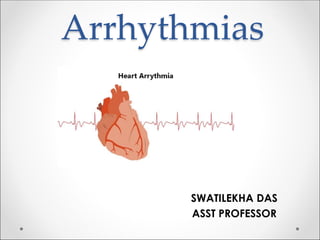
Understanding Cardiac Arrhythmias
•
28 likes•16,636 views

Arrhythmias- Easy ppt for Nurses normal pathophysiology ECG Cardiac Arrhythmias

Recommended
More Related Content
What's hot
What's hot (20)
Similar to Understanding Cardiac Arrhythmias
Similar to Understanding Cardiac Arrhythmias (20)
More from Swatilekha Das
More from Swatilekha Das (20)
Recently uploaded
Recently uploaded (20)
Understanding Cardiac Arrhythmias
- 2. Normal Pathophysiology SAN AVN Impulses originate regularly at a frequency of 60-100 beat/ min SA node – AV node – Bundle of His – Purkinje fibres
- 3. ECG • Normal PR interval- 0.12s to 0.2 s (3 to 5 small square) • Normal QRS complex- <0.12s (< 3 small squares)
- 5. Cardiac Arrhythmias ●An abnormality of the cardiac rhythm is called a cardiac arrhythmia. ● Arrhythmias may cause sudden death, syncope, heart failure, dizziness, palpitations or no symptoms at all. ● There are two main types of arrhythmia: BRADYCARDIA: THE HEART RATE IS SLOW (< 60 B.P.M). TACHYCARDIA: THE HEART RATE IS FAST (> 100 B.P.M).
- 6. Bradyarrhythmias Sinus Bradycardia • Physiological variant due to strong vagal tone or atheletic training. Common causes: • Extrinsic causes; Hypothermia, hypothyroidism, raised intracranial pressure. Drug therapy with beta-blockers, digitalis and other antiarrhythmic drugs. • Intrinsic causes; Acute ischaemia and infarction of the sinus node (as a complication of acute myocardial infarction). Chronic degenerative changes such as fibrosis of the atrium and sinus node (sick sinus syndrome).
- 7. AF (Atrial fibrillation) Atrial fibrillation (AF) - • Electrical signals come from the atria at a very fast & erratic rate. Ventricles contract in an irregular manner because of the erratic signals coming from the atria. • The ECG shows normal but irregular QRS complexes and no P waves. • Common causes include CAD, valvular heart disease, hypertension, hyperthyroidism and others. In some patients no cause can be found 'lone' atrial fibrillation.
- 8. Atrial flutter HR200-350/min • Electrical signals come from the atria at a fast but even rate, causing ventricles to contract faster and increase the heart rate. • When the signals from the atria are coming at a faster rate than the ventricles can respond to, the ECG pattern develops a signature "sawtooth" pattern, showing two or more flutter waves between each QRS complex.
- 9. Atrioventricular reciprocating tachycardia (AVRT) • Large circuit comprising the AV node, the His bundle, the ventricle and an abnormal connection from the ventricle back to the atrium. This abnormal connection is called an accessory pathway or bypass tract. • Bypass tracts result from incomplete separation of the atria and the ventricles during fetal development. • Atrial activation occurs after ventricular activation and the P wave is usually clearly seen between the QRS and T complexes
- 10. Management Acute Management • Associated haemodynamic instability require emergency cardioversion. • If haemodynamically stable, vagal manoeuvres, including right carotid massage, Valsalva manoeuvre and facial immersion in cold water. • If not successful, IV adenosine, verapamil, diltiazem, or beta-blockers should be tried. Long-term management • Ablation of an accessory pathway. • Verapamil, diltiazem & β-blockers - effective in 60-80% of patients.
- 11. The Wolf Parkinson White Syndrome (WPW) ►An abnormal band of atrial tissue connects the atria and ventricles and can electrically bypass the normal pathways of conduction; a re-entry circuit can develop causing paroxysms of tachycardia. ►ECG: - Short PR interval - Delta wave - upstroke of QRS complex ►Drug treatment - flecainamide, amiodarone or disopyramide. ►Digoxin & verapamil are contraindicated - enhance antegrade conduction through the AP by increasing the refractory period in the AV node ►Treatment - transvenous catheter radiofrequency ablation
- 13. VF • A condition in which many electrical signals are sent from the ventricles at a very fast and erratic rate. As a result, the ventricles are unable to fill with blood and pump. • Life-threatening - no pulse and complete LOC. • ECG - shapeless, rapid oscillations, no hint of organized complexes • Requires prompt defibrillation to restore the normal rhythm and function of the heart. • It may cause sudden cardiac death. Basic and advanced cardiac life support is needed • Survivors of these ventricular tachyarrhythmias are, in the absence of an identifiable reversible cause (e.g. acute myocardial infarction, severe metabolic disturbance), at high risk of sudden death. Implantable cardioverter-defibrillators (ICDs) are first-line therapy
- 15. VT • An electrical signal is sent from the ventricles at a very fast but often regular rate. • ECG - rapid ventricular rhythm with broad (often 0.14 s or more), abnormal QRS complexes. • Treatment: emergency DC cardioversion • intravenous therapy with class I drugs or amiodarone
- 17. Bundle branch block • Interruption of the right or left branch of the bundle of Hiss delays activation of the corresponding ventricle leading to broadening of the QRS complex • Both RT and LT BBB show wide deformed QRS complex. • Treatment – may require pacemaker
- 19. DIAGNOSTIC STUDIES • FBC • U&E • Glucose • Ca • Mg • TSH • 24 hr ECG/ exercise ECG • Electrophysiology • Echo • Cardiac catheterisation
- 20. Management • Conservative – smoking cessation, alcohol cessation, lifestyle (diet, weight loss, exercise) • Pharmacological therapy.
- 21. Treatment • Cardioversion. • Pacemaker therapy. • Surgical therapy e.g. aneurysmal excision. • Interventional therapy “ablation”
- 22. Thank you