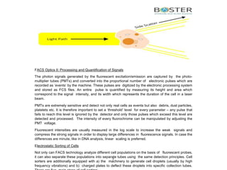
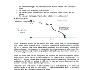
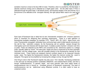
Flow Cytometry Principle
By BosterBio
https://www.bosterbio.com/
Principles of Flow Cytometry
This section deals with the different aspects of FACS instrumentation and the basic system of cell
detection. The diagram below illustrates the experimental setup and general procedure of a typical
FACS experiment. A population of mixed cells is sorted into a negative sample and a positive
sample containing cells of interest by the flow cytometer.
4
A. The Flow Cytometer
The flow cytometer instrument consists of three core systems: fluidics, optics, and electronics. The
fluidics system includes a flow cell, where the sample fluid is injected. The optics system consists of
various filters, light detectors, and the light source, which is usually a laser line producing a single
wavelength of light at a specific frequency. This is where the particles are passed through at least
one laser beam. Lasers are available at different wavelengths ranging from ultraviolet to far red and
have a variable range of power levels as well (photon output/time). A detector in front of the light
beam measures forward scatter light signals (FSC) and detectors to the side measure side scatter
light signals (SSC). Fluorescence detectors measure the fluorescence signal intensity emitted from
positively stained cells and particles. Within the flow cytometer, all of these different light signals are
split into defined wavelengths and channeled by a set of filters and mirrors so that each sensor will
detect fluorescence only at a specified wavelength. These sensors are called photomultiplying tubes
(PMTs). Various filters are used in the flow cytometer to direct photons of the correct wavelength to
each PMT. Short pass (SP) filters allow transmission of photons below a specific wavelength while
long pass (LP) filters allow transmission above a specific wavelength. Band pass (BP) filters allow
transmission of photons that have wavelengths within a narrow range. Each PMT will also detect
any other fluorophores emitting at a similar wavelength to the fluorophore it is detecting. These light
signals are converted by the electronics system to data that can be visualized and interpreted by
software. The diagram below illustrates the parts and setup of a typical flow cytometer instrument.
5
FACS Fluidics System
The objective of the fluidic system is to order the cell suspension into a uniform stream of single cells
so that they can be efficiently detected. The fluidics system consists of a central core through which
the sheath fluid flows under high pressure and encloses the sample fluid. Since the sheath fluid is
under higher pressure, it moves faster than the sample and thus creates a drag on the sample fluid.
It is this hydrodynamic force on the sample fluid that creates the single stream and allows the cells
to be analyzed individually as they pass through the illumination source.
FACS Optics I: Detection - Lasers and Types of Optical Filters
After a single stream is formed, each cell passes through the different laser beams and is sorted
according to its light scattering and, if labelled with a fluorophore (fluorochrome), its fluorescence
emission properties. Each laser produces a single wavelength of light at a specific frequency and
depending on the wavelength, they are classified as ultraviolet, violet, blue, yellow/green, red and
infra-red. Most of the modern flow cytometers are equipped with 3-5 lasers and can therefore
simultaneously detect multiple signals.
Once a laser beam excites the fluorophores of the suitable excitation range, the latter emit signals at
different wavelengths which pass through specific optical filters - each filter allowing only specific
wavelengths to pass through. The wavelengths passing through every filter are then captured by
their corresponding detectors and quantified. Every laser channel has multiple filters and their
respective detectors are each specific for a different fluorochrome. With an optimum combination of
lasers and filters, an instrument can detect as many as 20 different fluorochromes!
Apart from the fluorescent emission signal, the direction of light scatter also provides information
about a cell type. When light is scattered at an angle of 20° from the excitation beam, it is called
forward scatter (FSC) and roughly equates to a cell’s size. When the scatter occurs at an angle of
90° relative to the excitation beam, it is called side scatter (SSC) and is a measure of cellular
granularity.
6
FACS Optics II: Processing and Quantification of Signals
The photon signals generated by the fluorescent excitation/emission are captured by the photo-
multiplier tubes (PMTs) and converted into the proportional number of electronic pulses which are
recorded as ‘events’ by the machine. These pulses are digitized by the electronic processing system
and stored as FCS files. An entire pulse is quantified by measuring its height and area which
correspond to the signal intensity, and its width which represents the duration of the cell in a laser
beam.
PMTs are extremely sensitive and detect not only real cells as events but also debris, dust particles,
platelets etc. It is therefore important to set a ‘threshold’ level for every parameter – any pulse that
fails to reach this level is ignored by the detector and only those pulses which exceed this level are
detected and processed. The intensity of every fluorochrome can be manipulated by adjusting the
PMT voltage.
Fluorescent intensities are usually measured in the log scale to increase the weak signals and
compress the strong signals in order to display large differences in fluorescence signals. In case the
differences are minute, like in DNA analysis, linear scaling is preferred.
Electrostatic Sorting of Cells
Not only can FACS technology analyze different cell populations on the basis of fluorescent probes,
it can also separate these populations into separate tubes using the same detection principles. Cell
sorters are additionally equipped with a) the machinery to generate cell droplets (usually by high
frequency vibrations) and b) charged plates to deflect these droplets into specific collection tubes.
7
- The stream containing the cells is broken down into droplets and each cell is captured in a
droplet.
- The droplets pass between the deflection plates.
- The charged droplets (those enclosing cells) are deflected in the electrostatic field and
collected.
- The uncharged droplets pass though and are collected in the waste chamber.
B. The Fluorophores
The Basis of Fluorescence
When a fluorophore absorbs light, its electrons move from a resting state to a maximum energy
state – this is termed excitation. It then undergoes a conformational change wherein the electrons
fall back to the resting state and the excess energy is released as the fluorescence – emission. The
difference between the wavelengths of the excitation and emission maxima is called stokes shift.
The cycle of excitation and emission is repeated several times for each fluorochrome moiety and is
read out as the fluorescent signal.
Since the emitted light has less energy than the excitation, the emission wavelength of any
fluorochrome is longer than its excitation wavelength and therefore has a different color. For
example, FITC absorbs between 400-530 nm with peak absorption or excitation maxima at 490 nm
and emits from 475-650 nm, peaking at 525 nm. It is important to excite flurochromes at their
excitation maxima to increase the intensity of emission.
The emitted color (and intensity) of a fluorochrome which is finally detected by an instrument
depends largely on the laser and filter settings. To take the example of FITC, its absorption
spectrum lies in both the blue and green spectra, but the
8
excitation maxima is close to the blue 488 nm laser. Therefore, when it is excited by the blue laser, it
absorbs maximum energy and is detected as a bright green color. On the other hand, if FITC is
detected through the green laser, it will absorb poorly and fluoresce weakly in the red channel. For a
flurochrome to be detected at the strongest intensity, the right optical filters need to be used which
only allow the specific wavelength to pass through.
Each type of fluorescent dye or label has its own characteristic excitation and emission spectrum
which is important for designing flow cytometry experiments. There is a wide selection of
fluorophores available nowadays; for example, FITC, PerCP, APC, PE, Cy5.5, Alexa Fluors, and
more. Fluorescently conjugated antibodies have commonly been used to label specific structures on
the cell for flow cytometric analysis. As the fluorescing cell (or particle) passes through the
interrogation point and interacts with the laser beam, it creates a pulse of photon emission over time
(a peak). These are detected by the PMTs and converted by the electronics system to a voltage
pulse, typically called an "event". The total pulse height and area is measured by the flow cytometer
instrument, and the voltage pulse area will correlate directly to the fluorescence intensity for that
individual event. These events are assigned channel numbers based on its measured intensity
(pulse area). The higher the fluorescence intensity, the higher the channel number the event is
assigned. This signal can be amplified by increasing the voltage running through the PMT.
One thing to note is that fluorescent signals may also occur from naturally fluorescing substances
in the cell such as reduced pyridine nucleotides (NAD(P)H) and oxidized flavins (FAD), and this is
termed “autofluorescence”. In general, larger and more granular cells have higher levels of
autofluorescence due to having an increased amount of fluorescent compounds. The level of
autofluorescence can be determined using unstained controls. It is often useful to have an
unstained control,
9
or an FMO (Fluorescence Minus One) control. The FMO control will help you to identify and gate
cells during data analysis.
Common Fluorophores and Spectral Characteristics
To design a multicolor panel, it is imperative to know the excitation and emission spectra of the
fluorochromes in use. In addition, the laser and filter configuration of the instrument is also vital for
the optimal detection and minimal spillover of the different fluorochromes. Here is a table of some
commonly used fluorophores and their spectral characteristics:
10
Fluorochrome Excitation (nm) Emission (nm)
7-AAD 546 647
APC 650 660
CFP 458 480
DAPI 358 461
FITC 494 520
GFP 395 508
Hoechst 33342 & 33258 352 461
Pacific Blue 410 455
PE 496, 565 578
PE-Cy7 480, 743 767
PerCP 482 678
PerCP-Cy5.5 482, 675 695
Rhodamine 123 507 529
Selecting the Right Fluorophore Conjugate
Knowing each conjugate label’s properties will be important in choosing the right fluorophores for
your experiment. Some key properties to know about fluorescent conjugate tags are listed here:
- Maximum Excitation Wavelength (λex) – The peak wavelength in the excitation (absorption)
spectra, measured in nanometers (nm)
- Maximum Emission Wavelength (λem) – The peak wavelength in the emission spectra,
measured in nanometers (nm)
- Extinction Coefficient (ε max ) – (also called molar absorptivity) The capacity for the
fluorochrome to absorb light at a given wavelength, usually measured at the maximum
excitation wavelength with the units M −1 cm −1
- Fluorescence Quantum Yield (Φf) – The number of photons emitted per absorbed photon. A
high quantum yield is important, and this number ranges between 0 and 1
- Fluorescence Decay Time (τfl) – The time interval after which the number of
excited fluorescent molecules is reduced to 1/e (approx. 37%) via the loss of energy, usually
measured in nanoseconds
- Brightness – The fluorescence output per fluorophore measured. Fluorophores with high
brightness values can be used to detect lower- abundance targets. Calculated as the product
of the extinction coefficient (at the relevant excitation wavelength) and the fluorescence
quantum yield divided by 1000, with the units M −1 cm −1
- Stokes Shift – The difference between the Maximum Emission Wavelength and Maximum
Excitation Wavelength, measured in nanometers
- Laser line – Which laser line(s) to use for detection using flow cytometry
- Common filter set – The standard microscope filter set that generates the best imaging
results
- Photostability – How resistant a substance is to change resulting from exposure to light
Single and tandem dyes
Single dyes such as FITC, PE, APC, and PerCP consist of a simple fluorophore moiety; tandem
dyes on the other hand consist of a small fluorophore covalently bound to a bigger fluorophore.
Examples include PE-Cy7, APC-Cy7, PerCP-Cy5.5 etc.
The tandem dyes offer the advantage of brighter fluorescence as compared to single fluorophores.
After the smaller dye is activated and reaches its maximum intensity, it transfers its energy to the
second dye (in tandem) which in turn produces the fluorescent emission. This process, known as
fluorescence resonance energy transfer (FRET), achieves higher stokes shift and thus increases the
number of colors that can be analyzed from a single laser wavelength. For example, although PE
and PerCP-Cy5.5 absorb at 488 nm, they emit in the yellow and red channels respectively.
The disadvantage of tandem dyes is that they are highly unstable during the fixation process and
are bleached out in the presence of methanol, a common fix-perm agent in intracellular staining.
Spectral Overlap and Compensation
When working with multiple colors, there is always a high chance of the overlap of the emission
spectra of different fluorochromes – with the result that one fluorochrome is detected in the channel
for another, also known as spillover. To subtract the signal of a fluorochrome from all the ‘incorrect’
channels, a mathematical process called compensation is used.
The basis of compensation control is to use single stain samples and detect the spillover of each
fluorochrome into every channel but its own. By the compensation calculation inbuilt in the detection
and analysis software, any spillover can be minimized. For every single color in the panel, the
compensation control must consist of a negative and a positive population. In order to compensate
correctly, there are two main points to consider. One, the PMTs should be adjusted so that both
the positive and negative populations have similar auto-fluorescence. Second, the maximum
fluorescent intensities (MFIs) of the positive and negative populations of a control should be aligned
in the other channels. Comparing MFIs is a much more accurate way to compensate rather than just
by ‘eyeballing’ it.
The End.

Flow Cytometry Principle

  • 1.
    Flow Cytometry Principle ByBosterBio https://www.bosterbio.com/
  • 2.
    Principles of FlowCytometry This section deals with the different aspects of FACS instrumentation and the basic system of cell detection. The diagram below illustrates the experimental setup and general procedure of a typical FACS experiment. A population of mixed cells is sorted into a negative sample and a positive sample containing cells of interest by the flow cytometer. 4
  • 3.
    A. The FlowCytometer The flow cytometer instrument consists of three core systems: fluidics, optics, and electronics. The fluidics system includes a flow cell, where the sample fluid is injected. The optics system consists of various filters, light detectors, and the light source, which is usually a laser line producing a single wavelength of light at a specific frequency. This is where the particles are passed through at least one laser beam. Lasers are available at different wavelengths ranging from ultraviolet to far red and have a variable range of power levels as well (photon output/time). A detector in front of the light beam measures forward scatter light signals (FSC) and detectors to the side measure side scatter light signals (SSC). Fluorescence detectors measure the fluorescence signal intensity emitted from positively stained cells and particles. Within the flow cytometer, all of these different light signals are split into defined wavelengths and channeled by a set of filters and mirrors so that each sensor will detect fluorescence only at a specified wavelength. These sensors are called photomultiplying tubes (PMTs). Various filters are used in the flow cytometer to direct photons of the correct wavelength to each PMT. Short pass (SP) filters allow transmission of photons below a specific wavelength while long pass (LP) filters allow transmission above a specific wavelength. Band pass (BP) filters allow transmission of photons that have wavelengths within a narrow range. Each PMT will also detect any other fluorophores emitting at a similar wavelength to the fluorophore it is detecting. These light signals are converted by the electronics system to data that can be visualized and interpreted by software. The diagram below illustrates the parts and setup of a typical flow cytometer instrument. 5
  • 4.
    FACS Fluidics System Theobjective of the fluidic system is to order the cell suspension into a uniform stream of single cells so that they can be efficiently detected. The fluidics system consists of a central core through which the sheath fluid flows under high pressure and encloses the sample fluid. Since the sheath fluid is under higher pressure, it moves faster than the sample and thus creates a drag on the sample fluid. It is this hydrodynamic force on the sample fluid that creates the single stream and allows the cells to be analyzed individually as they pass through the illumination source. FACS Optics I: Detection - Lasers and Types of Optical Filters After a single stream is formed, each cell passes through the different laser beams and is sorted according to its light scattering and, if labelled with a fluorophore (fluorochrome), its fluorescence emission properties. Each laser produces a single wavelength of light at a specific frequency and depending on the wavelength, they are classified as ultraviolet, violet, blue, yellow/green, red and infra-red. Most of the modern flow cytometers are equipped with 3-5 lasers and can therefore simultaneously detect multiple signals. Once a laser beam excites the fluorophores of the suitable excitation range, the latter emit signals at different wavelengths which pass through specific optical filters - each filter allowing only specific wavelengths to pass through. The wavelengths passing through every filter are then captured by their corresponding detectors and quantified. Every laser channel has multiple filters and their respective detectors are each specific for a different fluorochrome. With an optimum combination of lasers and filters, an instrument can detect as many as 20 different fluorochromes! Apart from the fluorescent emission signal, the direction of light scatter also provides information about a cell type. When light is scattered at an angle of 20° from the excitation beam, it is called forward scatter (FSC) and roughly equates to a cell’s size. When the scatter occurs at an angle of 90° relative to the excitation beam, it is called side scatter (SSC) and is a measure of cellular granularity. 6
  • 5.
    FACS Optics II:Processing and Quantification of Signals The photon signals generated by the fluorescent excitation/emission are captured by the photo- multiplier tubes (PMTs) and converted into the proportional number of electronic pulses which are recorded as ‘events’ by the machine. These pulses are digitized by the electronic processing system and stored as FCS files. An entire pulse is quantified by measuring its height and area which correspond to the signal intensity, and its width which represents the duration of the cell in a laser beam. PMTs are extremely sensitive and detect not only real cells as events but also debris, dust particles, platelets etc. It is therefore important to set a ‘threshold’ level for every parameter – any pulse that fails to reach this level is ignored by the detector and only those pulses which exceed this level are detected and processed. The intensity of every fluorochrome can be manipulated by adjusting the PMT voltage. Fluorescent intensities are usually measured in the log scale to increase the weak signals and compress the strong signals in order to display large differences in fluorescence signals. In case the differences are minute, like in DNA analysis, linear scaling is preferred. Electrostatic Sorting of Cells Not only can FACS technology analyze different cell populations on the basis of fluorescent probes, it can also separate these populations into separate tubes using the same detection principles. Cell sorters are additionally equipped with a) the machinery to generate cell droplets (usually by high frequency vibrations) and b) charged plates to deflect these droplets into specific collection tubes. 7
  • 6.
    - The streamcontaining the cells is broken down into droplets and each cell is captured in a droplet. - The droplets pass between the deflection plates. - The charged droplets (those enclosing cells) are deflected in the electrostatic field and collected. - The uncharged droplets pass though and are collected in the waste chamber. B. The Fluorophores The Basis of Fluorescence When a fluorophore absorbs light, its electrons move from a resting state to a maximum energy state – this is termed excitation. It then undergoes a conformational change wherein the electrons fall back to the resting state and the excess energy is released as the fluorescence – emission. The difference between the wavelengths of the excitation and emission maxima is called stokes shift. The cycle of excitation and emission is repeated several times for each fluorochrome moiety and is read out as the fluorescent signal. Since the emitted light has less energy than the excitation, the emission wavelength of any fluorochrome is longer than its excitation wavelength and therefore has a different color. For example, FITC absorbs between 400-530 nm with peak absorption or excitation maxima at 490 nm and emits from 475-650 nm, peaking at 525 nm. It is important to excite flurochromes at their excitation maxima to increase the intensity of emission. The emitted color (and intensity) of a fluorochrome which is finally detected by an instrument depends largely on the laser and filter settings. To take the example of FITC, its absorption spectrum lies in both the blue and green spectra, but the 8
  • 7.
    excitation maxima isclose to the blue 488 nm laser. Therefore, when it is excited by the blue laser, it absorbs maximum energy and is detected as a bright green color. On the other hand, if FITC is detected through the green laser, it will absorb poorly and fluoresce weakly in the red channel. For a flurochrome to be detected at the strongest intensity, the right optical filters need to be used which only allow the specific wavelength to pass through. Each type of fluorescent dye or label has its own characteristic excitation and emission spectrum which is important for designing flow cytometry experiments. There is a wide selection of fluorophores available nowadays; for example, FITC, PerCP, APC, PE, Cy5.5, Alexa Fluors, and more. Fluorescently conjugated antibodies have commonly been used to label specific structures on the cell for flow cytometric analysis. As the fluorescing cell (or particle) passes through the interrogation point and interacts with the laser beam, it creates a pulse of photon emission over time (a peak). These are detected by the PMTs and converted by the electronics system to a voltage pulse, typically called an "event". The total pulse height and area is measured by the flow cytometer instrument, and the voltage pulse area will correlate directly to the fluorescence intensity for that individual event. These events are assigned channel numbers based on its measured intensity (pulse area). The higher the fluorescence intensity, the higher the channel number the event is assigned. This signal can be amplified by increasing the voltage running through the PMT. One thing to note is that fluorescent signals may also occur from naturally fluorescing substances in the cell such as reduced pyridine nucleotides (NAD(P)H) and oxidized flavins (FAD), and this is termed “autofluorescence”. In general, larger and more granular cells have higher levels of autofluorescence due to having an increased amount of fluorescent compounds. The level of autofluorescence can be determined using unstained controls. It is often useful to have an unstained control, 9
  • 8.
    or an FMO(Fluorescence Minus One) control. The FMO control will help you to identify and gate cells during data analysis. Common Fluorophores and Spectral Characteristics To design a multicolor panel, it is imperative to know the excitation and emission spectra of the fluorochromes in use. In addition, the laser and filter configuration of the instrument is also vital for the optimal detection and minimal spillover of the different fluorochromes. Here is a table of some commonly used fluorophores and their spectral characteristics: 10 Fluorochrome Excitation (nm) Emission (nm) 7-AAD 546 647 APC 650 660 CFP 458 480 DAPI 358 461 FITC 494 520 GFP 395 508 Hoechst 33342 & 33258 352 461 Pacific Blue 410 455 PE 496, 565 578 PE-Cy7 480, 743 767 PerCP 482 678 PerCP-Cy5.5 482, 675 695 Rhodamine 123 507 529 Selecting the Right Fluorophore Conjugate Knowing each conjugate label’s properties will be important in choosing the right fluorophores for your experiment. Some key properties to know about fluorescent conjugate tags are listed here: - Maximum Excitation Wavelength (λex) – The peak wavelength in the excitation (absorption) spectra, measured in nanometers (nm) - Maximum Emission Wavelength (λem) – The peak wavelength in the emission spectra, measured in nanometers (nm) - Extinction Coefficient (ε max ) – (also called molar absorptivity) The capacity for the fluorochrome to absorb light at a given wavelength, usually measured at the maximum excitation wavelength with the units M −1 cm −1
  • 9.
    - Fluorescence QuantumYield (Φf) – The number of photons emitted per absorbed photon. A high quantum yield is important, and this number ranges between 0 and 1 - Fluorescence Decay Time (τfl) – The time interval after which the number of excited fluorescent molecules is reduced to 1/e (approx. 37%) via the loss of energy, usually measured in nanoseconds - Brightness – The fluorescence output per fluorophore measured. Fluorophores with high brightness values can be used to detect lower- abundance targets. Calculated as the product of the extinction coefficient (at the relevant excitation wavelength) and the fluorescence quantum yield divided by 1000, with the units M −1 cm −1 - Stokes Shift – The difference between the Maximum Emission Wavelength and Maximum Excitation Wavelength, measured in nanometers - Laser line – Which laser line(s) to use for detection using flow cytometry - Common filter set – The standard microscope filter set that generates the best imaging results - Photostability – How resistant a substance is to change resulting from exposure to light Single and tandem dyes Single dyes such as FITC, PE, APC, and PerCP consist of a simple fluorophore moiety; tandem dyes on the other hand consist of a small fluorophore covalently bound to a bigger fluorophore. Examples include PE-Cy7, APC-Cy7, PerCP-Cy5.5 etc.
  • 10.
    The tandem dyesoffer the advantage of brighter fluorescence as compared to single fluorophores. After the smaller dye is activated and reaches its maximum intensity, it transfers its energy to the second dye (in tandem) which in turn produces the fluorescent emission. This process, known as fluorescence resonance energy transfer (FRET), achieves higher stokes shift and thus increases the number of colors that can be analyzed from a single laser wavelength. For example, although PE and PerCP-Cy5.5 absorb at 488 nm, they emit in the yellow and red channels respectively. The disadvantage of tandem dyes is that they are highly unstable during the fixation process and are bleached out in the presence of methanol, a common fix-perm agent in intracellular staining. Spectral Overlap and Compensation When working with multiple colors, there is always a high chance of the overlap of the emission spectra of different fluorochromes – with the result that one fluorochrome is detected in the channel for another, also known as spillover. To subtract the signal of a fluorochrome from all the ‘incorrect’ channels, a mathematical process called compensation is used. The basis of compensation control is to use single stain samples and detect the spillover of each fluorochrome into every channel but its own. By the compensation calculation inbuilt in the detection and analysis software, any spillover can be minimized. For every single color in the panel, the compensation control must consist of a negative and a positive population. In order to compensate correctly, there are two main points to consider. One, the PMTs should be adjusted so that both the positive and negative populations have similar auto-fluorescence. Second, the maximum fluorescent intensities (MFIs) of the positive and negative populations of a control should be aligned in the other channels. Comparing MFIs is a much more accurate way to compensate rather than just by ‘eyeballing’ it.
  • 11.