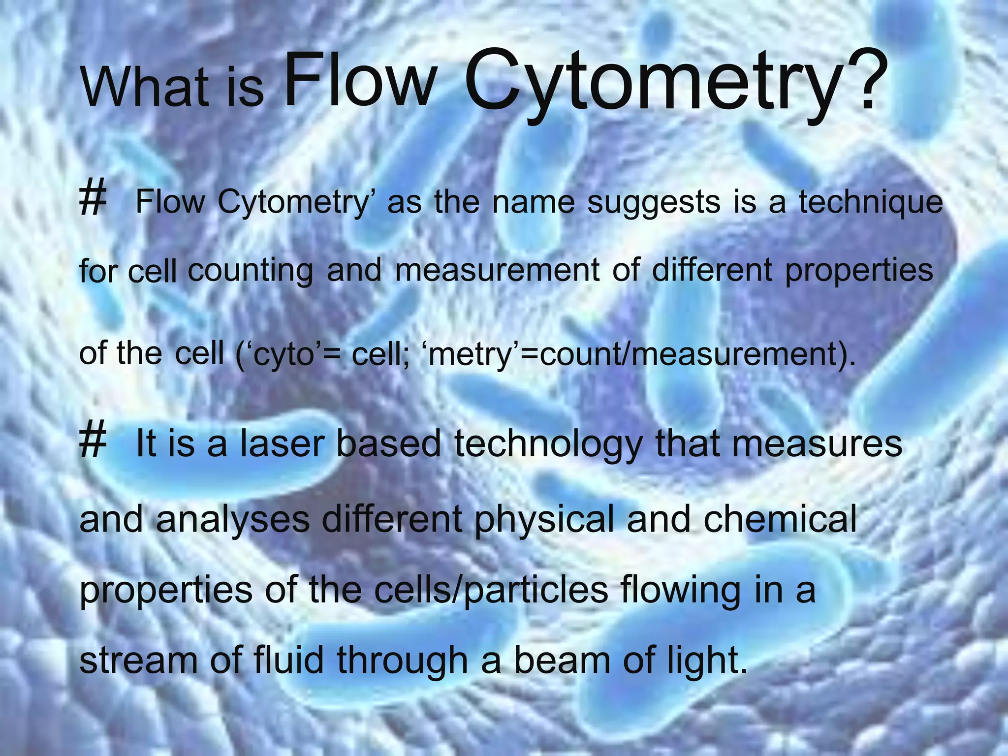
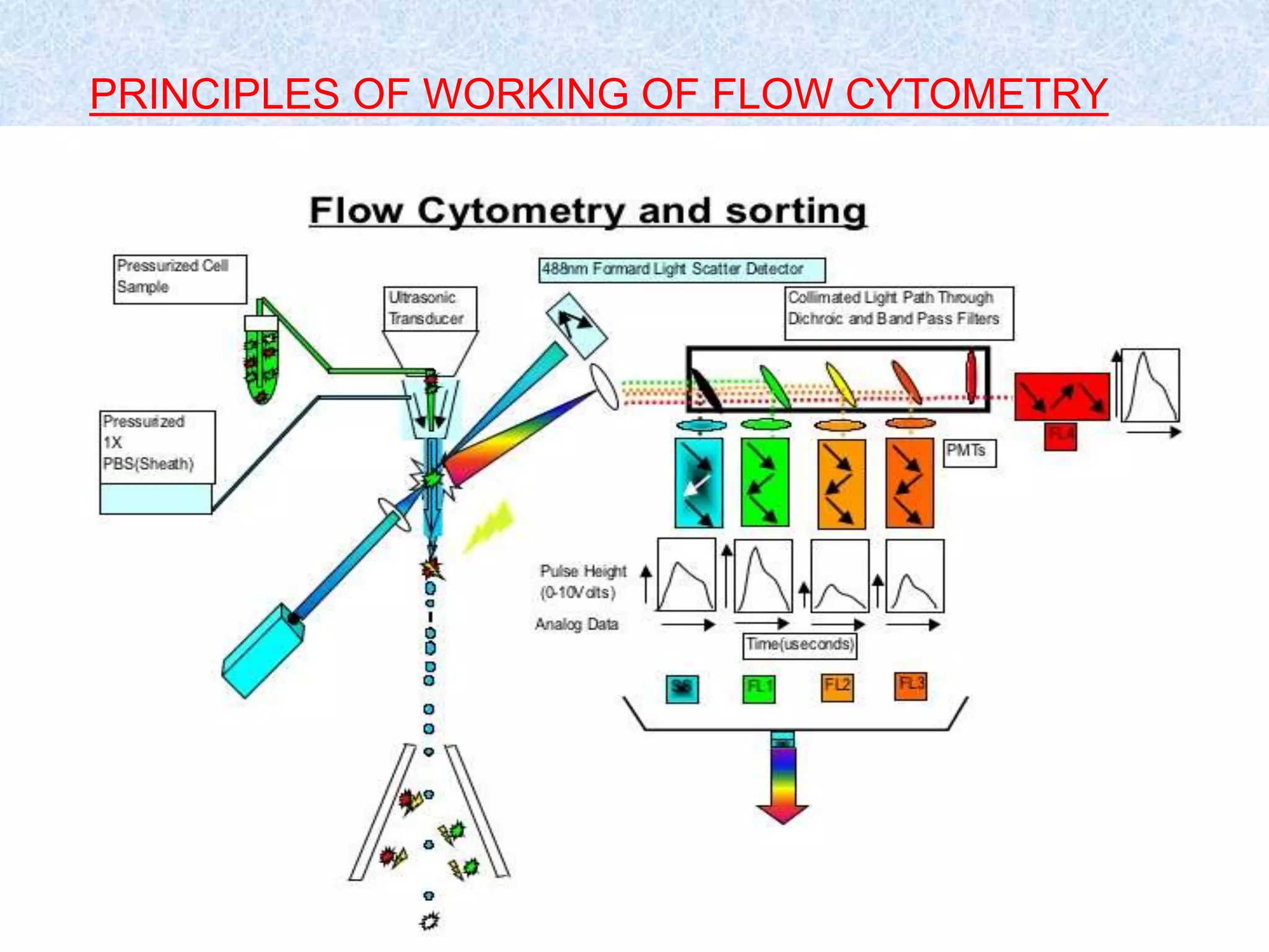
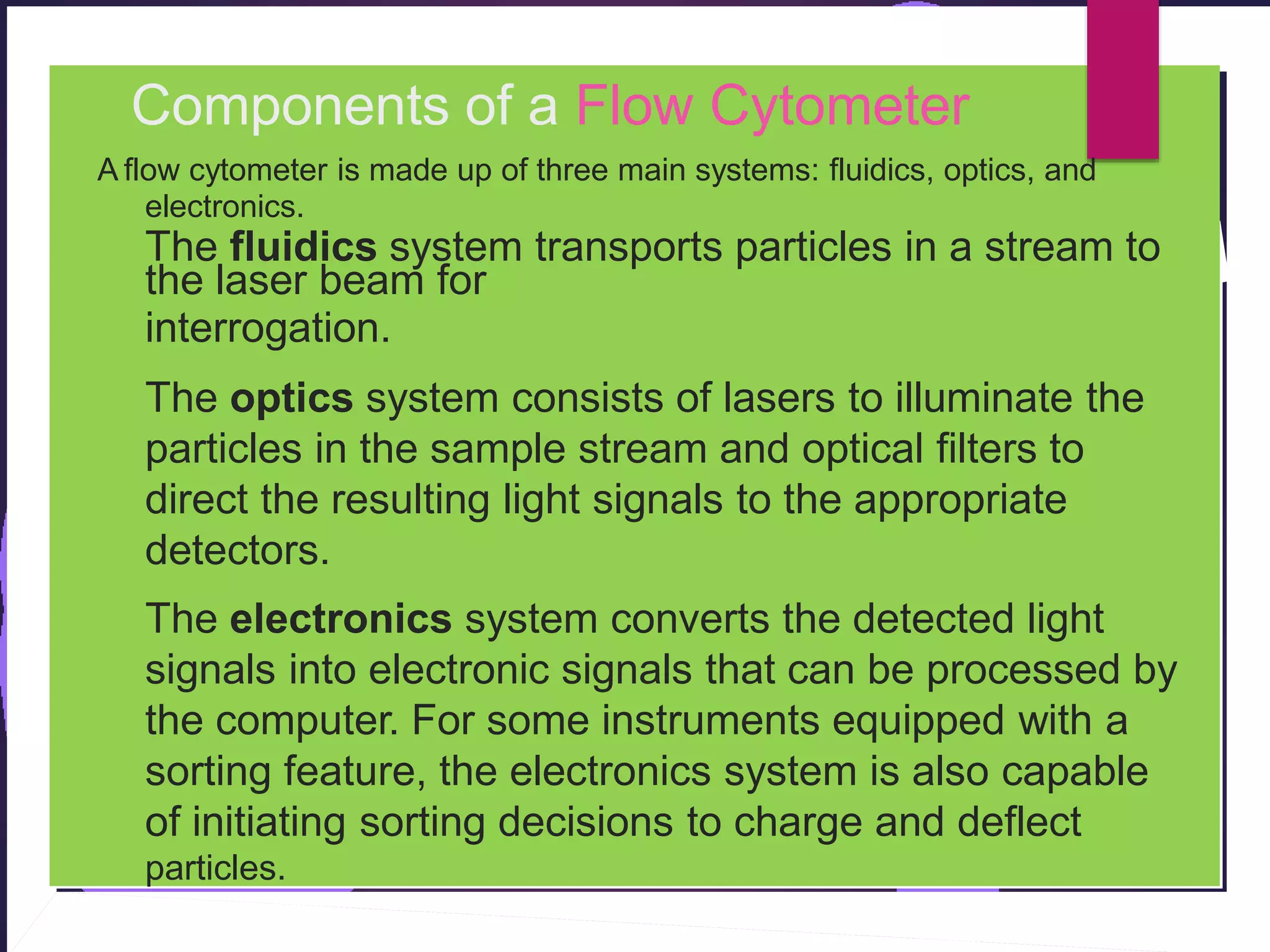
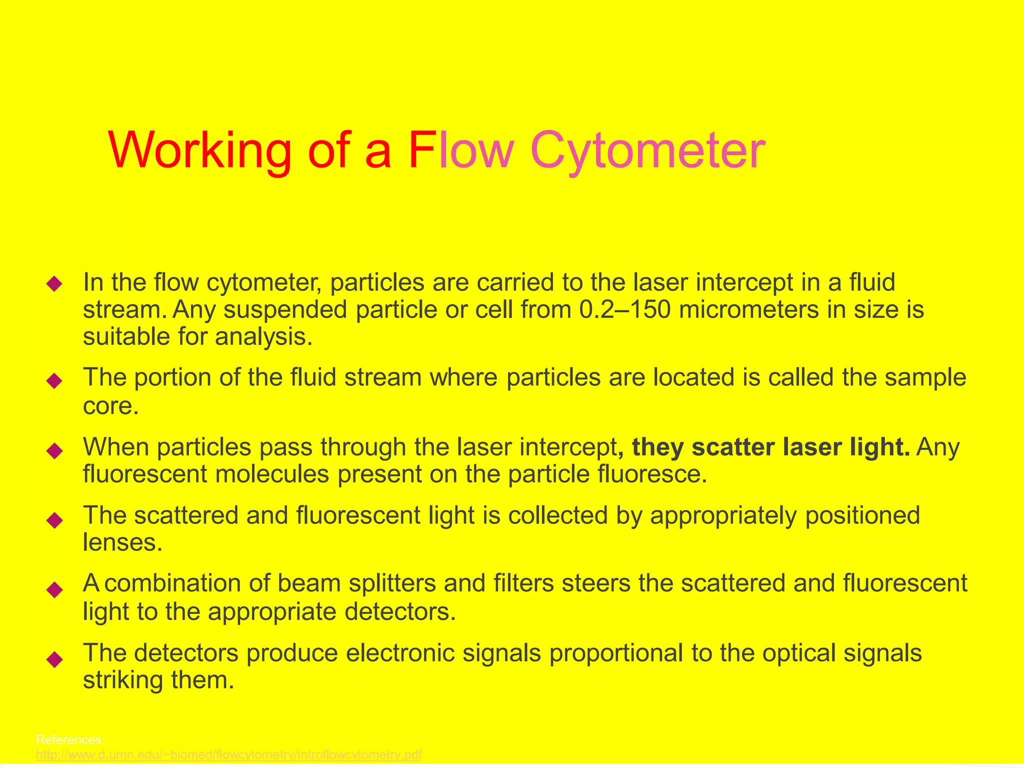
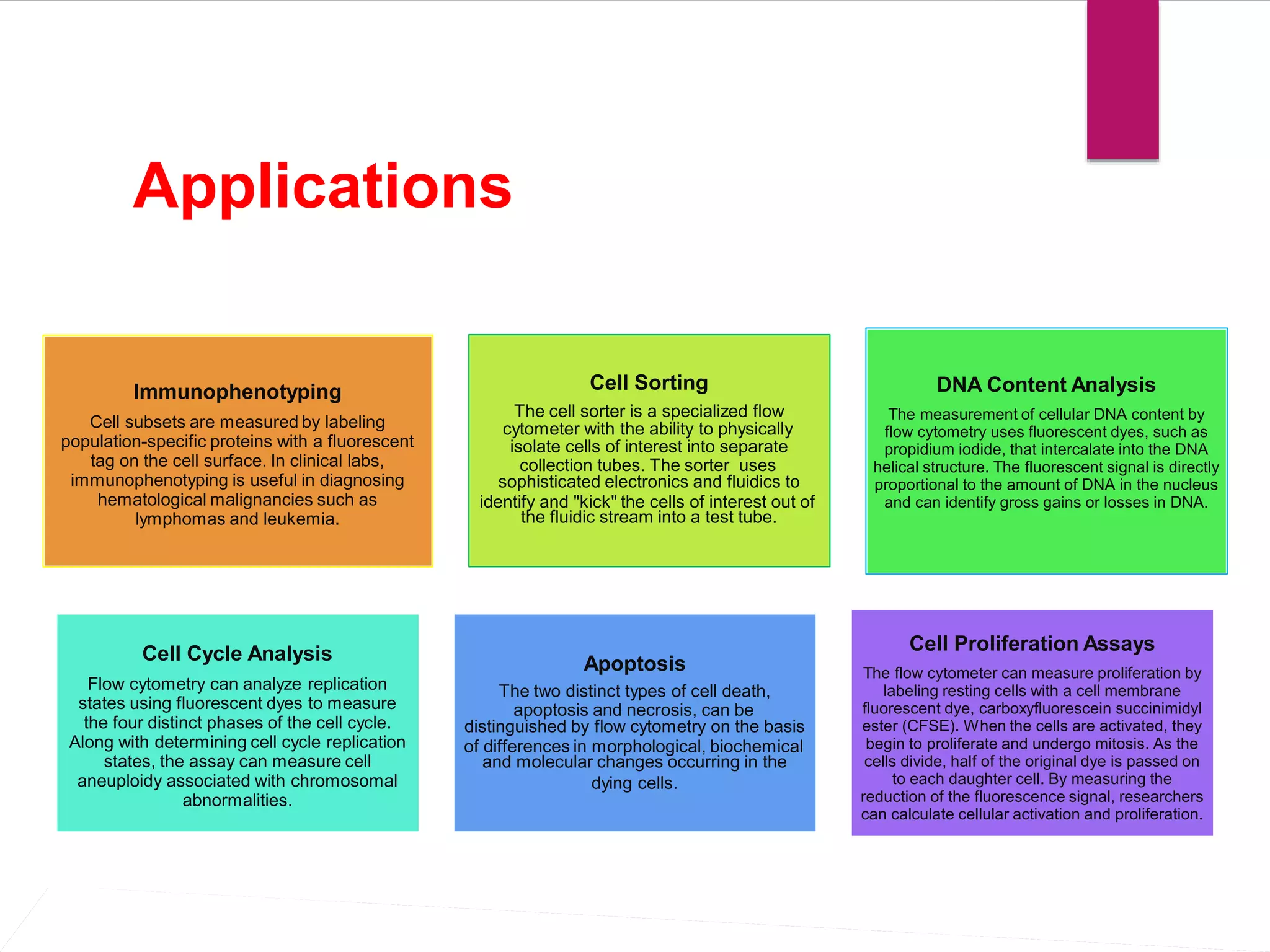
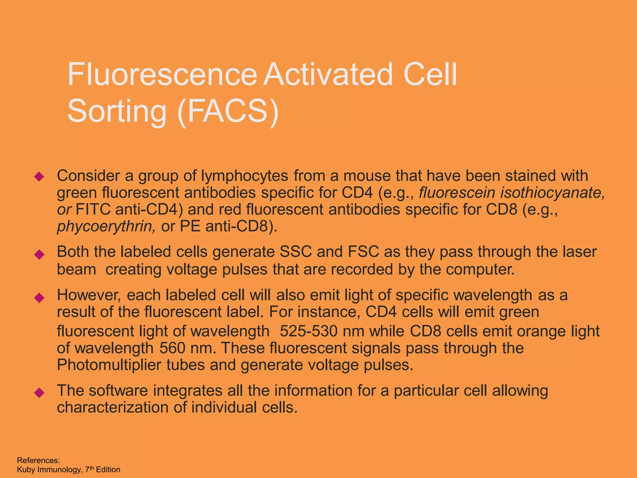
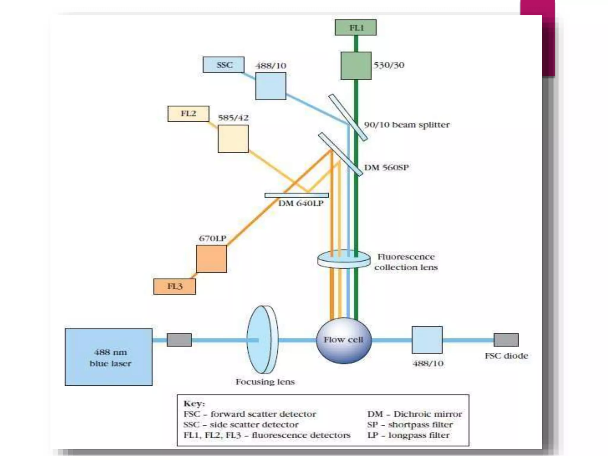
2. What is Flow Cytometry?
# Flow Cytometry’ as the name suggests is a technique
for cell counting and measurement of different properties
of the cell (‘cyto’= cell; ‘metry’=count/measurement).
# It is a laser based technology that measures
and analyses different physical and chemical
properties of the cells/particles flowing in a
stream of fluid through a beam of light.
4. Components of a Flow Cytometer
A flow cytometer is made up of three main systems: fluidics, optics, and
electronics.
The fluidics system transports particles in a stream to
the laser beam for
interrogation.
The optics system consists of lasers to illuminate the
particles in the sample stream and optical filters to
direct the resulting light signals to the appropriate
detectors.
The electronics system converts the detected light
signals into electronic signals that can be processed by
the computer. For some instruments equipped with a
sorting feature, the electronics system is also capable
of initiating sorting decisions to charge and deflect
particles.
5. Working of a Flow Cytometer
In the flow cytometer, particles are carried to the laser intercept in a fluid
stream. Any suspended particle or cell from 0.2–150 micrometers in size is
suitable for analysis.
The portion of the fluid stream where particles are located is called the sample
core.
When particles pass through the laser intercept, they scatter laser light. Any
fluorescent molecules present on the particle fluoresce.
The scattered and fluorescent light is collected by appropriately positioned
lenses.
A combination of beam splitters and filters steers the scattered and fluorescent
light to the appropriate detectors.
The detectors produce electronic signals proportional to the optical signals
striking them.
References:
http://www.d.umn.edu/~biomed/flowcytometry/introflowcytometry.pdf
6. Applications
cytometer with the ability to physically
collection tubes. The sorter uses
identify and "kick" the cells of interest out of
apoptosis and necrosis, can be
of differences in morphological, biochemical
dying cells.
Apoptosis
The two distinct types of cell death,
distinguished by flow cytometry on the basis
and molecular changes occurring in the
Cell Cycle Analysis
Flow cytometry can analyze replication
states using fluorescent dyes to measure
the four distinct phases of the cell cycle.
Along with determining cell cycle replication
states, the assay can measure cell
aneuploidy associated with chromosomal
abnormalities.
Cell Proliferation Assays
The flow cytometer can measure proliferation by
labeling resting cells with a cell membrane
fluorescent dye, carboxyfluorescein succinimidyl
ester (CFSE). When the cells are activated, they
begin to proliferate and undergo mitosis. As the
cells divide, half of the original dye is passed on
to each daughter cell. By measuring the
reduction of the fluorescence signal, researchers
can calculate cellular activation and proliferation.
Cell Sorting
The cell sorter is a specialized flow
isolate cells of interest into separate
sophisticated electronics and fluidics to
the fluidic stream into a test tube.
Immunophenotyping
Cell subsets are measured by labeling
population-specific proteins with a fluorescent
tag on the cell surface. In clinical labs,
immunophenotyping is useful in diagnosing
hematological malignancies such as
lymphomas and leukemia.
DNA Content Analysis
The measurement of cellular DNA content by
flow cytometry uses fluorescent dyes, such as
propidium iodide, that intercalate into the DNA
helical structure. The fluorescent signal is directly
proportional to the amount of DNA in the nucleus
and can identify gross gains or losses in DNA.
7. Fluorescence Activated Cell
Sorting (FACS)
Consider a group of lymphocytes from a mouse that have been stained with
green fluorescent antibodies specific for CD4 (e.g., fluorescein isothiocyanate,
or FITC anti-CD4) and red fluorescent antibodies specific for CD8 (e.g.,
phycoerythrin, or PE anti-CD8).
Both the labeled cells generate SSC and FSC as they pass through the laser
beam creating voltage pulses that are recorded by the computer.
However, each labeled cell will also emit light of specific wavelength as a
result of the fluorescent label. For instance, CD4 cells will emit green
fluorescent light of wavelength 525-530 nm while CD8 cells emit orange
of wavelength 560 nm. These fluorescent signals pass through the
Photomultiplier tubes and generate voltage pulses.
The software integrates all the information for a particular cell allowing
characterization of individual cells.
light
References:
Kuby Immunology, 7th Edition
8.
9. Clinical Applications:
DNA Content Analysis
Investigators are currently using techniques of DNA flow cytometry to
measure ploidy status (DNA content) and proliferative potential (S phase
fraction) in a wide variety of solid tumors. These measurements have shown
relevance for diagnosis, prognosis, and treatment for patients with cancer.
The measurement of cellular DNA content by flow cytometry uses
fluorescent dyes, such as propidium iodide, that intercalate into the DNA
helical structure. The fluorescent signal is
directly proportional to the amount of DNA in the nucleus and can identify gross
gains or losses in DNA.
Abnormal DNA content, also known as “DNA content aneuploidy”, can be
determined in a tumor cell population. DNA aneuploidy generally is associated
with malignancy; however, certain benign conditions may appear aneuploid.
Cell Cycle Analysis: This technique is based on the premise that cells in G0
or G1 phases of
the cell cycle possess a normal diploid chromosomal, and hence DNA content
(2n) whereas
cells in G2 and just prior to mitosis (M) contain exactly twice this amount (4n).
As DNA is
synthesized during S-phase, cells are found with a DNA content ranging
between 2n and 4n. A
histogram plot of DNA content against cell numbers gives the classical DNA
profile for a
proliferating cell culture.
References:
http://europepmc.org/abstract/med/2645625
http://www.clinchem.org/content/46/8/1221.full
http://www.icms.qmul.ac.uk/flowcytometry/uses/cellcycleanalysis/cellcycle/index.html
10. Flow Cytometry and Ecology
Assessments of diversity, abundance, and activity of water column
microorganisms are fundamental to studies in aquatic microbiology.
Currently, most applications of flow cytometry to environmental samples make
use of various morphological and physiological characteristics of the cells (e.g.,
size and pigment content of photosynthetic organisms).
These criteria generally are not sufficient for identification at the genus or
species level. Staining with DNA-specific fluorochromes offers information
about numbers of bacterial cells but not about their identity.
The combined use of dyes that bind preferentially to G- C or A. T base pairs
has been used to distinguish organisms of different G+C content
References:
Appl.%20Environ.%20Microbiol.-1990-Amann-1919-25.pdf
11. Flow Cytometry and Cancer
Research
The prognosis of patients with cancer is largely determined by the specific histological diagnosis,
tumor mass stage, and host performance status.
Quantitative cytology in the form of flow cytometry has greatly advanced the objective elucidation of
tumor cell heterogeneity by using probes that discriminate tumor and normal cells and assess
differentiate as well as proliferative tumor cell properties.
Both DNA content analysis and FACS can be utilised in cancer research.
Abnormal nuclear DMA content is a conclusive marker of malignancy and is found with increasing
frequency in leukemia (23% among 793 patients), in lymphoma (53% among 360 patients), and in
myeloma (76% among 177 patients), as well as in solid tumors (75% among 3611 patients), for an
overall incidence of 67% in 4941 patients.
Flow cytometric immunophenotyping (FCI) aids in the differentiation of chronic lymphocytic leukemia
(CLL) from mantle cell lymphoma (MCL); however, overlapping phenotypes may occur. CD11c
expression has been reported in up to 90% of CLL cases but has rarely been reported in MCL.
Whether CD11c can be used to exclude MCL has not been directly addressed. FCI reports were
reviewed for 90 MCL cases (44 patients) and 355 CLL/small lymphocytic lymphoma (SLL) cases (158
patients).
References:
http://ajcp.ascpjournals.org/content/134/2/271.full.pdf+html
http://cancerres.aacrjournals.org/content/43/9/3982.full.pdf+html