Flow cytometry allows the measurement of physical characteristics of single cells as they flow through a laser beam. It measures size, granularity, and fluorescence. Applications include immunophenotyping cancers and leukemias, monitoring transplant rejection and HIV, and determining DNA content and proliferation. Recent advances include improved instruments, new antibodies, and assessment of cytoplasmic/nuclear antigens and T-cell clonality.


![Light sources
Lasers are the light source of choice for clinical cytometry.
Alternative sources of excitation are available (mercury arc , halogen lamps)-
Less focused and broader spectrum of excitation –lead to decreased resolution and autoflorecence
when compared to lasers.
Laser used- Argon type-488 nm wavelength.
Florochromes used-
1. Florescin IsoThioCyanate[FITC] and
2. Phycoerythrin[PE]](https://image.slidesharecdn.com/flowcytometryraghuveer-170311081408/85/Flow-Cytometry-12-320.jpg)


![Data Analysis
Flow cytometric data is stored according to a standard format, the Flow Cytometry Standard
(FCS) format, developed by the Society for Analytical Cytology.
A single cell analyzed for four parameters :
FSC[Forward Scattered Light]
SSC[Side Scattered Light],
Fluorescence of Fluorescein isothiocyanate and Phycoerythrin.
Generate 8 bytes of data.](https://image.slidesharecdn.com/flowcytometryraghuveer-170311081408/85/Flow-Cytometry-18-320.jpg)


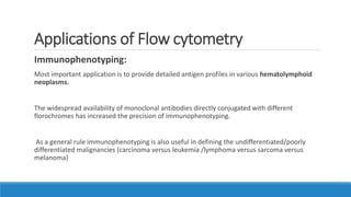
![Chronic lymphoid leukemia
Immunophenotyping by flow cytometry helps in differentiating between:
1. Early stage of lymphoid leukemia and a persistent reactive lymphocytosis
2. Clinically significant subtypes of mature lymphoid leukemias.
B cell clonality: Determined by population expression of single type of surface or cytoplasmic
immunoglobulin light chains ( kappa and lambda)
T cell clonality: Difficult: Currently , there is no reliable analog of kappa and lambda light chain
expression for the T cell receptor protein.
Hence indirect method: Lack of expression of Pan-T cell antigens[CD2,3,4,5,7]](https://image.slidesharecdn.com/flowcytometryraghuveer-170311081408/85/Flow-Cytometry-26-320.jpg)


![FlowBasics2[1]](https://cdn.slidesharecdn.com/ss_thumbnails/7f56678c-0f61-43d6-bbfe-d51ebe159eed-160219222349-thumbnail.jpg?width=640&height=640&fit=bounds)


