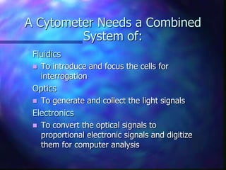
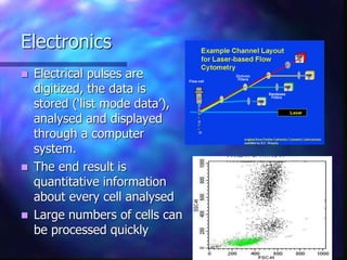
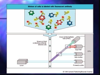
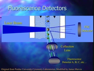
Basics of Flow Cytometry
Dr. Sobia Khalid
What Is Flow Cytometry?
 Flow ~ cells in motion
 Cyto ~ cell
 Metry ~ measure
 Measuring properties of cells while in a fluid
stream
Learning Objectives
 Basic principles of flow cytometry
 Separation of cells on appearance
 Separation based on light emitting dyes
 Studying the cells of interest
A Cytometer Needs a Combined
System of:
Fluidics
 To introduce and focus the cells for
interrogation
Optics
 To generate and collect the light signals
Electronics
 To convert the optical signals to
proportional electronic signals and digitize
them for computer analysis
Basic Principles of Flow Cytometry
 Single cell or particle suspension
 Fluorescent dyes or Abs that can be attached to
an antigen or protein of interest
 Flow cell, sheath fluid and a focused laser beam
The introduction of a large volume into a small volume in such a
way that it becomes “focused” along an axis is called
Hydrodynamic Focusing.
Basic Principles cont’d
 Light is either scattered
or absorbed when it
strikes a cell
 Light scatter is dependent
on the internal structure,
size and shape.
 Forward scatter = size of
the cell
 Side Scatter = complexity
of the cell
Basic Principles cont’d
 Fluorescent dyes
absorb light of a
specific wavelength and
re-emit light of a
different wavelength
 Fluorescent signals are
detected by PMT and
amplified
 Optical filters are used
to steer light of specific
wavelengths to the
photo dector
Reflected
Dichroic
Filter
Passed
Short or Long Pass
Filter
Band Pass Filter
Adsorbed
Absorption
Filter
Electronics
 Electrical pulses are
digitized, the data is
stored (‘list mode data’),
analysed and displayed
through a computer
system.
 The end result is
quantitative information
about every cell analysed
 Large numbers of cells can
be processed quickly
Applications of flow cytometry
 Cell Sorting
 Immunophenotyping (leukaemia/lymphoma)
 Immunemonitoring (T-cell subsets in HIV)
 Leukocyte function assays to determine the
functional status of cells within the immune
system
 DNA Content cell cycle analysis
 Detection of PNH (CD59 and CD55)
 CD34 Enumeration
 Apoptosis
 Foetal haemoglobin
 T-cell subsets analysis
SCOPE OF FLOWCYTOMETRY
 diagnosis and monitoring of therapy for
hematological malignancies
 This scope can be achieved in five different
steps:
1)Assignment of cellular lineage of the malignant
cell
2)Analysis of clonality
3)Analysis of cellular maturation
4)Aberrant features of the malignant cell
populations
5) Detection of minimal residual disease(MRD)
Specimen collection
Sample collection only after appointment with the
Lab
The date and time of specimen collection should be
recorded.
Specimen should be transported to the flow
cytometry laboratory as soon as possible (within
hours).
Information about age,sex,presumptive diagnosis,
differential blood count and status of lymph nodes
and spleen should be provided.
Anticoagulant
EDTA; preferred anticoagulant (2-3ml is enough)
Cells can be analyzed by morphology and automated hematology
analyzers using the same specimen.
Reduced cell aggregation
Heparin - Ficoll density gradient preparations of
mononuclear cells.
Sample storage
Samples storage at room temperature
(18 to 22 C) until staining & analysis is recommended.
Storage at temperatures below 10 C may lead to adsorption of
immunoglobulins to cells and to a selective loss of cells or
antigens.
Flowcytometry procedure :
 Steps:
1. Sample Preparation:
• Dilution with buffer (if required)
• Addition of fluorochrome-conjugated mAbs
• Lyzing
2. Washing
3. Addition of permeable sol. (if required)
4. Cell fixation
Light Scatter
 When light from a laser interrogates a cell, that
cell scatters light in all directions.
 The scattered light can travel from the
interrogation point down a path to a detector.
Forward Scatter
 Light that is scattered in the forward direction (along the
same axis the laser is traveling) is detected in the
Forward Scatter Channel.
 The intensity of this signal has been attributed to cell
size, refractive index (membrane permeability)
 Forward Scatter=FSC=FALS=LALS
Forward Scatter
FSC
Detector
Laser Beam
Original from Purdue University Cytometry Laboratories
Side Scatter
 Laser light that is scattered at 90 degrees to the
axis of the laser path is detected in the Side
Scatter Channel
 The intensity of this signal is proportional to the
amount of cytosolic structure in the cell (eg.
granules, cell inclusions, etc.)
 Side Scatter=SSC=RALS=90 degree Scatter
Side Scatter
FSC
Detector
Collection
Lens
SSC
Detector
Laser Beam
Original from Purdue University Cytometry Laboratories
Why Look at FSC v. SSC
 Since FSC ~ size and SSC ~ internal structure, a
correlated measurement between them can allow
for differentiation of cell types in a heterogenous
cell population
FSC
SSC
Lymphocytes
Monocytes
Granulocytes
RBCs, Debris,
Dead Cells
Forward and Side Scatter
Side Scatter (granularity)
Forward
Scatter
(Size)
Neutrophils
Monocytes
Lymphocytes
Fluorescence Channels
 As the laser interrogates the cell, fluorochromes
on/in the cell (intrinsic or extrinsic) may absorb
some of the light and become excited
 As those fluorochromes leave their excited state,
they release energy in the form of a photon with
a specific wavelength, longer than the excitation
wavelength
 Those photons pass through the collection lens
and are split and steered down specific channels
with the use of filters.
Fluorescence Detectors
FSC
Detector
Collection
Lens
Laser Beam
Fluorescence
Detector A, B, C, etc…
Original from Purdue University Cytometry Laboratories, Modified by James Marvin
Filters
 Many wavelengths of light will be scattered from
a cell, we need a way to split the light into its
specific wavelengths in order to detect them
independently. This is done with filters
 Optical filters are designed such that they absorb
or reflect some wavelengths of light, while
transmitting other.
 3 types of filters
 Long Pass filter
 Short Pass filter
 Band Pass filter
Long Pass Filters
 Transmit all wavelengths greater than specified
wavelength
 Example: 500LP will transmit all wavelengths
greater than 500nm
400nm 500nm 600nm 700nm
Transmittance
Original from Cytomation Training Manual, Modified by James Marvin
Short Pass Filter
 Transmits all wavelengths less than specified
wavelength
 Example: 600SP will transmit all wavelengths
less than 600nm.
400nm 500nm 600nm 700nm
Transmittance
Original from Cytomation Training Manual, Modified by James Marvin
Band Pass Filter
 Transmits a specific band of wavelengths
 Example: 550/20BP Filter will transmit
wavelengths of light between 540nm and
560nm (550/20 = 550+/-10, not 550+/-20)
400nm 500nm 600nm 700nm
Transmittance
Original from Cytomation Training Manual, Modified by James Marvin
Dichroic Filters
 Can be a long pass or short pass filter
 Filter is placed at a 45º angle to the incident light
 Part of the light is reflected at 90º to the incident
light, and part of the light is transmitted and
continues on.
Dichroic
Filter
Detector 1
Detector 2
Detectors
 There are two main types of photo detectors
used in flow cytometry
 Photodiodes
o Used for strong signals, when saturation is
a potential problem (eg. FSC detector)
 Photomultiplier tubes (PMT)
o More sensitive than a Photodiode, a PMT is
used for detecting small amounts of
fluorescence emitted from fluorochromes.
Electronics
 Detectors basically collect photons of light and
convert them to current
 The electronics must process that light signal and
convert the current to a digitized value/# that
the computer can graph
Size and Granularity
Size
 Fluorochromes Are Molecules That Emit
Fluorescence Upon Excitation With Light
 Ex. FITC (Fluorescein Isothiocyanate)
 PE (Phycoerythrin)
 PerCP (Peridinin Chlorophyll Protein)
 APC (Allophycocyanin)
 Some Fluorochromes Are Proteins, Some Are
Small Organic Compounds
 Ex. PE (Phycoerythrin)-Protein
 Ex. FITC (Fluorescein Isothiocyanate)
One Parameter Histogram
 A one-parameter histogram is a graph of cell
count on the y-axis and the measurement
parameter on x-axis.
FITC
E
v
e
n
t
s
Two Parameter Histograms
 A graph
representing
two
measurement
parameters, on
the x- and y-
axes, and cell
count height on
a density
gradient. This is
similar to a
topographical
map.
+/+
+/-
-/-
-/+
Interpretation
 Once the values for each parameter are in a list
mode file, specialized software can graphically
represent it.
 The data can be displayed in 1, 2, or 3
dimensional format
 Common programs include…
 CellQuest
 Flowjo
 WinMDI
 FCS Express
 Kaluza
Gating
 Allow you to view cells of interest
 lets you decide which data to view and which data to
ignore or discard.
 Gating can subsequently be changed when you
analyze your data without any loss of information.
Side Scatter (granularity)
Forward
Scatter
(Size)
Neutrophils
Monocytes
Lymphocytes
 THANKS

Flow Cytometry

  • 1.
    Basics of FlowCytometry Dr. Sobia Khalid
  • 2.
    What Is FlowCytometry?  Flow ~ cells in motion  Cyto ~ cell  Metry ~ measure  Measuring properties of cells while in a fluid stream
  • 4.
    Learning Objectives  Basicprinciples of flow cytometry  Separation of cells on appearance  Separation based on light emitting dyes  Studying the cells of interest
  • 5.
    A Cytometer Needsa Combined System of: Fluidics  To introduce and focus the cells for interrogation Optics  To generate and collect the light signals Electronics  To convert the optical signals to proportional electronic signals and digitize them for computer analysis
  • 6.
    Basic Principles ofFlow Cytometry  Single cell or particle suspension  Fluorescent dyes or Abs that can be attached to an antigen or protein of interest  Flow cell, sheath fluid and a focused laser beam
  • 7.
    The introduction ofa large volume into a small volume in such a way that it becomes “focused” along an axis is called Hydrodynamic Focusing.
  • 8.
    Basic Principles cont’d Light is either scattered or absorbed when it strikes a cell  Light scatter is dependent on the internal structure, size and shape.  Forward scatter = size of the cell  Side Scatter = complexity of the cell
  • 9.
    Basic Principles cont’d Fluorescent dyes absorb light of a specific wavelength and re-emit light of a different wavelength  Fluorescent signals are detected by PMT and amplified  Optical filters are used to steer light of specific wavelengths to the photo dector Reflected Dichroic Filter Passed Short or Long Pass Filter Band Pass Filter Adsorbed Absorption Filter
  • 10.
    Electronics  Electrical pulsesare digitized, the data is stored (‘list mode data’), analysed and displayed through a computer system.  The end result is quantitative information about every cell analysed  Large numbers of cells can be processed quickly
  • 11.
    Applications of flowcytometry  Cell Sorting  Immunophenotyping (leukaemia/lymphoma)  Immunemonitoring (T-cell subsets in HIV)  Leukocyte function assays to determine the functional status of cells within the immune system  DNA Content cell cycle analysis  Detection of PNH (CD59 and CD55)  CD34 Enumeration  Apoptosis  Foetal haemoglobin  T-cell subsets analysis
  • 12.
    SCOPE OF FLOWCYTOMETRY diagnosis and monitoring of therapy for hematological malignancies  This scope can be achieved in five different steps: 1)Assignment of cellular lineage of the malignant cell 2)Analysis of clonality 3)Analysis of cellular maturation 4)Aberrant features of the malignant cell populations 5) Detection of minimal residual disease(MRD)
  • 13.
    Specimen collection Sample collectiononly after appointment with the Lab The date and time of specimen collection should be recorded. Specimen should be transported to the flow cytometry laboratory as soon as possible (within hours). Information about age,sex,presumptive diagnosis, differential blood count and status of lymph nodes and spleen should be provided.
  • 14.
    Anticoagulant EDTA; preferred anticoagulant(2-3ml is enough) Cells can be analyzed by morphology and automated hematology analyzers using the same specimen. Reduced cell aggregation Heparin - Ficoll density gradient preparations of mononuclear cells.
  • 15.
    Sample storage Samples storageat room temperature (18 to 22 C) until staining & analysis is recommended. Storage at temperatures below 10 C may lead to adsorption of immunoglobulins to cells and to a selective loss of cells or antigens.
  • 16.
    Flowcytometry procedure : Steps: 1. Sample Preparation: • Dilution with buffer (if required) • Addition of fluorochrome-conjugated mAbs • Lyzing 2. Washing 3. Addition of permeable sol. (if required) 4. Cell fixation
  • 18.
    Light Scatter  Whenlight from a laser interrogates a cell, that cell scatters light in all directions.  The scattered light can travel from the interrogation point down a path to a detector.
  • 19.
    Forward Scatter  Lightthat is scattered in the forward direction (along the same axis the laser is traveling) is detected in the Forward Scatter Channel.  The intensity of this signal has been attributed to cell size, refractive index (membrane permeability)  Forward Scatter=FSC=FALS=LALS
  • 20.
    Forward Scatter FSC Detector Laser Beam Originalfrom Purdue University Cytometry Laboratories
  • 21.
    Side Scatter  Laserlight that is scattered at 90 degrees to the axis of the laser path is detected in the Side Scatter Channel  The intensity of this signal is proportional to the amount of cytosolic structure in the cell (eg. granules, cell inclusions, etc.)  Side Scatter=SSC=RALS=90 degree Scatter
  • 22.
  • 23.
    Why Look atFSC v. SSC  Since FSC ~ size and SSC ~ internal structure, a correlated measurement between them can allow for differentiation of cell types in a heterogenous cell population FSC SSC Lymphocytes Monocytes Granulocytes RBCs, Debris, Dead Cells
  • 24.
    Forward and SideScatter Side Scatter (granularity) Forward Scatter (Size) Neutrophils Monocytes Lymphocytes
  • 25.
    Fluorescence Channels  Asthe laser interrogates the cell, fluorochromes on/in the cell (intrinsic or extrinsic) may absorb some of the light and become excited  As those fluorochromes leave their excited state, they release energy in the form of a photon with a specific wavelength, longer than the excitation wavelength  Those photons pass through the collection lens and are split and steered down specific channels with the use of filters.
  • 26.
    Fluorescence Detectors FSC Detector Collection Lens Laser Beam Fluorescence DetectorA, B, C, etc… Original from Purdue University Cytometry Laboratories, Modified by James Marvin
  • 27.
    Filters  Many wavelengthsof light will be scattered from a cell, we need a way to split the light into its specific wavelengths in order to detect them independently. This is done with filters  Optical filters are designed such that they absorb or reflect some wavelengths of light, while transmitting other.  3 types of filters  Long Pass filter  Short Pass filter  Band Pass filter
  • 28.
    Long Pass Filters Transmit all wavelengths greater than specified wavelength  Example: 500LP will transmit all wavelengths greater than 500nm 400nm 500nm 600nm 700nm Transmittance Original from Cytomation Training Manual, Modified by James Marvin
  • 29.
    Short Pass Filter Transmits all wavelengths less than specified wavelength  Example: 600SP will transmit all wavelengths less than 600nm. 400nm 500nm 600nm 700nm Transmittance Original from Cytomation Training Manual, Modified by James Marvin
  • 30.
    Band Pass Filter Transmits a specific band of wavelengths  Example: 550/20BP Filter will transmit wavelengths of light between 540nm and 560nm (550/20 = 550+/-10, not 550+/-20) 400nm 500nm 600nm 700nm Transmittance Original from Cytomation Training Manual, Modified by James Marvin
  • 31.
    Dichroic Filters  Canbe a long pass or short pass filter  Filter is placed at a 45º angle to the incident light  Part of the light is reflected at 90º to the incident light, and part of the light is transmitted and continues on. Dichroic Filter Detector 1 Detector 2
  • 32.
    Detectors  There aretwo main types of photo detectors used in flow cytometry  Photodiodes o Used for strong signals, when saturation is a potential problem (eg. FSC detector)  Photomultiplier tubes (PMT) o More sensitive than a Photodiode, a PMT is used for detecting small amounts of fluorescence emitted from fluorochromes.
  • 33.
    Electronics  Detectors basicallycollect photons of light and convert them to current  The electronics must process that light signal and convert the current to a digitized value/# that the computer can graph
  • 34.
  • 35.
     Fluorochromes AreMolecules That Emit Fluorescence Upon Excitation With Light  Ex. FITC (Fluorescein Isothiocyanate)  PE (Phycoerythrin)  PerCP (Peridinin Chlorophyll Protein)  APC (Allophycocyanin)  Some Fluorochromes Are Proteins, Some Are Small Organic Compounds  Ex. PE (Phycoerythrin)-Protein  Ex. FITC (Fluorescein Isothiocyanate)
  • 36.
    One Parameter Histogram A one-parameter histogram is a graph of cell count on the y-axis and the measurement parameter on x-axis. FITC E v e n t s
  • 37.
    Two Parameter Histograms A graph representing two measurement parameters, on the x- and y- axes, and cell count height on a density gradient. This is similar to a topographical map. +/+ +/- -/- -/+
  • 38.
    Interpretation  Once thevalues for each parameter are in a list mode file, specialized software can graphically represent it.  The data can be displayed in 1, 2, or 3 dimensional format  Common programs include…  CellQuest  Flowjo  WinMDI  FCS Express  Kaluza
  • 39.
    Gating  Allow youto view cells of interest  lets you decide which data to view and which data to ignore or discard.  Gating can subsequently be changed when you analyze your data without any loss of information. Side Scatter (granularity) Forward Scatter (Size) Neutrophils Monocytes Lymphocytes
  • 40.