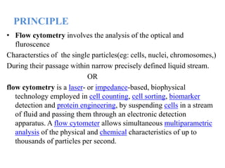
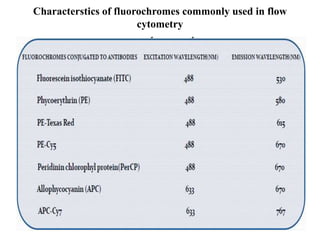
SlideShare a Scribd company logo
BY P.RAJGOPAL
PHARMACEUTICAL ANALYSIS
GITAM INSTITUTE OF PHARMACY
Reg No:121725201007
CONTENTS
• INTRODUCTION
• PRINCIPLE
• CONCEPT OF SCATTRING
• IMMUNO PHENOTYPING ANALYSIS
• ANTIBODY
• FLUOROCHROMES
• PROCESSING OF SPECIMEN FOR ANALYSIS
• APPLICATIONS OF CYTOMETRY
INTRODUCTION
• The concept of flow cytometry has been in existence for more
than five decades.
• Flow cytometric immunophenotyping first appeared in (FCI) first
appeared in clinical laboratories in the 1980s,in the wake of the
AIDS epidemics.
• Intially utilized to assess CD4 T-Cells,the technique was soon
applied to lymphoid and eventually myeloid and neoplasm
• Current flow cytometry have the capability of simultanously
measuring of multiple parameters of individual cells in a cell
suspension.
• Thus, a large number of cell specimens can be processed with a
quick turn around time
• In addition, flow cytometry is also highly sensitive and detect
immunophenotype of cells in a specimen with a thousand of
cells
The parameters analyzed by flowcytometry include
• Physical properties of cells:the size cytoplasmic granularity, and
amount of DNA contents and
• Cell antigens/markers(surface, cytoplasmic and nuclear) that
can be recognized by a specific antibodies.
By using appropriate antibody panel,flow cytometry can
be reveal
• the cell type(hematopoietic,lymphoid,or non hematopoietic)
• Cell linkage(B- and T cells natural killer cells,
myeloid,monocytic cells, neuro/neuro endocrine cells and
epithelial cells)
• Cell maturation stage( precursors vs matured cells).
PRINCIPLE
• Flow cytometry involves the analysis of the optical and
fluroscence
Characterstics of the single particles(eg: cells, nuclei, chromosomes,)
During their passage within narrow precisely defined liquid stream.
OR
flow cytometry is a laser- or impedance-based, biophysical
technology employed in cell counting, cell sorting, biomarker
detection and protein engineering, by suspending cells in a stream
of fluid and passing them through an electronic detection
apparatus. A flow cytometer allows simultaneous multiparametric
analysis of the physical and chemical characteristics of up to
thousands of particles per second.
For cell analysis the basic components of a flowcytometer includes
FLOW SYSTEM
OPTICAL
SYSTEM
ELECTRONIC
SYSTEM
COMPUTER
SYSTEM
Schematic diagram of flow cytometry
CONCEPT OF SCATTERING
• Physical properties, such as size( represented by forward
angle light scatter) and internal complexity( represented by
left- angle scatter) can resolve in certain cell populations.
•
•
•
•
•
• FSC collects light at 180º from the point at the point intersects the
cells, usually on a linear scale it is correlated with the cell size,
and thus can distinguish normal lympocytes (small), monocytes(
intermediate) and neoplastic cells(generally they are larger in size)
• SSC collects right angle light at 90º and is correlated with
cytoplasmic granularity and nuclear configuration
• The combination of both SSCand FSC can distinguish normal
lymphocytes , granulocytes and monocytes.
• The detection of lymphocytes and monocytes provides a reliable
internal control to evalute the size of the cell of interest.
IMMUNOPHENOTYPING ANALYSIS
• Requires
 Antibody
 Fluorochromes
• Antibody:
• Highly specific monoclonal antibodies are used that are
produced by cloned antibody secreting cells
• Antibody are based on the cluster differentiation (CD)- a
protocol used for differentation and distinction of cell surface
antigens.
• Using CD system we can identify cells by the presence or the
absence of particular surface marker for e.g. CD3+ or CD20-
etc...,
FLUOROCHROMES
• Fluorochromes are substances that can be exited by certain light
source(such as laser) and emit a fluoroscent signal at a single
wavelength.
• Fluroscent dyes can directly bind to certain cellular content, such as
DNA and DNA, and allows us to perform quantitative analysis on
individual cells.
• However, in most cases fluorochromes are conjugated with mono
clonal antibodies, which specifically target cellular antigen
/markers
Characterstics of fluorochromes commonly used in flow
cytometry
IMMUNOPHENOTYPING ANAYSIS
• Antibodies conjugated to flouroscent dyes can bind specific on cell
membrance or inside cells. When labelled cells are passed to light
source, the fluoroscent molecules are excited to higher energy state.
Upon returning to their resting states, the flurochromes emits light
light energy at higher wave lengths,the use of multiple
flurochromes,each with similar excitation wavelength and different
emission wavelength (colours), allows several properties to be
measured simulatiously.
Simultaneous detection of multiple cells
antigen/markers
• Multiple cell antigens(Ag)are recognised by flourochromo
conjugated specific antibodies(Ab) Because different
fluorochromes have different emission wavelength/colours, they
can be simultaneously determined detected by flow cytometer.
Abnormal/aberrant antigenic expression can be
grouped into four categories
• Abnormal increased or decreased level of antigenic expression
(aberrant expression)
• Gain of antigen not normally expressed in cell type
• Expression of antigens not synchornized with normal
development and maturation stage of the celltype or linkage .
• Homogenous expression of the antigen(s) by a cell poplation
that normally show more heterogenous expression.
Processing of specimen for flow cytometry
• Specimen suitable for flow cytometry
• Theortically, any specimen from which a single cell suspension can be
generated are suitable for flow cytometry analysis.
• However,a lack of distinct antigen / markers in the cells of the interest or
tissues limit the diagonistic valuesof flow cytometry.
• Common specimen suitables for flow cytometry include:
peripheral blood
bone marrow
body fluids
Cerebrospinal fluid
Urine
Lymph nodes( cell or fresh tissus)
Any fine needle aspiration
Fresh tissue suspension for hematopoietic and lymphoid tissues
SPECIMENSTORAGE
• For blood and bone marrow specimens, anticoagulants such as
EDTA, heparin, or acid citrate dextrose are needed.
Fresh tissue specimens are best transported and stored in sterile
tissue culture medium
Although specimens may be stored at room
temperature,refrigeration is prefered, particularly when there is
delay for flow cytometric analysis.
For flow cytometric analysis , single cell suspensions of the fresh
tissues can be acheived by mechanical dissolution.
General notes on cell preparation
• Single cell suspension are required for optimal staining of
samples for flow cytometry.
• The narrow boresof the sample injection needle and tubing on
a flow cytometry will be easily clogged by aggregated cells
and debris.
• Preparation of single cell suspensionfrom solid tissue requires
Mechanical dissociation and / or enzymatic digestion for optimal
Recovery of cells from the tissue.
Processing of solid tissues
• Tissue is weighed and mechanically and enzymatic disaggregated
Into a single cell suspension
• Collengenase is the most commonly used enzyme, followed by
dispase and trypsin
• Enzymatic digestion is performed in an incubator or a shaking
water bath
• Mechanical disaggeration can be accomplished with paired
scalpels or scissors.
• The process often requirescentrifugation , harvest of single cells
and redigestion of tissues fragments.
• The sample should be visually inspected at all phases of tissue
digestion
• In the final stage ,cell suspensions are passed through a 70 to 200
micron filter to remove aggregates
• Cell suspensions are then counted and viability is determined by a
dye exclusion assay such as trypan blue
• Sample is finally incubated with requires antibodies attached with
Fluorochrome in optimal temperature and pH and analysed by
flowcytometer
Application of flow cytometry
DNA CONTENT Analysis
• The measurement of cellular DNA contents by flow cytometry
uses fluoroscent dyes such as propidium iodide,that intercalate
into DNA helical structure
• The fluoroscent signals is directly proportional to the amount of
DNA in the nucleus and can identify gross gains or losses in the
DNA
• Abnormal DNA content, also known as DNA content aneuploidy
can be determined in a tumor cell population.
• DNA aneuploidy generally is associated with malignancy
Erythrocyte analysis
• Detection and quantification of fetal red cells in maternal blood.
The use of flow cytometry for the detection of fetal cells is much
more objective reproducible , and sensitive than the kleihauer-
betke test.
Diagnosis of PNH
• Conventional laboratory tests for the diagnosis of PNH
Include the sugar water test and Ham’s acid hemolysis test.
• Antibodies to CD55 and CD59 are specific for decay-accelerating
factor and membrance- inhibitor of reactive lysis,respectively,can
be analyzed by flow cytometry to make a definitive diagnosis of
PNH
Reticulate analysis
• Reticulate count are based on identification of residual ribosomes and
RNA in immuture non nucleated red blood cells by using supravital stain.
The flow cytometric enumeration of reticulocytes uses fluoroscent dyes
that bind the residual RNA, such as thiazole orange.
• A region has been drawn on the red blood cells in the scatter plot. The
other major cluster in the scatter plot are the platlets .
• The histogram was gated on the red blood cells and the region on it
delinate cells with high(H), medium(M) and low (L) flouresence
corresponding to increasing reticulocyte maturity.N marks nucleated red
cells
• In the blood bank, the flow cytometry can be used as a complementary
or replacement test for red cell immunology,including RBC- bound
immunoglobilns and red cell antigens.Flow has been used to accurately
identify and phenotype the recepients’s red cells.
• Flow cytometry is being used increasingly in the blood bank to assess
leukocyte contamination in leukocyte- reduced blood products.
Leukocyte analysis:
Perhaps the best example of simultanous analysis of multiple characterstics
by flow cytometry invloves the immonophenotyping
Of leukemias and lymphomas
Immunophenotyping by flow ctyometry (FCM) is an essential aid for
accurately diagnosing and prognosticating leukamia and lymphoma.
• WHO classification has divided non-Hodgkin lymphoma into B-cell and
T/NK cell subtypes, whichare further subclassified into precursor and
peripheral lymhomas.
• The ability to analyze multiple cellular characteristics,along with new
antibodies and gating strategies, has substantially enhanced the utility
of flow cytometry inthe diagnosis of leukemias and lymphomas.
• Different leukemias and lymphomas often have subtle differences in
their antigen profiles that make them ideal for analysis by flow
cytometry.
Diagnosis of B-Cell Lymphomas by using
specific antibodies in flow cytometer
Identification of lineage specific antigens for
perfect
diagnosis
• Immunologic monitoring of HIV-infectedpatients is a
mainstay of the clinical flow cytometry
• and provides the best possible way for enumeration of CD4+ T
lymphocytes and HIV viral load.
• Flow cytometry can be used for lymphomaphenotyping of
fine needle aspirates, and is apowerful adjunct to cytologic
diagnosis.
• Neutropenia may be immune or nonimmune innature.
Immune neutropenia may result fromgranulocytespecific
autoantibodies, granulocytespecificalloantibodies, or
transfusion-related anti-HLA antibodies. Flow cytometry can
readily identifyanti-neutrophil antibodies that are either bound
togranulocytes or free in plasma and confirm the originof
neutropenia, possibly eliminating the need for abone marrow
procedure.
• Functional deficiencies of leukocytes can beassessed by flow
cytometry. Assays for oxidative burst,phagocytosis,
opsonization, adhesion, and structureare available.
• One of the clinical example is LAD type I is caused bya genetic
deficiency of β2 -integrins, which areheterodimers of CD11 and
CD18. This deficiency leadsto a loss of neutrophil and monocyte
migration.
• The high sensitivity and capacity for simultaneousanalysis of
multiple characteristics make flowcytometry useful for the detection
of minimalresidual disease, especially if abnormal patterns
ofantigen expression are present.
Platelet analysis
• Flow cytometry is an excellent method for directanalysis of
platelet-bound antibodies, and it hasalso been shown to be of
benefit in detection of freeplasma antibodies in ITP.
• The reticulated platelet count can be quantified by flow
cytometry in order to assess the rate of thrombopoiesis. This
measurement can separateunexplained thrombocytopenias into
those withincreased destruction and those with defects in platelet
production.
• The pathogenesis and molecular defects of manyprimary
thrombocytopathies are well known andrelate to defects in
structural or functional glycoproteins,such as the abnormal
expression of gpIIb/IIIa inGlanzmann thrombasthenia and gpIb in
Bernard-Soulier disease.
• Flow cytometry is a rapid and useful method ofobtaining a
diagnosis.
Other applications
• Flow cytometry is indicated in the evaluation of serous
effusionsand CSF, including aqueous or vitreous
humor of patientswith a history of hematolymphoid
neoplasia.
• Flow cytometry assists in the differential diagnosis
betweenplasma cell myeloma and monoclonal
gammopathies ofundetermined significance by
determining the percentage ofaberrant or clonal plasma
cells of all bone marrow plasma cells.
• Flow cytometry is useful in diagnostic evaluation of
unexplainedmarrow plasmacytosis by assessing
phenotypically aberrant orclonal plasma cells and its
ability to detect other underlyingmonoclonal B-cell
process.
• Tissue-based lymphoid neoplasias commonly affect lymph
nodes, spleen, mucosa-associated lymphoid tissue, skin, or
nonlymphoid solid organs resulting inmasses or organomegaly.
• Flow cytometry is extremely useful in the diagnosis and
subclassification of tissue-based lymphoid
neoplasias,,organomegaly and tissue infiltrates
THANK YOU

More Related Content

What's hot

Flow Cytometry
Flow CytometryFlow Cytometry
Flow Cytometry
Dr. Sobia Khalid
 
Flow Cytometry - basics, principles and applications
Flow Cytometry - basics, principles and applicationsFlow Cytometry - basics, principles and applications
Flow Cytometry - basics, principles and applications
Ankit Raiyani
 
Flow Cytometry (FCM)
Flow Cytometry (FCM) Flow Cytometry (FCM)
Flow Cytometry (FCM)
Dr. Amer Ali Khaleel /HMU
 
Flowcytometry by asif
Flowcytometry by asifFlowcytometry by asif
Flowcytometry by asif
Asif Zeb
 
Flow Cytometry- Presentation
Flow Cytometry- PresentationFlow Cytometry- Presentation
Flow Cytometry- Presentation
BIDISHA MANDAL
 
flow cytometry presentation
flow cytometry  presentationflow cytometry  presentation
flow cytometry presentation
Jamile Saberzade
 
Flow cytometry
Flow cytometryFlow cytometry
Flow cytometry
Pradeep Singh Narwat
 
Kumc introduction to flow cytometry
Kumc introduction to flow cytometryKumc introduction to flow cytometry
Kumc introduction to flow cytometry
Richard Hastings
 
Tissue microarray
Tissue microarrayTissue microarray
Tissue microarray
MAHMOUD IBRAHIM
 
Flow cytometry
Flow cytometry Flow cytometry
Flow cytometry
RatnakumariP
 
FlowCytometry Basics
FlowCytometry BasicsFlowCytometry Basics
FlowCytometry BasicsAnna Öberg
 
Automated cell counter & its quality control
Automated cell counter & its quality controlAutomated cell counter & its quality control
Automated cell counter & its quality control
Saikat Mandal
 
Presentation on flow cytometry1
Presentation on flow cytometry1Presentation on flow cytometry1
Presentation on flow cytometry1
Nagendra sharma
 
Flow cytometry
Flow cytometryFlow cytometry
Flow cytometry
tashagarwal
 
HPLC in Pathology
HPLC in PathologyHPLC in Pathology
HPLC in Pathology
Aseem Jain
 
Automation in hematology
Automation in hematologyAutomation in hematology
Automation in hematology
Dr Siddartha
 
Flow cytometry
Flow cytometryFlow cytometry
Flow cytometry
RajanPaudel9
 
Flow cytometry
Flow cytometryFlow cytometry
Flow cytometry
Sanjeev Kumar
 
Cell block and its utility
Cell block and its utilityCell block and its utility
Cell block and its utility
Dr Prakriti Shukla
 
Flow cytometry
Flow cytometryFlow cytometry
Flow cytometry
Anushi Jain
 

What's hot (20)

Flow Cytometry
Flow CytometryFlow Cytometry
Flow Cytometry
 
Flow Cytometry - basics, principles and applications
Flow Cytometry - basics, principles and applicationsFlow Cytometry - basics, principles and applications
Flow Cytometry - basics, principles and applications
 
Flow Cytometry (FCM)
Flow Cytometry (FCM) Flow Cytometry (FCM)
Flow Cytometry (FCM)
 
Flowcytometry by asif
Flowcytometry by asifFlowcytometry by asif
Flowcytometry by asif
 
Flow Cytometry- Presentation
Flow Cytometry- PresentationFlow Cytometry- Presentation
Flow Cytometry- Presentation
 
flow cytometry presentation
flow cytometry  presentationflow cytometry  presentation
flow cytometry presentation
 
Flow cytometry
Flow cytometryFlow cytometry
Flow cytometry
 
Kumc introduction to flow cytometry
Kumc introduction to flow cytometryKumc introduction to flow cytometry
Kumc introduction to flow cytometry
 
Tissue microarray
Tissue microarrayTissue microarray
Tissue microarray
 
Flow cytometry
Flow cytometry Flow cytometry
Flow cytometry
 
FlowCytometry Basics
FlowCytometry BasicsFlowCytometry Basics
FlowCytometry Basics
 
Automated cell counter & its quality control
Automated cell counter & its quality controlAutomated cell counter & its quality control
Automated cell counter & its quality control
 
Presentation on flow cytometry1
Presentation on flow cytometry1Presentation on flow cytometry1
Presentation on flow cytometry1
 
Flow cytometry
Flow cytometryFlow cytometry
Flow cytometry
 
HPLC in Pathology
HPLC in PathologyHPLC in Pathology
HPLC in Pathology
 
Automation in hematology
Automation in hematologyAutomation in hematology
Automation in hematology
 
Flow cytometry
Flow cytometryFlow cytometry
Flow cytometry
 
Flow cytometry
Flow cytometryFlow cytometry
Flow cytometry
 
Cell block and its utility
Cell block and its utilityCell block and its utility
Cell block and its utility
 
Flow cytometry
Flow cytometryFlow cytometry
Flow cytometry
 

Similar to Flow cytometry for cell componenet analysis

FLOW CYTOMETRY -SUNANDA ARYA.pptx
FLOW CYTOMETRY -SUNANDA ARYA.pptxFLOW CYTOMETRY -SUNANDA ARYA.pptx
FLOW CYTOMETRY -SUNANDA ARYA.pptx
SunandaArya
 
Flow cytometry
Flow cytometryFlow cytometry
Flow cytometry
Sreenivasa Reddy Thalla
 
flow cytometry.pptx
flow cytometry.pptxflow cytometry.pptx
flow cytometry.pptx
jasveer15
 
Immunoflowcytometry, the basics and applications.pptx
Immunoflowcytometry, the basics and applications.pptxImmunoflowcytometry, the basics and applications.pptx
Immunoflowcytometry, the basics and applications.pptx
AmirRaziq1
 
Applications of Flow Cytometry | Cell Analysis
Applications of Flow Cytometry | Cell AnalysisApplications of Flow Cytometry | Cell Analysis
Applications of Flow Cytometry | Cell Analysis
University of The Punjab
 
Flow cytometry in cell biology
Flow cytometry in cell biologyFlow cytometry in cell biology
Flow cytometry in cell biology
Amani Riyadh
 
Monoclonal antibodies dr. asm
Monoclonal antibodies dr. asmMonoclonal antibodies dr. asm
Monoclonal antibodies dr. asm
SNJBs SSDJ College of Pharmacy, Chandwad
 
Laser scanning cytometry and liquid based cytology
Laser scanning cytometry and liquid based cytologyLaser scanning cytometry and liquid based cytology
Laser scanning cytometry and liquid based cytologyanaonline
 
MOLECULAR AND CYTOGENETIC ANALYSIS -BMLS GENERAL &HBT-1.pptx
MOLECULAR AND CYTOGENETIC ANALYSIS -BMLS GENERAL &HBT-1.pptxMOLECULAR AND CYTOGENETIC ANALYSIS -BMLS GENERAL &HBT-1.pptx
MOLECULAR AND CYTOGENETIC ANALYSIS -BMLS GENERAL &HBT-1.pptx
AmosiRichard
 
Prep and Modern Bio Techniques
Prep and Modern Bio TechniquesPrep and Modern Bio Techniques
Prep and Modern Bio Techniquesihmcbiology1213
 
Laser scanning cytometry and liquid based cytology
Laser scanning cytometry and liquid based cytologyLaser scanning cytometry and liquid based cytology
Laser scanning cytometry and liquid based cytologyanaonline
 
Monoclonal antibodies production(1)
Monoclonal antibodies production(1)Monoclonal antibodies production(1)
Monoclonal antibodies production(1)
Yenda Manishankar
 
Flow cytometry
Flow cytometryFlow cytometry
Flow cytometry
Saranya Ganesh
 
Immunocompetence tests
Immunocompetence testsImmunocompetence tests
Immunocompetence tests
Malathi Murugesan
 
CdC CROSSMATCH(CDCXM)
CdC CROSSMATCH(CDCXM)CdC CROSSMATCH(CDCXM)
Advanced techniques in animal cell culture
Advanced techniques in animal cell cultureAdvanced techniques in animal cell culture
Advanced techniques in animal cell culture
Rajpal Choudhary
 
CHARACTERISATION OF CELLS LINES
CHARACTERISATION OF CELLS LINESCHARACTERISATION OF CELLS LINES
CHARACTERISATION OF CELLS LINES
AishwaryaDande1
 
Biology and characterization of the cell culture
Biology and characterization of the cell cultureBiology and characterization of the cell culture
Biology and characterization of the cell culture
KAUSHAL SAHU
 
Virus detection identification
Virus detection identificationVirus detection identification
Virus detection identification
vivekbps
 
Flow cytometry
Flow cytometryFlow cytometry
Flow cytometry
Noorahmad Shaik
 

Similar to Flow cytometry for cell componenet analysis (20)

FLOW CYTOMETRY -SUNANDA ARYA.pptx
FLOW CYTOMETRY -SUNANDA ARYA.pptxFLOW CYTOMETRY -SUNANDA ARYA.pptx
FLOW CYTOMETRY -SUNANDA ARYA.pptx
 
Flow cytometry
Flow cytometryFlow cytometry
Flow cytometry
 
flow cytometry.pptx
flow cytometry.pptxflow cytometry.pptx
flow cytometry.pptx
 
Immunoflowcytometry, the basics and applications.pptx
Immunoflowcytometry, the basics and applications.pptxImmunoflowcytometry, the basics and applications.pptx
Immunoflowcytometry, the basics and applications.pptx
 
Applications of Flow Cytometry | Cell Analysis
Applications of Flow Cytometry | Cell AnalysisApplications of Flow Cytometry | Cell Analysis
Applications of Flow Cytometry | Cell Analysis
 
Flow cytometry in cell biology
Flow cytometry in cell biologyFlow cytometry in cell biology
Flow cytometry in cell biology
 
Monoclonal antibodies dr. asm
Monoclonal antibodies dr. asmMonoclonal antibodies dr. asm
Monoclonal antibodies dr. asm
 
Laser scanning cytometry and liquid based cytology
Laser scanning cytometry and liquid based cytologyLaser scanning cytometry and liquid based cytology
Laser scanning cytometry and liquid based cytology
 
MOLECULAR AND CYTOGENETIC ANALYSIS -BMLS GENERAL &HBT-1.pptx
MOLECULAR AND CYTOGENETIC ANALYSIS -BMLS GENERAL &HBT-1.pptxMOLECULAR AND CYTOGENETIC ANALYSIS -BMLS GENERAL &HBT-1.pptx
MOLECULAR AND CYTOGENETIC ANALYSIS -BMLS GENERAL &HBT-1.pptx
 
Prep and Modern Bio Techniques
Prep and Modern Bio TechniquesPrep and Modern Bio Techniques
Prep and Modern Bio Techniques
 
Laser scanning cytometry and liquid based cytology
Laser scanning cytometry and liquid based cytologyLaser scanning cytometry and liquid based cytology
Laser scanning cytometry and liquid based cytology
 
Monoclonal antibodies production(1)
Monoclonal antibodies production(1)Monoclonal antibodies production(1)
Monoclonal antibodies production(1)
 
Flow cytometry
Flow cytometryFlow cytometry
Flow cytometry
 
Immunocompetence tests
Immunocompetence testsImmunocompetence tests
Immunocompetence tests
 
CdC CROSSMATCH(CDCXM)
CdC CROSSMATCH(CDCXM)CdC CROSSMATCH(CDCXM)
CdC CROSSMATCH(CDCXM)
 
Advanced techniques in animal cell culture
Advanced techniques in animal cell cultureAdvanced techniques in animal cell culture
Advanced techniques in animal cell culture
 
CHARACTERISATION OF CELLS LINES
CHARACTERISATION OF CELLS LINESCHARACTERISATION OF CELLS LINES
CHARACTERISATION OF CELLS LINES
 
Biology and characterization of the cell culture
Biology and characterization of the cell cultureBiology and characterization of the cell culture
Biology and characterization of the cell culture
 
Virus detection identification
Virus detection identificationVirus detection identification
Virus detection identification
 
Flow cytometry
Flow cytometryFlow cytometry
Flow cytometry
 

Recently uploaded

TEST BANK for Operations Management, 14th Edition by William J. Stevenson, Ve...
TEST BANK for Operations Management, 14th Edition by William J. Stevenson, Ve...TEST BANK for Operations Management, 14th Edition by William J. Stevenson, Ve...
TEST BANK for Operations Management, 14th Edition by William J. Stevenson, Ve...
kevinkariuki227
 
Report Back from SGO 2024: What’s the Latest in Cervical Cancer?
Report Back from SGO 2024: What’s the Latest in Cervical Cancer?Report Back from SGO 2024: What’s the Latest in Cervical Cancer?
Report Back from SGO 2024: What’s the Latest in Cervical Cancer?
bkling
 
24 Upakrama.pptx class ppt useful in all
24 Upakrama.pptx class ppt useful in all24 Upakrama.pptx class ppt useful in all
24 Upakrama.pptx class ppt useful in all
DrSathishMS1
 
basicmodesofventilation2022-220313203758.pdf
basicmodesofventilation2022-220313203758.pdfbasicmodesofventilation2022-220313203758.pdf
basicmodesofventilation2022-220313203758.pdf
aljamhori teaching hospital
 
POST OPERATIVE OLIGURIA and its management
POST OPERATIVE OLIGURIA and its managementPOST OPERATIVE OLIGURIA and its management
POST OPERATIVE OLIGURIA and its management
touseefaziz1
 
For Better Surat #ℂall #Girl Service ❤85270-49040❤ Surat #ℂall #Girls
For Better Surat #ℂall #Girl Service ❤85270-49040❤ Surat #ℂall #GirlsFor Better Surat #ℂall #Girl Service ❤85270-49040❤ Surat #ℂall #Girls
For Better Surat #ℂall #Girl Service ❤85270-49040❤ Surat #ℂall #Girls
Savita Shen $i11
 
263778731218 Abortion Clinic /Pills In Harare ,
263778731218 Abortion Clinic /Pills In Harare ,263778731218 Abortion Clinic /Pills In Harare ,
263778731218 Abortion Clinic /Pills In Harare ,
sisternakatoto
 
Superficial & Deep Fascia of the NECK.pptx
Superficial & Deep Fascia of the NECK.pptxSuperficial & Deep Fascia of the NECK.pptx
Superficial & Deep Fascia of the NECK.pptx
Dr. Rabia Inam Gandapore
 
How to Give Better Lectures: Some Tips for Doctors
How to Give Better Lectures: Some Tips for DoctorsHow to Give Better Lectures: Some Tips for Doctors
How to Give Better Lectures: Some Tips for Doctors
LanceCatedral
 
New Drug Discovery and Development .....
New Drug Discovery and Development .....New Drug Discovery and Development .....
New Drug Discovery and Development .....
NEHA GUPTA
 
Tom Selleck Health: A Comprehensive Look at the Iconic Actor’s Wellness Journey
Tom Selleck Health: A Comprehensive Look at the Iconic Actor’s Wellness JourneyTom Selleck Health: A Comprehensive Look at the Iconic Actor’s Wellness Journey
Tom Selleck Health: A Comprehensive Look at the Iconic Actor’s Wellness Journey
greendigital
 
Ozempic: Preoperative Management of Patients on GLP-1 Receptor Agonists
Ozempic: Preoperative Management of Patients on GLP-1 Receptor Agonists  Ozempic: Preoperative Management of Patients on GLP-1 Receptor Agonists
Ozempic: Preoperative Management of Patients on GLP-1 Receptor Agonists
Saeid Safari
 
HOT NEW PRODUCT! BIG SALES FAST SHIPPING NOW FROM CHINA!! EU KU DB BK substit...
HOT NEW PRODUCT! BIG SALES FAST SHIPPING NOW FROM CHINA!! EU KU DB BK substit...HOT NEW PRODUCT! BIG SALES FAST SHIPPING NOW FROM CHINA!! EU KU DB BK substit...
HOT NEW PRODUCT! BIG SALES FAST SHIPPING NOW FROM CHINA!! EU KU DB BK substit...
GL Anaacs
 
ARTIFICIAL INTELLIGENCE IN HEALTHCARE.pdf
ARTIFICIAL INTELLIGENCE IN  HEALTHCARE.pdfARTIFICIAL INTELLIGENCE IN  HEALTHCARE.pdf
ARTIFICIAL INTELLIGENCE IN HEALTHCARE.pdf
Anujkumaranit
 
Pulmonary Thromboembolism - etilogy, types, medical- Surgical and nursing man...
Pulmonary Thromboembolism - etilogy, types, medical- Surgical and nursing man...Pulmonary Thromboembolism - etilogy, types, medical- Surgical and nursing man...
Pulmonary Thromboembolism - etilogy, types, medical- Surgical and nursing man...
VarunMahajani
 
Prix Galien International 2024 Forum Program
Prix Galien International 2024 Forum ProgramPrix Galien International 2024 Forum Program
Prix Galien International 2024 Forum Program
Levi Shapiro
 
ANATOMY AND PHYSIOLOGY OF URINARY SYSTEM.pptx
ANATOMY AND PHYSIOLOGY OF URINARY SYSTEM.pptxANATOMY AND PHYSIOLOGY OF URINARY SYSTEM.pptx
ANATOMY AND PHYSIOLOGY OF URINARY SYSTEM.pptx
Swetaba Besh
 
Charaka Samhita Sutra sthana Chapter 15 Upakalpaniyaadhyaya
Charaka Samhita Sutra sthana Chapter 15 UpakalpaniyaadhyayaCharaka Samhita Sutra sthana Chapter 15 Upakalpaniyaadhyaya
Charaka Samhita Sutra sthana Chapter 15 Upakalpaniyaadhyaya
Dr KHALID B.M
 
Evaluation of antidepressant activity of clitoris ternatea in animals
Evaluation of antidepressant activity of clitoris ternatea in animalsEvaluation of antidepressant activity of clitoris ternatea in animals
Evaluation of antidepressant activity of clitoris ternatea in animals
Shweta
 
Physiology of Chemical Sensation of smell.pdf
Physiology of Chemical Sensation of smell.pdfPhysiology of Chemical Sensation of smell.pdf
Physiology of Chemical Sensation of smell.pdf
MedicoseAcademics
 

Recently uploaded (20)

TEST BANK for Operations Management, 14th Edition by William J. Stevenson, Ve...
TEST BANK for Operations Management, 14th Edition by William J. Stevenson, Ve...TEST BANK for Operations Management, 14th Edition by William J. Stevenson, Ve...
TEST BANK for Operations Management, 14th Edition by William J. Stevenson, Ve...
 
Report Back from SGO 2024: What’s the Latest in Cervical Cancer?
Report Back from SGO 2024: What’s the Latest in Cervical Cancer?Report Back from SGO 2024: What’s the Latest in Cervical Cancer?
Report Back from SGO 2024: What’s the Latest in Cervical Cancer?
 
24 Upakrama.pptx class ppt useful in all
24 Upakrama.pptx class ppt useful in all24 Upakrama.pptx class ppt useful in all
24 Upakrama.pptx class ppt useful in all
 
basicmodesofventilation2022-220313203758.pdf
basicmodesofventilation2022-220313203758.pdfbasicmodesofventilation2022-220313203758.pdf
basicmodesofventilation2022-220313203758.pdf
 
POST OPERATIVE OLIGURIA and its management
POST OPERATIVE OLIGURIA and its managementPOST OPERATIVE OLIGURIA and its management
POST OPERATIVE OLIGURIA and its management
 
For Better Surat #ℂall #Girl Service ❤85270-49040❤ Surat #ℂall #Girls
For Better Surat #ℂall #Girl Service ❤85270-49040❤ Surat #ℂall #GirlsFor Better Surat #ℂall #Girl Service ❤85270-49040❤ Surat #ℂall #Girls
For Better Surat #ℂall #Girl Service ❤85270-49040❤ Surat #ℂall #Girls
 
263778731218 Abortion Clinic /Pills In Harare ,
263778731218 Abortion Clinic /Pills In Harare ,263778731218 Abortion Clinic /Pills In Harare ,
263778731218 Abortion Clinic /Pills In Harare ,
 
Superficial & Deep Fascia of the NECK.pptx
Superficial & Deep Fascia of the NECK.pptxSuperficial & Deep Fascia of the NECK.pptx
Superficial & Deep Fascia of the NECK.pptx
 
How to Give Better Lectures: Some Tips for Doctors
How to Give Better Lectures: Some Tips for DoctorsHow to Give Better Lectures: Some Tips for Doctors
How to Give Better Lectures: Some Tips for Doctors
 
New Drug Discovery and Development .....
New Drug Discovery and Development .....New Drug Discovery and Development .....
New Drug Discovery and Development .....
 
Tom Selleck Health: A Comprehensive Look at the Iconic Actor’s Wellness Journey
Tom Selleck Health: A Comprehensive Look at the Iconic Actor’s Wellness JourneyTom Selleck Health: A Comprehensive Look at the Iconic Actor’s Wellness Journey
Tom Selleck Health: A Comprehensive Look at the Iconic Actor’s Wellness Journey
 
Ozempic: Preoperative Management of Patients on GLP-1 Receptor Agonists
Ozempic: Preoperative Management of Patients on GLP-1 Receptor Agonists  Ozempic: Preoperative Management of Patients on GLP-1 Receptor Agonists
Ozempic: Preoperative Management of Patients on GLP-1 Receptor Agonists
 
HOT NEW PRODUCT! BIG SALES FAST SHIPPING NOW FROM CHINA!! EU KU DB BK substit...
HOT NEW PRODUCT! BIG SALES FAST SHIPPING NOW FROM CHINA!! EU KU DB BK substit...HOT NEW PRODUCT! BIG SALES FAST SHIPPING NOW FROM CHINA!! EU KU DB BK substit...
HOT NEW PRODUCT! BIG SALES FAST SHIPPING NOW FROM CHINA!! EU KU DB BK substit...
 
ARTIFICIAL INTELLIGENCE IN HEALTHCARE.pdf
ARTIFICIAL INTELLIGENCE IN  HEALTHCARE.pdfARTIFICIAL INTELLIGENCE IN  HEALTHCARE.pdf
ARTIFICIAL INTELLIGENCE IN HEALTHCARE.pdf
 
Pulmonary Thromboembolism - etilogy, types, medical- Surgical and nursing man...
Pulmonary Thromboembolism - etilogy, types, medical- Surgical and nursing man...Pulmonary Thromboembolism - etilogy, types, medical- Surgical and nursing man...
Pulmonary Thromboembolism - etilogy, types, medical- Surgical and nursing man...
 
Prix Galien International 2024 Forum Program
Prix Galien International 2024 Forum ProgramPrix Galien International 2024 Forum Program
Prix Galien International 2024 Forum Program
 
ANATOMY AND PHYSIOLOGY OF URINARY SYSTEM.pptx
ANATOMY AND PHYSIOLOGY OF URINARY SYSTEM.pptxANATOMY AND PHYSIOLOGY OF URINARY SYSTEM.pptx
ANATOMY AND PHYSIOLOGY OF URINARY SYSTEM.pptx
 
Charaka Samhita Sutra sthana Chapter 15 Upakalpaniyaadhyaya
Charaka Samhita Sutra sthana Chapter 15 UpakalpaniyaadhyayaCharaka Samhita Sutra sthana Chapter 15 Upakalpaniyaadhyaya
Charaka Samhita Sutra sthana Chapter 15 Upakalpaniyaadhyaya
 
Evaluation of antidepressant activity of clitoris ternatea in animals
Evaluation of antidepressant activity of clitoris ternatea in animalsEvaluation of antidepressant activity of clitoris ternatea in animals
Evaluation of antidepressant activity of clitoris ternatea in animals
 
Physiology of Chemical Sensation of smell.pdf
Physiology of Chemical Sensation of smell.pdfPhysiology of Chemical Sensation of smell.pdf
Physiology of Chemical Sensation of smell.pdf
 

Flow cytometry for cell componenet analysis

  • 1. BY P.RAJGOPAL PHARMACEUTICAL ANALYSIS GITAM INSTITUTE OF PHARMACY Reg No:121725201007
  • 2. CONTENTS • INTRODUCTION • PRINCIPLE • CONCEPT OF SCATTRING • IMMUNO PHENOTYPING ANALYSIS • ANTIBODY • FLUOROCHROMES • PROCESSING OF SPECIMEN FOR ANALYSIS • APPLICATIONS OF CYTOMETRY
  • 3. INTRODUCTION • The concept of flow cytometry has been in existence for more than five decades. • Flow cytometric immunophenotyping first appeared in (FCI) first appeared in clinical laboratories in the 1980s,in the wake of the AIDS epidemics. • Intially utilized to assess CD4 T-Cells,the technique was soon applied to lymphoid and eventually myeloid and neoplasm • Current flow cytometry have the capability of simultanously measuring of multiple parameters of individual cells in a cell suspension. • Thus, a large number of cell specimens can be processed with a quick turn around time
  • 4. • In addition, flow cytometry is also highly sensitive and detect immunophenotype of cells in a specimen with a thousand of cells The parameters analyzed by flowcytometry include • Physical properties of cells:the size cytoplasmic granularity, and amount of DNA contents and • Cell antigens/markers(surface, cytoplasmic and nuclear) that can be recognized by a specific antibodies. By using appropriate antibody panel,flow cytometry can be reveal • the cell type(hematopoietic,lymphoid,or non hematopoietic) • Cell linkage(B- and T cells natural killer cells, myeloid,monocytic cells, neuro/neuro endocrine cells and epithelial cells) • Cell maturation stage( precursors vs matured cells).
  • 5. PRINCIPLE • Flow cytometry involves the analysis of the optical and fluroscence Characterstics of the single particles(eg: cells, nuclei, chromosomes,) During their passage within narrow precisely defined liquid stream. OR flow cytometry is a laser- or impedance-based, biophysical technology employed in cell counting, cell sorting, biomarker detection and protein engineering, by suspending cells in a stream of fluid and passing them through an electronic detection apparatus. A flow cytometer allows simultaneous multiparametric analysis of the physical and chemical characteristics of up to thousands of particles per second.
  • 6. For cell analysis the basic components of a flowcytometer includes FLOW SYSTEM OPTICAL SYSTEM ELECTRONIC SYSTEM COMPUTER SYSTEM
  • 7. Schematic diagram of flow cytometry
  • 8. CONCEPT OF SCATTERING • Physical properties, such as size( represented by forward angle light scatter) and internal complexity( represented by left- angle scatter) can resolve in certain cell populations. • • • • •
  • 9. • FSC collects light at 180º from the point at the point intersects the cells, usually on a linear scale it is correlated with the cell size, and thus can distinguish normal lympocytes (small), monocytes( intermediate) and neoplastic cells(generally they are larger in size) • SSC collects right angle light at 90º and is correlated with cytoplasmic granularity and nuclear configuration • The combination of both SSCand FSC can distinguish normal lymphocytes , granulocytes and monocytes. • The detection of lymphocytes and monocytes provides a reliable internal control to evalute the size of the cell of interest.
  • 10.
  • 11. IMMUNOPHENOTYPING ANALYSIS • Requires  Antibody  Fluorochromes • Antibody: • Highly specific monoclonal antibodies are used that are produced by cloned antibody secreting cells • Antibody are based on the cluster differentiation (CD)- a protocol used for differentation and distinction of cell surface antigens. • Using CD system we can identify cells by the presence or the absence of particular surface marker for e.g. CD3+ or CD20- etc...,
  • 12. FLUOROCHROMES • Fluorochromes are substances that can be exited by certain light source(such as laser) and emit a fluoroscent signal at a single wavelength. • Fluroscent dyes can directly bind to certain cellular content, such as DNA and DNA, and allows us to perform quantitative analysis on individual cells. • However, in most cases fluorochromes are conjugated with mono clonal antibodies, which specifically target cellular antigen /markers
  • 13. Characterstics of fluorochromes commonly used in flow cytometry
  • 14. IMMUNOPHENOTYPING ANAYSIS • Antibodies conjugated to flouroscent dyes can bind specific on cell membrance or inside cells. When labelled cells are passed to light source, the fluoroscent molecules are excited to higher energy state. Upon returning to their resting states, the flurochromes emits light light energy at higher wave lengths,the use of multiple flurochromes,each with similar excitation wavelength and different emission wavelength (colours), allows several properties to be measured simulatiously.
  • 15. Simultaneous detection of multiple cells antigen/markers • Multiple cell antigens(Ag)are recognised by flourochromo conjugated specific antibodies(Ab) Because different fluorochromes have different emission wavelength/colours, they can be simultaneously determined detected by flow cytometer.
  • 16. Abnormal/aberrant antigenic expression can be grouped into four categories • Abnormal increased or decreased level of antigenic expression (aberrant expression) • Gain of antigen not normally expressed in cell type • Expression of antigens not synchornized with normal development and maturation stage of the celltype or linkage . • Homogenous expression of the antigen(s) by a cell poplation that normally show more heterogenous expression.
  • 17.
  • 18. Processing of specimen for flow cytometry • Specimen suitable for flow cytometry • Theortically, any specimen from which a single cell suspension can be generated are suitable for flow cytometry analysis. • However,a lack of distinct antigen / markers in the cells of the interest or tissues limit the diagonistic valuesof flow cytometry. • Common specimen suitables for flow cytometry include: peripheral blood bone marrow body fluids Cerebrospinal fluid Urine Lymph nodes( cell or fresh tissus) Any fine needle aspiration Fresh tissue suspension for hematopoietic and lymphoid tissues
  • 19. SPECIMENSTORAGE • For blood and bone marrow specimens, anticoagulants such as EDTA, heparin, or acid citrate dextrose are needed. Fresh tissue specimens are best transported and stored in sterile tissue culture medium Although specimens may be stored at room temperature,refrigeration is prefered, particularly when there is delay for flow cytometric analysis. For flow cytometric analysis , single cell suspensions of the fresh tissues can be acheived by mechanical dissolution.
  • 20. General notes on cell preparation • Single cell suspension are required for optimal staining of samples for flow cytometry. • The narrow boresof the sample injection needle and tubing on a flow cytometry will be easily clogged by aggregated cells and debris. • Preparation of single cell suspensionfrom solid tissue requires Mechanical dissociation and / or enzymatic digestion for optimal Recovery of cells from the tissue.
  • 21. Processing of solid tissues • Tissue is weighed and mechanically and enzymatic disaggregated Into a single cell suspension • Collengenase is the most commonly used enzyme, followed by dispase and trypsin • Enzymatic digestion is performed in an incubator or a shaking water bath • Mechanical disaggeration can be accomplished with paired scalpels or scissors. • The process often requirescentrifugation , harvest of single cells and redigestion of tissues fragments. • The sample should be visually inspected at all phases of tissue digestion
  • 22. • In the final stage ,cell suspensions are passed through a 70 to 200 micron filter to remove aggregates • Cell suspensions are then counted and viability is determined by a dye exclusion assay such as trypan blue • Sample is finally incubated with requires antibodies attached with Fluorochrome in optimal temperature and pH and analysed by flowcytometer
  • 23. Application of flow cytometry DNA CONTENT Analysis • The measurement of cellular DNA contents by flow cytometry uses fluoroscent dyes such as propidium iodide,that intercalate into DNA helical structure • The fluoroscent signals is directly proportional to the amount of DNA in the nucleus and can identify gross gains or losses in the DNA • Abnormal DNA content, also known as DNA content aneuploidy can be determined in a tumor cell population. • DNA aneuploidy generally is associated with malignancy
  • 24.
  • 25. Erythrocyte analysis • Detection and quantification of fetal red cells in maternal blood. The use of flow cytometry for the detection of fetal cells is much more objective reproducible , and sensitive than the kleihauer- betke test.
  • 26. Diagnosis of PNH • Conventional laboratory tests for the diagnosis of PNH Include the sugar water test and Ham’s acid hemolysis test. • Antibodies to CD55 and CD59 are specific for decay-accelerating factor and membrance- inhibitor of reactive lysis,respectively,can be analyzed by flow cytometry to make a definitive diagnosis of PNH
  • 27. Reticulate analysis • Reticulate count are based on identification of residual ribosomes and RNA in immuture non nucleated red blood cells by using supravital stain. The flow cytometric enumeration of reticulocytes uses fluoroscent dyes that bind the residual RNA, such as thiazole orange. • A region has been drawn on the red blood cells in the scatter plot. The other major cluster in the scatter plot are the platlets . • The histogram was gated on the red blood cells and the region on it delinate cells with high(H), medium(M) and low (L) flouresence corresponding to increasing reticulocyte maturity.N marks nucleated red cells
  • 28. • In the blood bank, the flow cytometry can be used as a complementary or replacement test for red cell immunology,including RBC- bound immunoglobilns and red cell antigens.Flow has been used to accurately identify and phenotype the recepients’s red cells. • Flow cytometry is being used increasingly in the blood bank to assess leukocyte contamination in leukocyte- reduced blood products. Leukocyte analysis: Perhaps the best example of simultanous analysis of multiple characterstics by flow cytometry invloves the immonophenotyping Of leukemias and lymphomas Immunophenotyping by flow ctyometry (FCM) is an essential aid for accurately diagnosing and prognosticating leukamia and lymphoma. • WHO classification has divided non-Hodgkin lymphoma into B-cell and T/NK cell subtypes, whichare further subclassified into precursor and peripheral lymhomas.
  • 29. • The ability to analyze multiple cellular characteristics,along with new antibodies and gating strategies, has substantially enhanced the utility of flow cytometry inthe diagnosis of leukemias and lymphomas. • Different leukemias and lymphomas often have subtle differences in their antigen profiles that make them ideal for analysis by flow cytometry.
  • 30.
  • 31. Diagnosis of B-Cell Lymphomas by using specific antibodies in flow cytometer
  • 32. Identification of lineage specific antigens for perfect diagnosis
  • 33. • Immunologic monitoring of HIV-infectedpatients is a mainstay of the clinical flow cytometry • and provides the best possible way for enumeration of CD4+ T lymphocytes and HIV viral load.
  • 34. • Flow cytometry can be used for lymphomaphenotyping of fine needle aspirates, and is apowerful adjunct to cytologic diagnosis. • Neutropenia may be immune or nonimmune innature. Immune neutropenia may result fromgranulocytespecific autoantibodies, granulocytespecificalloantibodies, or transfusion-related anti-HLA antibodies. Flow cytometry can readily identifyanti-neutrophil antibodies that are either bound togranulocytes or free in plasma and confirm the originof neutropenia, possibly eliminating the need for abone marrow procedure. • Functional deficiencies of leukocytes can beassessed by flow cytometry. Assays for oxidative burst,phagocytosis, opsonization, adhesion, and structureare available.
  • 35. • One of the clinical example is LAD type I is caused bya genetic deficiency of β2 -integrins, which areheterodimers of CD11 and CD18. This deficiency leadsto a loss of neutrophil and monocyte migration.
  • 36. • The high sensitivity and capacity for simultaneousanalysis of multiple characteristics make flowcytometry useful for the detection of minimalresidual disease, especially if abnormal patterns ofantigen expression are present.
  • 37. Platelet analysis • Flow cytometry is an excellent method for directanalysis of platelet-bound antibodies, and it hasalso been shown to be of benefit in detection of freeplasma antibodies in ITP. • The reticulated platelet count can be quantified by flow cytometry in order to assess the rate of thrombopoiesis. This measurement can separateunexplained thrombocytopenias into those withincreased destruction and those with defects in platelet production. • The pathogenesis and molecular defects of manyprimary thrombocytopathies are well known andrelate to defects in structural or functional glycoproteins,such as the abnormal expression of gpIIb/IIIa inGlanzmann thrombasthenia and gpIb in Bernard-Soulier disease.
  • 38. • Flow cytometry is a rapid and useful method ofobtaining a diagnosis.
  • 39. Other applications • Flow cytometry is indicated in the evaluation of serous effusionsand CSF, including aqueous or vitreous humor of patientswith a history of hematolymphoid neoplasia. • Flow cytometry assists in the differential diagnosis betweenplasma cell myeloma and monoclonal gammopathies ofundetermined significance by determining the percentage ofaberrant or clonal plasma cells of all bone marrow plasma cells. • Flow cytometry is useful in diagnostic evaluation of unexplainedmarrow plasmacytosis by assessing phenotypically aberrant orclonal plasma cells and its ability to detect other underlyingmonoclonal B-cell process.
  • 40. • Tissue-based lymphoid neoplasias commonly affect lymph nodes, spleen, mucosa-associated lymphoid tissue, skin, or nonlymphoid solid organs resulting inmasses or organomegaly. • Flow cytometry is extremely useful in the diagnosis and subclassification of tissue-based lymphoid neoplasias,,organomegaly and tissue infiltrates

Editor's Notes

  1. When labelled cells passed