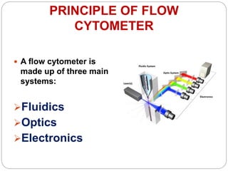
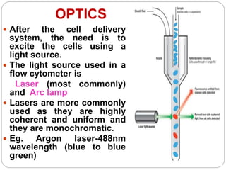
SlideShare a Scribd company logo
Dr. POOSAPATI RATNA KUMARI
PROFESSOR
DEPT OF MICROBIOLOGY
FLOW CYTOMETRY
INTRODUCTION
Flow cytometry is the science of
measuring physical and chemical
properties of live cells or other
biological particles as they pass in
a fluid, single-cell stream through
a measuring apparatus.
 Flow ~ cells in motion
 Cyto ~ cell
 Metry ~ measure
Measuring properties of cells while
in a fluid stream
HISTORY
 16th century - Leeuwenhoek (simple microscope) –
Father of Cytometry
 1742 - Lomonosov - dark field illumination and
performed light scatter measurements
 1934 – Moldavan - photoelectric technique for
counting cells flowing through a capillary tube, and
flow cytometry was born
 1938 - Caspersson built a crude flow cytometer to
measure cell properties in the UV and visible
regions
 1940 - Crosland and Taylor developed a blood cell
counter using the sheath flow principle, light scatter
and darkfield illumination
 1949 - Wallace Coulter patented the first non-
optical electronic blood cell counter
 1957 - The first Model A Coulter Counter was
introduced primarily to count erythrocytes and
leukocytes from blood
 1973 - Crissman and Steinkamp introduced
propidium iodide
 Mid 1970s - Leonard Herzenberg at Stanford
coined the term, Fluorescence Activated Cell
Sorter, or FACS
PURPOSE OF FLOW
CYTOMETRY
 Flow cytometry detects and measures
- a particle’s relative size,
- relative granularity or internal
complexity
- relative fluorescence intensity
 These characteristics are determined using
an optical-to-electronic coupling system that
records how the cell or particle scatters
incident laser light and emits fluorescence.
PRINCIPLE OF FLOW
CYTOMETER
 A flow cytometer is
made up of three main
systems:
Fluidics
Optics
Electronics
 Fluidic system -
transports particles in a
stream to the laser beam
for interrogation.
 Optics system
consists of lasers to
illuminate the particles in
the sample stream and
optical filters to direct the
resulting light signals to
the appropriate detectors.
 Electronics system
converts the detected light
signals into electronic
signals that can be
processed by the
THE FLUIDICS – FLOW
SYSTEM
 One of the fundamentals of flow cytometry is the
ability to measure the properties of individual
particles, which is managed by the fluidics system
 The fluidics system consists of FLOW CELL
(Quartz chamber)
 Central channel / core – through which the sample
is injected
 Outer sheath – contains faster flowing fluid known
as Sheath fluid (0.9% Saline), enclosing the central
core.
 As the sheath fluid moves, it creates a massive drag
effect on the narrowing central chamber.
 Under optimal conditions (laminar flow) the fluid in
the central chamber will not mix with the sheath
fluid.
Incoming Laser
Sample
Sheath Sheath
Sheath
Sample
Sample
Core
Stream
Low Differential High Differential
Laser Focal Point
OPTICS
 After the cell delivery
system, the need is to
excite the cells using a
light source.
 The light source used in a
flow cytometer is
Laser (most commonly)
and Arc lamp
 Lasers are more commonly
used as they are highly
coherent and uniform and
they are monochromatic.
 Eg. Argon laser-488nm
wavelength (blue to blue
green)
 Forward scatter channel (FSC) - equates to the
particle’s size and can also be used to distinguish
between cellular debris and living cells.
 Side scatter channel (SSC) provides information
about the granular content within a particle.
 Both FSC and SSC are unique for every particle, and
a combination of the two may be used to differentiate
different cell types in a heterogeneous sample.
 Neutrophils and
eosinophils produce a
great deal of side scatter
due to their cytoplasmic
granules.
 Large cells such as
monocytes and
neutrophils produce
more forward scatter
than normal RBCs, and
normal lymphocytes
Why Look at FSC v. SSC
 Since FSC ~ size and SSC ~ internal
structure, a correlated measurement
between them can allow for differentiation
of cell types in a heterogeneous cell
population
FSC
SSC
Lymphocytes
Monocytes
Granulocytes
RBCs, Debris,
Dead Cells
FLUORESCENT ABs
- Cells are labeled with fluorescent
antibodies directed against cell
surface molecules
- Using different color
fluorochromes allows counting of
many markers simultaneously and
allows identification of several
markers on the same cell (
Multiparameter Flow)
- In the instrument, cells pass one-
by-one past a laser to excite the
fluorochromes and there are
detectors for each type of
OPTICAL FILTERS
 Once the fluorescence light from a
cell has been captured by the
collection optics, the spectral
component of interest for each stain
must be separated spatially for
detection.
 Optical filters are designed such
that they absorb or reflect a
wavelength of light, while
transmitting other.
 4 types of filters are used in
flow cytometry
 Long Pass filter
 Short Pass filter
 Band Pass filter
 Dichoric filter
OPTICAL DETECTORS
 The two main photo detector types used in flow cytometry
are
 silicon photodiodes - to detect forward scatter
detection
 Photomultiplier tubes (PMTs) - high sensitivity and
are, therefore, assigned to side scatter and fluorescence
detection.
FLUOROCHROMES
 Fluorochromes are
essentially dyes,
which accept light
energy (e.g. from a
laser) at a given
wavelength and re-
emit it at a longer
wavelength.
 These two processes
are called excitation
and emission.
 The process of
emission follows
extremely rapidly,
commonly in the
order of
nanoseconds, and is
GATING
 An important principle of flow cytometry data
analysis is to selectively visualize the cells of
interest while eliminating results from unwanted
particles e.g. dead cells and debris. This procedure
is called GATING.
 Cells have traditionally been gated according to
physical characteristics.
 Lysed whole blood cell analysis is the most
common application of gating.
DETECTION OF FLUOROCHROMES
1. Laser beam excites
fluorochrome
attached to particle at
one wavelength
2. Fluorochrome
emits light at
different
wavelength
3. Light is directed to the
photomultiplier tube
that detects and
quantifies the light.
4. Computer
software analyzes
data and generates
scatterplot
Photomultiplier
Tube
C
P
U
Cell
ELECTRONICS
 As a particle of interest passes through the
focus, fluoresces and is detected by a photo
detector, an electrical pulse is generated and
presented to the signal processing electronics.
 The instrument is triggered when this signal
exceeds a predefined threshold level.
 Electrical pulses are digitized, the data is stored
(‘list mode data’), analysed and displayed through
a computer system.
 The end result is quantitative information about
every cell analysed
 Large numbers of cells can be processed quickly
ELECTRONICS
QUALITY CONTROL IN
FLOWCYTOMETRY
Divided into 2 parts
 Internal quality control
 External quality control
INTERNAL QUALITY CONTROL IN
FLOWCYTOMETRY
 Internal quality control consists of a series of
activities that are performed by the laboratory
to ensure the instrument, reagents and staff are
performing within the limits set by the
laboratory.
 Quality control of the instrument can be divided into
three separate procedures
 First procedure is usually carried out once or twice
a year by qualified service personnel who check
the performance of components such as the lasers,
photomultiplier tubes (pmts), optical filters, and log
and linear amplifiers.
 Second procedure is performed by the operator
with each start-up of the instrument. Using
appropriate reference microbeads with dedicated
plots and instrument settings, parameters such as
cvs and mean or median channel number can be
recorded.
 Third procedure consists of the calibration of the
fluorescence channels
EXTERNAL QUALITY CONTROL IN
FLOWCYTOMETRY
 Samples, either fresh or stabilized, are distributed on
a regular basis, and the laboratory is asked to
process them as they would any other sample.
 Results are submitted back to a central agency for
analysis, and a report indicating the relative
performance of each participating laboratory is
issued.
 External quality control schemes check the complete
process, from the technical side of sample
preparation (staining, data analysis, interpretation)
ADVANTAGES DISADVANTAGES
 High speed analyses
depending on the flow rate;
 Measures single cells and
a large number of cells;
 Simultaneous analysis
multiple parameters;
 Identifies small
populations;
 Quantification of
florescence intensities;
 Sorting of predefined cells
populations (up to
70.000/s);
 Portable equipment's.
 Very expensive and
sophisticated instruments;
 Requires management by
a highly trained specialist
and on-going
maintenance by service
engineers.
 Complex instruments are
prone to problems with
the microfluidics system
(blockages) and also
require warm-up, laser
calibration and cleaning
for each use.
 Needs single cell particle;
 Little information on intra-
Common specimens suitable for
flowcytometry analysis include
Peripheral blood,
Bone marrow,
Body fluids,
Cerebrospinal fluid,
Urine,
Lymph node (cells or fresh tissues),
Any fine-needle aspirates,
Fresh tissues suspicious for
hematopoietic and lymphoid disorders.
APPLICATIONS OF FLOW
CYTOMETRY
FLUORESCENCE-ACTIVATED CELL SORTING
(FACS)
 Fluorescence-
activated cell
sorting (FACS) is a
development of flow
cytometry that enables
sorting of a mixture of
cells into two or more
fractions, cell-by-cell,
utilising the scatter and
fluorescence signals of
each cell.
APPLICATIONS IN CLINICAL
MICROBIOLOGY
DETECTION OF BACTERIA
 Flow cytometry is a sensitive analytical technique that
can be readily applied to the enumeration of viable
bacteria in a biological sample.
 The possibility of using fluorochrome-labeled
antibodies to specific antigens render them one of the
most powerful tools in the identification of pathogens.
 Flow Cytometry in conjunction with fluorescent
antibodies has been used to detect surface antigens
in -
 Hemophilus, Salmonella, Mycobacterium, Brucella,
Mycoplasma, Pseudomonas, Bacteroides and
DETECTION OF FUNGI
 Surface antigens of Candida albicans can be
detected by flow cytometry in conjunction with
available antibodies.
 Using FCM and antibodies directed against yeasts
, Pierard et al identified fungal pathogens and
differentiated them from non-pathogenic ones.
DETECTION OF PARASITES
 FCM has been used to detect intracellular
parasites such as Plasmodium.
 The absence of DNA in erythrocytes has enabled
this intracellular parasite’s DNA to be stained
with specific fluorochromes and detected by
FCM.
 The multiparameter analysis permitted by FCM
can be used to study other characteristics, such
as parasite antigens expressed by erythrocyte or
viability state of parasitized cell.
DETECTION OF VIRUSES
 FCM can detect viral antigens either on the surface
of or within infected cells.
 It can rapidly detect and quantify virus infected cells
using antibodies that specifically recognise surface
or internal antigens.
Leukocyte Analysis
CD3
 A normal person has a significant proportion of CD3-
positive lymphocytes. In the patient with leukaemia,
staining for CD3 is absent.
CD20
 In the leukaemia patient there are a large number of
cells staining positive for CD20. In the healthy person
only a few stain positive.
HLA-DR
 The leukaemia patient is HLA-DR-positive. In the
normal person only a small number of cells stain
positive.
 Being CD3-negative, CD20-positive and HLA-DR-
positive, a clinician could diagnose with certainty that
this patient is suffering from a B cell lineage
leukaemia or lymphoma
DNA analysis (Malignancy)
 DNA aneuploidy generally is associated with
malignancy; often correlates with many types of
cancer such as multiple myeloma, and childhood
acute lymphoblastic leukemia (ALL).
 Flow Cytometry used to differentiate malignant
cells from their normal counterparts. The
distinction between normal and leukemic bone
marrow precursors is essential for the diagnosis
and treatment monitoring of acute lymphoblastic
leukemia.
 Although conventional cytogenetics can detect
smaller DNA content differences, flow cytometry
allows more rapid analysis of a larger number of
cells.
Enzymatic deficiencies
 Gaucher disease is caused by a deficiency of the
enzyme glucocerebrosidase.
 Macrophages transform into pathogenic Gaucher cells
following the phagocytosis of red blood cells (RBCs)
and subsequent accumulation of glucosylceramide.
 Flow cytometry is a tool for measuring
Bglucocerebrosidase activity in Gauchers disease and
to study the abnormalities RBCs shape.
IMMUNOLOGY
 DIAGNOSIS AND MONITORING OF HIV
PROGRESSION
 Immunologic monitoring of HIV-infected patients is a
mainstay of the clinical flow cytometry laboratory.
 HIV infects helper/inducer T lymphocytes via the CD4
antigen.
 As HIV disease progresses, CD4-positive T
lymphocytes decrease in total number.
 The absolute CD4 count provides a powerful laboratory
measurement for predicting, staging, and monitoring
disease progression and response to treatment in HIV-
infected individuals
Immunophenotyping Applications
 Flow cytometry is used in “Immunophenotyping” -
Immunophenotyping is a technique used to study the
protein expressed by cells such as erythrocytes,
leukocytes, and platelets.
 Example:
 Immunophenotyping for HLA-B27.HLA-B27 is one of the
multiple major histocompatibility complex (MHC) class I-
specific antigens. Detection of HLA-B27 (positive) may
associate the patient with several other disorders, such
as Reiter’s syndrome, psoriatic arthritis, and inflammatory
bowel disease.
 Immunophenotyping of leukemias and lymphomas by
differentiating their antigen profiles. Flow cytometry can
also be used to identify leukemias that may be resistant
to therapy .
 Flow cytometry not only is can detect the presence or
Genetic disease and carriage
 Flow cytometry can be useful in discriminating
heterozygous gene mutations from normal
phenotypes.
 It may also identify heterozygous family members
who are carriers of a mutation for the purpose of
genetic counseling.
APPLICATIONS IN HEMATOLOGY
Detection of fetal Hb
 The use of flow cytometry for the detection of fetal
cells is much more objective, reproducible, and
sensitive than the Kleihauer-Betke test .
 Fluorescently labelled antibodies to the rhesus (D)
antigen can be used, or more recently, antibodies
directed against haemoglobin F
 This method has the ability to distinguish fetal cells
from F-cells (adult red cells with small amounts of
haemoglobin F).
PAROXYSMAL NOCTURNAL
HEMOGLOBINURIA
 Paroxysmal nocturnal hemoglobinuria (PNH) is an
acquired clonal stem cell disorder that leads to
intravascular hemolysis with associated thrombotic and
infectious complications.
 Antibodies to CD55 and CD59 are specific for decay-
accelerating factor and membrane-inhibitor of reactive
lysis, respectively, and can be analysed by flow
cytometry to make a definitive diagnosis of PNH
BIOLOGICAL APPLICATIONS FOR
CELL SORTING
Protein Engineering & Development
 Screening of peptide libraries for binding
 Selecting antibody mutants
 Screening for enzymatic activity
 Screening for over-producing cells
Cell Engineering
Disease Identification/Characterization
Isolating cells to characterize them based on multiple
modalities
Nucleic acid
Protein expression
Cellular function
FUTURE DIRECTIONS-
NEW TESTS
ICP 11 (1969) Distributed by Phywe,
Göttingen The first commercial flow
cytometer PDP 11 computer
TPS 1974 - 1979,
Designed by Bob Auer
Epics II 1975,
Designed by Mack Fulwyler and Jim
Corell Delivered to NCI/NIH
Early instruments
Modern Instruments
POLYCHROMATIC FLOW CYTOMETRY
In many instances the small volume of a
sample obtained from the patient is a limiting
factor for the number of tests that can be
performed .
 Hence the need for development of
polychromatic flow cytometric tests.(upto 6
fluorochromes)
 This will reduce the requirement for large
volume of specimen ,the time needed for
sample processing and total no. of antibodies
APPLICATIONS IN DIAGNOSIS OF
ALLERGY
 FCM is used to assess the reaction of basophils
to allergens .
 CD63 molecules in resting basophils are located
in the membranes of intracellular granules and
are not detectable on the cell surface.
 If basophils are sensitized in vivo with allergen
specific IgE , then exposure to allergen in vitro
will result in their degranulation, and therefore
the expression of CD63 on the cell surface of
activated basophils will increase in less than
10min.
ANTIFUNGAL SUSCEPTIBILITY
TESTING
 One advantage of FCM AST over conventional
methods is the 4 –hour incubation time compared
with current methods that require an incubation time
of 24 to 48 hrs.
 After incubation with antifungal agent, the fungal
cells are stained with a specific fluorescent dye and
the changes in fluorescence intensity of stained cells
due to efforts of antifungal agents is measured by
FCM.
 Fungal cells become more fluorescent if they are
susceptible to the antifungal agent
C0NCLUSION
Flow cytometry is a powerful technique for
correlating multiple characteristics on single cells.
This qualitative and quantitative technique has made
the transition from a research tool to standard clinical
testing.
Smaller, less expensive instruments and an
increasing number of clinically useful antibodies are
creating more opportunities for routine clinical
laboratories to use flow cytometry in the diagnosis and
management of disease.

More Related Content

What's hot

Flow Cytometry
Flow CytometryFlow Cytometry
Flow Cytometry
Dr. Sobia Khalid
 
Flow Cytometry - basics, principles and applications
Flow Cytometry - basics, principles and applicationsFlow Cytometry - basics, principles and applications
Flow Cytometry - basics, principles and applications
Ankit Raiyani
 
Flow cytometry
Flow cytometryFlow cytometry
Flow cytometry
bilal musharaf
 
Flow cytometry and fluorescence activated cell sorting (FACS)
Flow cytometry and fluorescence activated cell sorting (FACS)Flow cytometry and fluorescence activated cell sorting (FACS)
Flow cytometry and fluorescence activated cell sorting (FACS)
Abu Sufiyan Chhipa
 
Flow cytometry definition, principle, parts, steps, types, uses
Flow cytometry definition, principle, parts, steps, types, usesFlow cytometry definition, principle, parts, steps, types, uses
Flow cytometry definition, principle, parts, steps, types, uses
Gayathri Devi S
 
Flow cytometry
Flow cytometryFlow cytometry
Flow cytometry
prachann
 
An introduction to flow cytometry- Ashwini.R
An introduction to flow cytometry- Ashwini.RAn introduction to flow cytometry- Ashwini.R
An introduction to flow cytometry- Ashwini.R
Ashwini R
 
Flowcytometry by asif
Flowcytometry by asifFlowcytometry by asif
Flowcytometry by asif
Asif Zeb
 
Fluorescence- Activated Cell Sorter (FACS)
Fluorescence- Activated Cell Sorter (FACS)Fluorescence- Activated Cell Sorter (FACS)
Fluorescence- Activated Cell Sorter (FACS)
Nidhi Parikh
 
Flow cytometry
Flow cytometryFlow cytometry
Flow cytometry
Tapeshwar Yadav
 
Flow cytometry for cell componenet analysis
Flow cytometry for cell componenet analysisFlow cytometry for cell componenet analysis
Flow cytometry for cell componenet analysis
RAJA GOPAL
 
Principle and applications of flow cytometry
Principle and applications of flow cytometryPrinciple and applications of flow cytometry
Principle and applications of flow cytometry
Dinesh Gangoda
 
Flow Cytometry- Presentation
Flow Cytometry- PresentationFlow Cytometry- Presentation
Flow Cytometry- Presentation
BIDISHA MANDAL
 
Flow cytometry
Flow cytometryFlow cytometry
Flow cytometry
Altamash Ali
 
Introduction to Flow Cytometry
Introduction to Flow CytometryIntroduction to Flow Cytometry
Introduction to Flow Cytometry
Robert (Rob) Salomon
 
Flow cytometry
Flow cytometry Flow cytometry
Flow cytometry
imrana tanvir
 
Flow basics2.ppt2
Flow basics2.ppt2Flow basics2.ppt2
Flow basics2.ppt2
viviansareno
 
Flow cytometry
Flow cytometryFlow cytometry
Flow cytometry
Sanjeev Kumar
 
Western blotting
Western blottingWestern blotting
Western blotting
Ashfaq Ahmad
 
FACS and MACS with their applications in biological research.
FACS and MACS with their applications in biological research.FACS and MACS with their applications in biological research.
FACS and MACS with their applications in biological research.
Deepak Agarwal
 

What's hot (20)

Flow Cytometry
Flow CytometryFlow Cytometry
Flow Cytometry
 
Flow Cytometry - basics, principles and applications
Flow Cytometry - basics, principles and applicationsFlow Cytometry - basics, principles and applications
Flow Cytometry - basics, principles and applications
 
Flow cytometry
Flow cytometryFlow cytometry
Flow cytometry
 
Flow cytometry and fluorescence activated cell sorting (FACS)
Flow cytometry and fluorescence activated cell sorting (FACS)Flow cytometry and fluorescence activated cell sorting (FACS)
Flow cytometry and fluorescence activated cell sorting (FACS)
 
Flow cytometry definition, principle, parts, steps, types, uses
Flow cytometry definition, principle, parts, steps, types, usesFlow cytometry definition, principle, parts, steps, types, uses
Flow cytometry definition, principle, parts, steps, types, uses
 
Flow cytometry
Flow cytometryFlow cytometry
Flow cytometry
 
An introduction to flow cytometry- Ashwini.R
An introduction to flow cytometry- Ashwini.RAn introduction to flow cytometry- Ashwini.R
An introduction to flow cytometry- Ashwini.R
 
Flowcytometry by asif
Flowcytometry by asifFlowcytometry by asif
Flowcytometry by asif
 
Fluorescence- Activated Cell Sorter (FACS)
Fluorescence- Activated Cell Sorter (FACS)Fluorescence- Activated Cell Sorter (FACS)
Fluorescence- Activated Cell Sorter (FACS)
 
Flow cytometry
Flow cytometryFlow cytometry
Flow cytometry
 
Flow cytometry for cell componenet analysis
Flow cytometry for cell componenet analysisFlow cytometry for cell componenet analysis
Flow cytometry for cell componenet analysis
 
Principle and applications of flow cytometry
Principle and applications of flow cytometryPrinciple and applications of flow cytometry
Principle and applications of flow cytometry
 
Flow Cytometry- Presentation
Flow Cytometry- PresentationFlow Cytometry- Presentation
Flow Cytometry- Presentation
 
Flow cytometry
Flow cytometryFlow cytometry
Flow cytometry
 
Introduction to Flow Cytometry
Introduction to Flow CytometryIntroduction to Flow Cytometry
Introduction to Flow Cytometry
 
Flow cytometry
Flow cytometry Flow cytometry
Flow cytometry
 
Flow basics2.ppt2
Flow basics2.ppt2Flow basics2.ppt2
Flow basics2.ppt2
 
Flow cytometry
Flow cytometryFlow cytometry
Flow cytometry
 
Western blotting
Western blottingWestern blotting
Western blotting
 
FACS and MACS with their applications in biological research.
FACS and MACS with their applications in biological research.FACS and MACS with their applications in biological research.
FACS and MACS with their applications in biological research.
 

Similar to Flow cytometry

FLOWCYTOMETRY........................pptx
FLOWCYTOMETRY........................pptxFLOWCYTOMETRY........................pptx
FLOWCYTOMETRY........................pptx
HarishankarSharma27
 
flow cytometry presentation
flow cytometry  presentationflow cytometry  presentation
flow cytometry presentation
Jamile Saberzade
 
Flow cytometry
Flow cytometryFlow cytometry
Flow cytometry
VishaliChandel
 
Flow cytometry by kk sahu
Flow cytometry by kk sahuFlow cytometry by kk sahu
Flow cytometry by kk sahu
KAUSHAL SAHU
 
flowcytometry-150403122734-conversion-gate01.pdf
flowcytometry-150403122734-conversion-gate01.pdfflowcytometry-150403122734-conversion-gate01.pdf
flowcytometry-150403122734-conversion-gate01.pdf
dmfrmicro
 
Flow cytometry-converted biology
 Flow cytometry-converted biology Flow cytometry-converted biology
Flow cytometry-converted biology
SourabYadav1
 
AUTOMATION IN HEMATOLOGY
AUTOMATION IN HEMATOLOGYAUTOMATION IN HEMATOLOGY
AUTOMATION IN HEMATOLOGY
Dr. Ajit Surya Singh
 
flow_cytometry.pdf
flow_cytometry.pdfflow_cytometry.pdf
flow_cytometry.pdf
SuruthiMichael
 
Flow cytometry
Flow cytometryFlow cytometry
Flow cytometry
Flow cytometryFlow cytometry
Flow cytometry
DRx Priya Shukla
 
FLOW CYTOMETRYDRM Overview from university
FLOW CYTOMETRYDRM  Overview from universityFLOW CYTOMETRYDRM  Overview from university
FLOW CYTOMETRYDRM Overview from university
barzanbiotech
 
Flowcytometry.pptx
Flowcytometry.pptxFlowcytometry.pptx
Flowcytometry.pptx
DEBASISSEN10
 
Flowcytometry.pptx
Flowcytometry.pptxFlowcytometry.pptx
Flowcytometry.pptx
DEBASISSEN9
 
Kumc introduction to flow cytometry
Kumc introduction to flow cytometryKumc introduction to flow cytometry
Kumc introduction to flow cytometry
Richard Hastings
 
Flowcytometry
FlowcytometryFlowcytometry
Flowcytometry
rameshkandimalla1723
 
Flow cytometry
Flow cytometryFlow cytometry
Flow cytometry
Saranya Ganesh
 
flow cytometry.pptx
flow cytometry.pptxflow cytometry.pptx
flow cytometry.pptx
jasveer15
 
Flow cytometry in cell biology
Flow cytometry in cell biologyFlow cytometry in cell biology
Flow cytometry in cell biology
Amani Riyadh
 
Flow Cytometry Principle
Flow Cytometry PrincipleFlow Cytometry Principle
Flow Cytometry Principle
Boster Biological Technology
 
Flow cytometry principle
Flow cytometry principleFlow cytometry principle
Flow cytometry principle
James Waita
 

Similar to Flow cytometry (20)

FLOWCYTOMETRY........................pptx
FLOWCYTOMETRY........................pptxFLOWCYTOMETRY........................pptx
FLOWCYTOMETRY........................pptx
 
flow cytometry presentation
flow cytometry  presentationflow cytometry  presentation
flow cytometry presentation
 
Flow cytometry
Flow cytometryFlow cytometry
Flow cytometry
 
Flow cytometry by kk sahu
Flow cytometry by kk sahuFlow cytometry by kk sahu
Flow cytometry by kk sahu
 
flowcytometry-150403122734-conversion-gate01.pdf
flowcytometry-150403122734-conversion-gate01.pdfflowcytometry-150403122734-conversion-gate01.pdf
flowcytometry-150403122734-conversion-gate01.pdf
 
Flow cytometry-converted biology
 Flow cytometry-converted biology Flow cytometry-converted biology
Flow cytometry-converted biology
 
AUTOMATION IN HEMATOLOGY
AUTOMATION IN HEMATOLOGYAUTOMATION IN HEMATOLOGY
AUTOMATION IN HEMATOLOGY
 
flow_cytometry.pdf
flow_cytometry.pdfflow_cytometry.pdf
flow_cytometry.pdf
 
Flow cytometry
Flow cytometryFlow cytometry
Flow cytometry
 
Flow cytometry
Flow cytometryFlow cytometry
Flow cytometry
 
FLOW CYTOMETRYDRM Overview from university
FLOW CYTOMETRYDRM  Overview from universityFLOW CYTOMETRYDRM  Overview from university
FLOW CYTOMETRYDRM Overview from university
 
Flowcytometry.pptx
Flowcytometry.pptxFlowcytometry.pptx
Flowcytometry.pptx
 
Flowcytometry.pptx
Flowcytometry.pptxFlowcytometry.pptx
Flowcytometry.pptx
 
Kumc introduction to flow cytometry
Kumc introduction to flow cytometryKumc introduction to flow cytometry
Kumc introduction to flow cytometry
 
Flowcytometry
FlowcytometryFlowcytometry
Flowcytometry
 
Flow cytometry
Flow cytometryFlow cytometry
Flow cytometry
 
flow cytometry.pptx
flow cytometry.pptxflow cytometry.pptx
flow cytometry.pptx
 
Flow cytometry in cell biology
Flow cytometry in cell biologyFlow cytometry in cell biology
Flow cytometry in cell biology
 
Flow Cytometry Principle
Flow Cytometry PrincipleFlow Cytometry Principle
Flow Cytometry Principle
 
Flow cytometry principle
Flow cytometry principleFlow cytometry principle
Flow cytometry principle
 

Recently uploaded

Physiology of Special Chemical Sensation of Taste
Physiology of Special Chemical Sensation of TastePhysiology of Special Chemical Sensation of Taste
Physiology of Special Chemical Sensation of Taste
MedicoseAcademics
 
BENIGN PROSTATIC HYPERPLASIA.BPH. BPHpdf
BENIGN PROSTATIC HYPERPLASIA.BPH. BPHpdfBENIGN PROSTATIC HYPERPLASIA.BPH. BPHpdf
BENIGN PROSTATIC HYPERPLASIA.BPH. BPHpdf
DR SETH JOTHAM
 
heat stroke and heat exhaustion in children
heat stroke and heat exhaustion in childrenheat stroke and heat exhaustion in children
heat stroke and heat exhaustion in children
SumeraAhmad5
 
basicmodesofventilation2022-220313203758.pdf
basicmodesofventilation2022-220313203758.pdfbasicmodesofventilation2022-220313203758.pdf
basicmodesofventilation2022-220313203758.pdf
aljamhori teaching hospital
 
Prix Galien International 2024 Forum Program
Prix Galien International 2024 Forum ProgramPrix Galien International 2024 Forum Program
Prix Galien International 2024 Forum Program
Levi Shapiro
 
ARTIFICIAL INTELLIGENCE IN HEALTHCARE.pdf
ARTIFICIAL INTELLIGENCE IN  HEALTHCARE.pdfARTIFICIAL INTELLIGENCE IN  HEALTHCARE.pdf
ARTIFICIAL INTELLIGENCE IN HEALTHCARE.pdf
Anujkumaranit
 
NVBDCP.pptx Nation vector borne disease control program
NVBDCP.pptx Nation vector borne disease control programNVBDCP.pptx Nation vector borne disease control program
NVBDCP.pptx Nation vector borne disease control program
Sapna Thakur
 
ARTHROLOGY PPT NCISM SYLLABUS AYURVEDA STUDENTS
ARTHROLOGY PPT NCISM SYLLABUS AYURVEDA STUDENTSARTHROLOGY PPT NCISM SYLLABUS AYURVEDA STUDENTS
ARTHROLOGY PPT NCISM SYLLABUS AYURVEDA STUDENTS
Dr. Vinay Pareek
 
Couples presenting to the infertility clinic- Do they really have infertility...
Couples presenting to the infertility clinic- Do they really have infertility...Couples presenting to the infertility clinic- Do they really have infertility...
Couples presenting to the infertility clinic- Do they really have infertility...
Sujoy Dasgupta
 
263778731218 Abortion Clinic /Pills In Harare ,
263778731218 Abortion Clinic /Pills In Harare ,263778731218 Abortion Clinic /Pills In Harare ,
263778731218 Abortion Clinic /Pills In Harare ,
sisternakatoto
 
KDIGO 2024 guidelines for diabetologists
KDIGO 2024 guidelines for diabetologistsKDIGO 2024 guidelines for diabetologists
KDIGO 2024 guidelines for diabetologists
د.محمود نجيب
 
Knee anatomy and clinical tests 2024.pdf
Knee anatomy and clinical tests 2024.pdfKnee anatomy and clinical tests 2024.pdf
Knee anatomy and clinical tests 2024.pdf
vimalpl1234
 
Ocular injury ppt Upendra pal optometrist upums saifai etawah
Ocular injury  ppt  Upendra pal  optometrist upums saifai etawahOcular injury  ppt  Upendra pal  optometrist upums saifai etawah
Ocular injury ppt Upendra pal optometrist upums saifai etawah
pal078100
 
Flu Vaccine Alert in Bangalore Karnataka
Flu Vaccine Alert in Bangalore KarnatakaFlu Vaccine Alert in Bangalore Karnataka
Flu Vaccine Alert in Bangalore Karnataka
addon Scans
 
Hemodialysis: Chapter 3, Dialysis Water Unit - Dr.Gawad
Hemodialysis: Chapter 3, Dialysis Water Unit - Dr.GawadHemodialysis: Chapter 3, Dialysis Water Unit - Dr.Gawad
Hemodialysis: Chapter 3, Dialysis Water Unit - Dr.Gawad
NephroTube - Dr.Gawad
 
HOT NEW PRODUCT! BIG SALES FAST SHIPPING NOW FROM CHINA!! EU KU DB BK substit...
HOT NEW PRODUCT! BIG SALES FAST SHIPPING NOW FROM CHINA!! EU KU DB BK substit...HOT NEW PRODUCT! BIG SALES FAST SHIPPING NOW FROM CHINA!! EU KU DB BK substit...
HOT NEW PRODUCT! BIG SALES FAST SHIPPING NOW FROM CHINA!! EU KU DB BK substit...
GL Anaacs
 
For Better Surat #ℂall #Girl Service ❤85270-49040❤ Surat #ℂall #Girls
For Better Surat #ℂall #Girl Service ❤85270-49040❤ Surat #ℂall #GirlsFor Better Surat #ℂall #Girl Service ❤85270-49040❤ Surat #ℂall #Girls
For Better Surat #ℂall #Girl Service ❤85270-49040❤ Surat #ℂall #Girls
Savita Shen $i11
 
New Drug Discovery and Development .....
New Drug Discovery and Development .....New Drug Discovery and Development .....
New Drug Discovery and Development .....
NEHA GUPTA
 
Phone Us ❤85270-49040❤ #ℂall #gIRLS In Surat By Surat @ℂall @Girls Hotel With...
Phone Us ❤85270-49040❤ #ℂall #gIRLS In Surat By Surat @ℂall @Girls Hotel With...Phone Us ❤85270-49040❤ #ℂall #gIRLS In Surat By Surat @ℂall @Girls Hotel With...
Phone Us ❤85270-49040❤ #ℂall #gIRLS In Surat By Surat @ℂall @Girls Hotel With...
Savita Shen $i11
 
Alcohol_Dr. Jeenal Mistry MD Pharmacology.pdf
Alcohol_Dr. Jeenal Mistry MD Pharmacology.pdfAlcohol_Dr. Jeenal Mistry MD Pharmacology.pdf
Alcohol_Dr. Jeenal Mistry MD Pharmacology.pdf
Dr Jeenal Mistry
 

Recently uploaded (20)

Physiology of Special Chemical Sensation of Taste
Physiology of Special Chemical Sensation of TastePhysiology of Special Chemical Sensation of Taste
Physiology of Special Chemical Sensation of Taste
 
BENIGN PROSTATIC HYPERPLASIA.BPH. BPHpdf
BENIGN PROSTATIC HYPERPLASIA.BPH. BPHpdfBENIGN PROSTATIC HYPERPLASIA.BPH. BPHpdf
BENIGN PROSTATIC HYPERPLASIA.BPH. BPHpdf
 
heat stroke and heat exhaustion in children
heat stroke and heat exhaustion in childrenheat stroke and heat exhaustion in children
heat stroke and heat exhaustion in children
 
basicmodesofventilation2022-220313203758.pdf
basicmodesofventilation2022-220313203758.pdfbasicmodesofventilation2022-220313203758.pdf
basicmodesofventilation2022-220313203758.pdf
 
Prix Galien International 2024 Forum Program
Prix Galien International 2024 Forum ProgramPrix Galien International 2024 Forum Program
Prix Galien International 2024 Forum Program
 
ARTIFICIAL INTELLIGENCE IN HEALTHCARE.pdf
ARTIFICIAL INTELLIGENCE IN  HEALTHCARE.pdfARTIFICIAL INTELLIGENCE IN  HEALTHCARE.pdf
ARTIFICIAL INTELLIGENCE IN HEALTHCARE.pdf
 
NVBDCP.pptx Nation vector borne disease control program
NVBDCP.pptx Nation vector borne disease control programNVBDCP.pptx Nation vector borne disease control program
NVBDCP.pptx Nation vector borne disease control program
 
ARTHROLOGY PPT NCISM SYLLABUS AYURVEDA STUDENTS
ARTHROLOGY PPT NCISM SYLLABUS AYURVEDA STUDENTSARTHROLOGY PPT NCISM SYLLABUS AYURVEDA STUDENTS
ARTHROLOGY PPT NCISM SYLLABUS AYURVEDA STUDENTS
 
Couples presenting to the infertility clinic- Do they really have infertility...
Couples presenting to the infertility clinic- Do they really have infertility...Couples presenting to the infertility clinic- Do they really have infertility...
Couples presenting to the infertility clinic- Do they really have infertility...
 
263778731218 Abortion Clinic /Pills In Harare ,
263778731218 Abortion Clinic /Pills In Harare ,263778731218 Abortion Clinic /Pills In Harare ,
263778731218 Abortion Clinic /Pills In Harare ,
 
KDIGO 2024 guidelines for diabetologists
KDIGO 2024 guidelines for diabetologistsKDIGO 2024 guidelines for diabetologists
KDIGO 2024 guidelines for diabetologists
 
Knee anatomy and clinical tests 2024.pdf
Knee anatomy and clinical tests 2024.pdfKnee anatomy and clinical tests 2024.pdf
Knee anatomy and clinical tests 2024.pdf
 
Ocular injury ppt Upendra pal optometrist upums saifai etawah
Ocular injury  ppt  Upendra pal  optometrist upums saifai etawahOcular injury  ppt  Upendra pal  optometrist upums saifai etawah
Ocular injury ppt Upendra pal optometrist upums saifai etawah
 
Flu Vaccine Alert in Bangalore Karnataka
Flu Vaccine Alert in Bangalore KarnatakaFlu Vaccine Alert in Bangalore Karnataka
Flu Vaccine Alert in Bangalore Karnataka
 
Hemodialysis: Chapter 3, Dialysis Water Unit - Dr.Gawad
Hemodialysis: Chapter 3, Dialysis Water Unit - Dr.GawadHemodialysis: Chapter 3, Dialysis Water Unit - Dr.Gawad
Hemodialysis: Chapter 3, Dialysis Water Unit - Dr.Gawad
 
HOT NEW PRODUCT! BIG SALES FAST SHIPPING NOW FROM CHINA!! EU KU DB BK substit...
HOT NEW PRODUCT! BIG SALES FAST SHIPPING NOW FROM CHINA!! EU KU DB BK substit...HOT NEW PRODUCT! BIG SALES FAST SHIPPING NOW FROM CHINA!! EU KU DB BK substit...
HOT NEW PRODUCT! BIG SALES FAST SHIPPING NOW FROM CHINA!! EU KU DB BK substit...
 
For Better Surat #ℂall #Girl Service ❤85270-49040❤ Surat #ℂall #Girls
For Better Surat #ℂall #Girl Service ❤85270-49040❤ Surat #ℂall #GirlsFor Better Surat #ℂall #Girl Service ❤85270-49040❤ Surat #ℂall #Girls
For Better Surat #ℂall #Girl Service ❤85270-49040❤ Surat #ℂall #Girls
 
New Drug Discovery and Development .....
New Drug Discovery and Development .....New Drug Discovery and Development .....
New Drug Discovery and Development .....
 
Phone Us ❤85270-49040❤ #ℂall #gIRLS In Surat By Surat @ℂall @Girls Hotel With...
Phone Us ❤85270-49040❤ #ℂall #gIRLS In Surat By Surat @ℂall @Girls Hotel With...Phone Us ❤85270-49040❤ #ℂall #gIRLS In Surat By Surat @ℂall @Girls Hotel With...
Phone Us ❤85270-49040❤ #ℂall #gIRLS In Surat By Surat @ℂall @Girls Hotel With...
 
Alcohol_Dr. Jeenal Mistry MD Pharmacology.pdf
Alcohol_Dr. Jeenal Mistry MD Pharmacology.pdfAlcohol_Dr. Jeenal Mistry MD Pharmacology.pdf
Alcohol_Dr. Jeenal Mistry MD Pharmacology.pdf
 

Flow cytometry

  • 1. Dr. POOSAPATI RATNA KUMARI PROFESSOR DEPT OF MICROBIOLOGY FLOW CYTOMETRY
  • 2. INTRODUCTION Flow cytometry is the science of measuring physical and chemical properties of live cells or other biological particles as they pass in a fluid, single-cell stream through a measuring apparatus.  Flow ~ cells in motion  Cyto ~ cell  Metry ~ measure Measuring properties of cells while in a fluid stream
  • 3. HISTORY  16th century - Leeuwenhoek (simple microscope) – Father of Cytometry  1742 - Lomonosov - dark field illumination and performed light scatter measurements  1934 – Moldavan - photoelectric technique for counting cells flowing through a capillary tube, and flow cytometry was born  1938 - Caspersson built a crude flow cytometer to measure cell properties in the UV and visible regions  1940 - Crosland and Taylor developed a blood cell counter using the sheath flow principle, light scatter and darkfield illumination
  • 4.  1949 - Wallace Coulter patented the first non- optical electronic blood cell counter  1957 - The first Model A Coulter Counter was introduced primarily to count erythrocytes and leukocytes from blood  1973 - Crissman and Steinkamp introduced propidium iodide  Mid 1970s - Leonard Herzenberg at Stanford coined the term, Fluorescence Activated Cell Sorter, or FACS
  • 5. PURPOSE OF FLOW CYTOMETRY  Flow cytometry detects and measures - a particle’s relative size, - relative granularity or internal complexity - relative fluorescence intensity  These characteristics are determined using an optical-to-electronic coupling system that records how the cell or particle scatters incident laser light and emits fluorescence.
  • 6. PRINCIPLE OF FLOW CYTOMETER  A flow cytometer is made up of three main systems: Fluidics Optics Electronics
  • 7.  Fluidic system - transports particles in a stream to the laser beam for interrogation.  Optics system consists of lasers to illuminate the particles in the sample stream and optical filters to direct the resulting light signals to the appropriate detectors.  Electronics system converts the detected light signals into electronic signals that can be processed by the
  • 8. THE FLUIDICS – FLOW SYSTEM  One of the fundamentals of flow cytometry is the ability to measure the properties of individual particles, which is managed by the fluidics system  The fluidics system consists of FLOW CELL (Quartz chamber)  Central channel / core – through which the sample is injected  Outer sheath – contains faster flowing fluid known as Sheath fluid (0.9% Saline), enclosing the central core.  As the sheath fluid moves, it creates a massive drag effect on the narrowing central chamber.  Under optimal conditions (laminar flow) the fluid in the central chamber will not mix with the sheath fluid.
  • 9. Incoming Laser Sample Sheath Sheath Sheath Sample Sample Core Stream Low Differential High Differential Laser Focal Point
  • 10. OPTICS  After the cell delivery system, the need is to excite the cells using a light source.  The light source used in a flow cytometer is Laser (most commonly) and Arc lamp  Lasers are more commonly used as they are highly coherent and uniform and they are monochromatic.  Eg. Argon laser-488nm wavelength (blue to blue green)
  • 11.  Forward scatter channel (FSC) - equates to the particle’s size and can also be used to distinguish between cellular debris and living cells.  Side scatter channel (SSC) provides information about the granular content within a particle.  Both FSC and SSC are unique for every particle, and a combination of the two may be used to differentiate different cell types in a heterogeneous sample.
  • 12.  Neutrophils and eosinophils produce a great deal of side scatter due to their cytoplasmic granules.  Large cells such as monocytes and neutrophils produce more forward scatter than normal RBCs, and normal lymphocytes
  • 13. Why Look at FSC v. SSC  Since FSC ~ size and SSC ~ internal structure, a correlated measurement between them can allow for differentiation of cell types in a heterogeneous cell population FSC SSC Lymphocytes Monocytes Granulocytes RBCs, Debris, Dead Cells
  • 14. FLUORESCENT ABs - Cells are labeled with fluorescent antibodies directed against cell surface molecules - Using different color fluorochromes allows counting of many markers simultaneously and allows identification of several markers on the same cell ( Multiparameter Flow) - In the instrument, cells pass one- by-one past a laser to excite the fluorochromes and there are detectors for each type of
  • 15. OPTICAL FILTERS  Once the fluorescence light from a cell has been captured by the collection optics, the spectral component of interest for each stain must be separated spatially for detection.  Optical filters are designed such that they absorb or reflect a wavelength of light, while transmitting other.  4 types of filters are used in flow cytometry  Long Pass filter  Short Pass filter  Band Pass filter  Dichoric filter
  • 16. OPTICAL DETECTORS  The two main photo detector types used in flow cytometry are  silicon photodiodes - to detect forward scatter detection  Photomultiplier tubes (PMTs) - high sensitivity and are, therefore, assigned to side scatter and fluorescence detection.
  • 17. FLUOROCHROMES  Fluorochromes are essentially dyes, which accept light energy (e.g. from a laser) at a given wavelength and re- emit it at a longer wavelength.  These two processes are called excitation and emission.  The process of emission follows extremely rapidly, commonly in the order of nanoseconds, and is
  • 18. GATING  An important principle of flow cytometry data analysis is to selectively visualize the cells of interest while eliminating results from unwanted particles e.g. dead cells and debris. This procedure is called GATING.  Cells have traditionally been gated according to physical characteristics.  Lysed whole blood cell analysis is the most common application of gating.
  • 19. DETECTION OF FLUOROCHROMES 1. Laser beam excites fluorochrome attached to particle at one wavelength 2. Fluorochrome emits light at different wavelength 3. Light is directed to the photomultiplier tube that detects and quantifies the light. 4. Computer software analyzes data and generates scatterplot Photomultiplier Tube C P U Cell
  • 20. ELECTRONICS  As a particle of interest passes through the focus, fluoresces and is detected by a photo detector, an electrical pulse is generated and presented to the signal processing electronics.  The instrument is triggered when this signal exceeds a predefined threshold level.  Electrical pulses are digitized, the data is stored (‘list mode data’), analysed and displayed through a computer system.  The end result is quantitative information about every cell analysed  Large numbers of cells can be processed quickly
  • 22. QUALITY CONTROL IN FLOWCYTOMETRY Divided into 2 parts  Internal quality control  External quality control
  • 23. INTERNAL QUALITY CONTROL IN FLOWCYTOMETRY  Internal quality control consists of a series of activities that are performed by the laboratory to ensure the instrument, reagents and staff are performing within the limits set by the laboratory.
  • 24.  Quality control of the instrument can be divided into three separate procedures  First procedure is usually carried out once or twice a year by qualified service personnel who check the performance of components such as the lasers, photomultiplier tubes (pmts), optical filters, and log and linear amplifiers.  Second procedure is performed by the operator with each start-up of the instrument. Using appropriate reference microbeads with dedicated plots and instrument settings, parameters such as cvs and mean or median channel number can be recorded.  Third procedure consists of the calibration of the fluorescence channels
  • 25. EXTERNAL QUALITY CONTROL IN FLOWCYTOMETRY  Samples, either fresh or stabilized, are distributed on a regular basis, and the laboratory is asked to process them as they would any other sample.  Results are submitted back to a central agency for analysis, and a report indicating the relative performance of each participating laboratory is issued.  External quality control schemes check the complete process, from the technical side of sample preparation (staining, data analysis, interpretation)
  • 26. ADVANTAGES DISADVANTAGES  High speed analyses depending on the flow rate;  Measures single cells and a large number of cells;  Simultaneous analysis multiple parameters;  Identifies small populations;  Quantification of florescence intensities;  Sorting of predefined cells populations (up to 70.000/s);  Portable equipment's.  Very expensive and sophisticated instruments;  Requires management by a highly trained specialist and on-going maintenance by service engineers.  Complex instruments are prone to problems with the microfluidics system (blockages) and also require warm-up, laser calibration and cleaning for each use.  Needs single cell particle;  Little information on intra-
  • 27. Common specimens suitable for flowcytometry analysis include Peripheral blood, Bone marrow, Body fluids, Cerebrospinal fluid, Urine, Lymph node (cells or fresh tissues), Any fine-needle aspirates, Fresh tissues suspicious for hematopoietic and lymphoid disorders.
  • 29. FLUORESCENCE-ACTIVATED CELL SORTING (FACS)  Fluorescence- activated cell sorting (FACS) is a development of flow cytometry that enables sorting of a mixture of cells into two or more fractions, cell-by-cell, utilising the scatter and fluorescence signals of each cell.
  • 30. APPLICATIONS IN CLINICAL MICROBIOLOGY DETECTION OF BACTERIA  Flow cytometry is a sensitive analytical technique that can be readily applied to the enumeration of viable bacteria in a biological sample.  The possibility of using fluorochrome-labeled antibodies to specific antigens render them one of the most powerful tools in the identification of pathogens.  Flow Cytometry in conjunction with fluorescent antibodies has been used to detect surface antigens in -  Hemophilus, Salmonella, Mycobacterium, Brucella, Mycoplasma, Pseudomonas, Bacteroides and
  • 31. DETECTION OF FUNGI  Surface antigens of Candida albicans can be detected by flow cytometry in conjunction with available antibodies.  Using FCM and antibodies directed against yeasts , Pierard et al identified fungal pathogens and differentiated them from non-pathogenic ones.
  • 32. DETECTION OF PARASITES  FCM has been used to detect intracellular parasites such as Plasmodium.  The absence of DNA in erythrocytes has enabled this intracellular parasite’s DNA to be stained with specific fluorochromes and detected by FCM.  The multiparameter analysis permitted by FCM can be used to study other characteristics, such as parasite antigens expressed by erythrocyte or viability state of parasitized cell.
  • 33. DETECTION OF VIRUSES  FCM can detect viral antigens either on the surface of or within infected cells.  It can rapidly detect and quantify virus infected cells using antibodies that specifically recognise surface or internal antigens.
  • 34. Leukocyte Analysis CD3  A normal person has a significant proportion of CD3- positive lymphocytes. In the patient with leukaemia, staining for CD3 is absent. CD20  In the leukaemia patient there are a large number of cells staining positive for CD20. In the healthy person only a few stain positive. HLA-DR  The leukaemia patient is HLA-DR-positive. In the normal person only a small number of cells stain positive.  Being CD3-negative, CD20-positive and HLA-DR- positive, a clinician could diagnose with certainty that this patient is suffering from a B cell lineage leukaemia or lymphoma
  • 35. DNA analysis (Malignancy)  DNA aneuploidy generally is associated with malignancy; often correlates with many types of cancer such as multiple myeloma, and childhood acute lymphoblastic leukemia (ALL).  Flow Cytometry used to differentiate malignant cells from their normal counterparts. The distinction between normal and leukemic bone marrow precursors is essential for the diagnosis and treatment monitoring of acute lymphoblastic leukemia.  Although conventional cytogenetics can detect smaller DNA content differences, flow cytometry allows more rapid analysis of a larger number of cells.
  • 36.
  • 37. Enzymatic deficiencies  Gaucher disease is caused by a deficiency of the enzyme glucocerebrosidase.  Macrophages transform into pathogenic Gaucher cells following the phagocytosis of red blood cells (RBCs) and subsequent accumulation of glucosylceramide.  Flow cytometry is a tool for measuring Bglucocerebrosidase activity in Gauchers disease and to study the abnormalities RBCs shape.
  • 38. IMMUNOLOGY  DIAGNOSIS AND MONITORING OF HIV PROGRESSION  Immunologic monitoring of HIV-infected patients is a mainstay of the clinical flow cytometry laboratory.  HIV infects helper/inducer T lymphocytes via the CD4 antigen.  As HIV disease progresses, CD4-positive T lymphocytes decrease in total number.  The absolute CD4 count provides a powerful laboratory measurement for predicting, staging, and monitoring disease progression and response to treatment in HIV- infected individuals
  • 39. Immunophenotyping Applications  Flow cytometry is used in “Immunophenotyping” - Immunophenotyping is a technique used to study the protein expressed by cells such as erythrocytes, leukocytes, and platelets.  Example:  Immunophenotyping for HLA-B27.HLA-B27 is one of the multiple major histocompatibility complex (MHC) class I- specific antigens. Detection of HLA-B27 (positive) may associate the patient with several other disorders, such as Reiter’s syndrome, psoriatic arthritis, and inflammatory bowel disease.  Immunophenotyping of leukemias and lymphomas by differentiating their antigen profiles. Flow cytometry can also be used to identify leukemias that may be resistant to therapy .  Flow cytometry not only is can detect the presence or
  • 40. Genetic disease and carriage  Flow cytometry can be useful in discriminating heterozygous gene mutations from normal phenotypes.  It may also identify heterozygous family members who are carriers of a mutation for the purpose of genetic counseling.
  • 41. APPLICATIONS IN HEMATOLOGY Detection of fetal Hb  The use of flow cytometry for the detection of fetal cells is much more objective, reproducible, and sensitive than the Kleihauer-Betke test .  Fluorescently labelled antibodies to the rhesus (D) antigen can be used, or more recently, antibodies directed against haemoglobin F  This method has the ability to distinguish fetal cells from F-cells (adult red cells with small amounts of haemoglobin F).
  • 42. PAROXYSMAL NOCTURNAL HEMOGLOBINURIA  Paroxysmal nocturnal hemoglobinuria (PNH) is an acquired clonal stem cell disorder that leads to intravascular hemolysis with associated thrombotic and infectious complications.  Antibodies to CD55 and CD59 are specific for decay- accelerating factor and membrane-inhibitor of reactive lysis, respectively, and can be analysed by flow cytometry to make a definitive diagnosis of PNH
  • 43. BIOLOGICAL APPLICATIONS FOR CELL SORTING Protein Engineering & Development  Screening of peptide libraries for binding  Selecting antibody mutants  Screening for enzymatic activity  Screening for over-producing cells Cell Engineering Disease Identification/Characterization Isolating cells to characterize them based on multiple modalities Nucleic acid Protein expression Cellular function
  • 45. ICP 11 (1969) Distributed by Phywe, Göttingen The first commercial flow cytometer PDP 11 computer TPS 1974 - 1979, Designed by Bob Auer Epics II 1975, Designed by Mack Fulwyler and Jim Corell Delivered to NCI/NIH Early instruments
  • 47. POLYCHROMATIC FLOW CYTOMETRY In many instances the small volume of a sample obtained from the patient is a limiting factor for the number of tests that can be performed .  Hence the need for development of polychromatic flow cytometric tests.(upto 6 fluorochromes)  This will reduce the requirement for large volume of specimen ,the time needed for sample processing and total no. of antibodies
  • 48. APPLICATIONS IN DIAGNOSIS OF ALLERGY  FCM is used to assess the reaction of basophils to allergens .  CD63 molecules in resting basophils are located in the membranes of intracellular granules and are not detectable on the cell surface.  If basophils are sensitized in vivo with allergen specific IgE , then exposure to allergen in vitro will result in their degranulation, and therefore the expression of CD63 on the cell surface of activated basophils will increase in less than 10min.
  • 49. ANTIFUNGAL SUSCEPTIBILITY TESTING  One advantage of FCM AST over conventional methods is the 4 –hour incubation time compared with current methods that require an incubation time of 24 to 48 hrs.  After incubation with antifungal agent, the fungal cells are stained with a specific fluorescent dye and the changes in fluorescence intensity of stained cells due to efforts of antifungal agents is measured by FCM.  Fungal cells become more fluorescent if they are susceptible to the antifungal agent
  • 50. C0NCLUSION Flow cytometry is a powerful technique for correlating multiple characteristics on single cells. This qualitative and quantitative technique has made the transition from a research tool to standard clinical testing. Smaller, less expensive instruments and an increasing number of clinically useful antibodies are creating more opportunities for routine clinical laboratories to use flow cytometry in the diagnosis and management of disease.