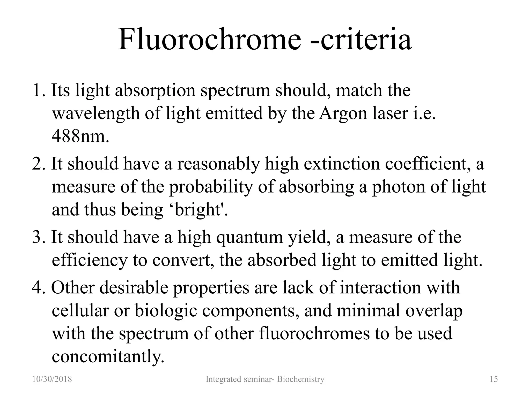
FLOW CYTOMETRY
Presenter: Dr G Santhi priya
Moderator: Dr Kavitha
10/30/2018 1Integrated seminar- Biochemistry
Contents
• Introduction
• Principle
• Fluorochromes
• Sample preparation and processing
• Antibody panel selection
• Gating
• Analysis of leukemia cells by flow cytometry
• Indications of flow cytometry
• References
10/30/2018 2Integrated seminar- Biochemistry
Introduction
• Flow cytometry is the study of physical and chemical
characteristics of a single cell in the fluid stream
10/30/2018 Integrated seminar- Biochemistry 3
• Flow cytometry provides
 valuable and reliable information in the diagnosis and
 classification of hematolymphoid malignancies,
 assessing prognosis and
 response to therapy,
 and for detection of minimal residual disease.
10/30/2018 Integrated seminar- Biochemistry 4
Compartments of Flow Cytometer
• Fluidics system
• Optics and detection
• Electronic system
510/30/2018 Integrated seminar- Biochemistry
Principle of Flow cytometry
10/30/2018 Integrated seminar- Biochemistry 6
• Monodisperse suspension
• Hydrodynamic focusing
• Only one cell or particle can
pass through the laser beam at
a given moment
• The sample pressure is always
higher than the sheath fluid
pressure
• A higher flow rate is generally
preferred for immuno
phenotypic analysis of cells,
while a lower flow rate is ideal
for DNA analysis
7
Fluidics
10/30/2018 Integrated seminar- Biochemistry
Optics & detection
• The light source used in a flow cytometer:
• Laser (more commonly)
• Arc lamp
810/30/2018 Integrated seminar- Biochemistry
• This ensures that the light
gets focused into a
volume
• That is small enough to
excite cells maximally,
• While allowing a
simultaneous passage of
cells in a single file at an
acceptable flow rate of
around 1000 cells per
second.
10/30/2018 Integrated seminar- Biochemistry 9
Signal Processing
• The electronic signals are converted to digital signals
with the help of convertors
• Digitalised data is stored as histograms or as list mode
data(LMD) files
1010/30/2018 Integrated seminar- Biochemistry
Graphic display of data
• Single histogram or cytogram
• Scatterogram or dot plot- FSC/ SSC and CD45/SSC
10/30/2018 Integrated seminar- Biochemistry 11
Histogram Scatterogram
10/30/2018 Integrated seminar- Biochemistry 12
Fluorochromes
• Fluorochromes are essentially dyes which accept energy
• From a laser, at any given wave length and re-emit, it at a
longer wavelength
• These two processes are called excitation and emission
respectively.
10/30/2018 Integrated seminar- Biochemistry 13
Commonly used fluorochromes in leukemia
immunophenotyping
Flurochrome Emission
maximum
Fluorescein isothiocynate(FITIC) 530 nm
Phycoerythrin(PE) 576 nm
Peridin-chlorophyll alpha complex (PerCP) 680 nm
Texas Red 620 nm
ECD (PE-Texas Red tandem) 615 nm
Allophycocyanin (APC) 660 nm
PC5 (PE-cyanine 5 dye tandem) 667 nm
10/30/2018 Integrated seminar- Biochemistry 14
Fluorochrome -criteria
1. Its light absorption spectrum should, match the
wavelength of light emitted by the Argon laser i.e.
488nm.
2. It should have a reasonably high extinction coefficient, a
measure of the probability of absorbing a photon of light
and thus being ‘bright'.
3. It should have a high quantum yield, a measure of the
efficiency to convert, the absorbed light to emitted light.
4. Other desirable properties are lack of interaction with
cellular or biologic components, and minimal overlap
with the spectrum of other fluorochromes to be used
concomitantly.
10/30/2018 Integrated seminar- Biochemistry 15
• When a light intersects a laser beam at the so
called‘interrogation point' two events occur:
A) Light scattering.
B) Emission of light (fluorescence).
10/30/2018 Integrated seminar- Biochemistry 16
17
A. Light Scatter
10/30/2018 Integrated seminar- Biochemistry
Forward scatter(FSC) Side scatter(SSC)
10/30/2018 18Integrated seminar- Biochemistry
B. Emission of fluorescent light(fluorescence)
• Cell subtypes can further be characterized using
fluorescent dyes conjugated to monoclonal antibodies.
• The fluorochrome conjugated to monoclonal antibodies
bind to the cell expressing the analogus antigen,& emit
fluorescent light, which can be measured.
10/30/2018 19Integrated seminar- Biochemistry
10/30/2018 Integrated seminar- Biochemistry 20
Sample preparation and processing
• Types of samples:- Can be performed on peripheral blood,
bone marrow aspirates, body fluids, lymphnode aspirates.
• Anticoagulants:- EDTA -24hrs,
Heparin – 48hrs
Acid citrate dextrose – 72hrs
• Sample storage:- Processed with in 48hrs
Temp – 18-22’ c
• Specimen integrity:- 1- Time elapsed between sample
collection and delivery to lab
2- Environmental conditions
10/30/2018 21Integrated seminar- Biochemistry
Sample processing
• Lysis of red cells :- With reagents like water, ammonium
chloride, hypotonic buffer.
• Removal of lysed red cells :- with an isotonic fluids like
phosphate buffered saline
• Staining with antibodies :- 1. Cell surface antigens
2. intracellular antigens
10/30/2018 22Integrated seminar- Biochemistry
• Intracytoplasmic markers :- some of the cytoplasmic
markers are used in lineage differentiation. Ex- in AML,
cMPO has the highest sensitivity & specificity.
• Membrane permeabilization :-Low concentration of non
ionic detergents like Saponin are used as permeabilizing
agents.
• Negative controls:- determines the level of non specific
binding & autofluoroscence.
10/30/2018 Integrated seminar- Biochemistry 23
Antibody panel selection
• Antibodies are assembled into panels for the
immmophenotypic analysis of leukemias and lymphomas.
• One step analysis
• Two step analysis- Primary panel
- secondary panel
10/30/2018 Integrated seminar- Biochemistry 24
Gating
• Process of identifing the abnormal cell population and to study
its characteristics
• Population of interest
• A gate is selected by defining a region on a cytogram
• Strategies: Conventional FSC/SSCgating
SSC/CD45 gating
CD19 gating for B cells
CD 3 gating for T cells
CD 38 gating for plasma cells
Reverse gating
Gating for exclusion of dead/ non- viable cells
Live gating
10/30/2018 Integrated seminar- Biochemistry 25
Conventional FSC/ SSC gating
10/30/2018 Integrated seminar- Biochemistry 26
SSC/ CD 45 gating
10/30/2018 Integrated seminar- Biochemistry 27
CD 19 gating CD 3 gating
10/30/2018 Integrated seminar- Biochemistry 28
CD 38 gating
10/30/2018 Integrated seminar- Biochemistry 29
Analysis of leukemia cells by flow cytometry
1. Detection of abnormal cell population
2. Lineage assignment
3. Detailed analysis of antigenic profile of abnormal cells and
its comparison with normal cells.
4. Establish neoplastic nature of the abnormality
5. Determination of maturity level of neoplastic cells
6. Accurate characterization of aberrant, antigenic profile of
the neoplastic cells for future tracking of residual disease
7. Diagnosis of the disease
8. Prognostication
9. Identification of targets for directed therapy
10/30/2018 Integrated seminar- Biochemistry 30
Indications of flow cytometry
10/30/2018 Integrated seminar- Biochemistry 31
10/30/2018 Integrated seminar- Biochemistry 32
10/30/2018 Integrated seminar- Biochemistry 33
10/30/2018 Integrated seminar- Biochemistry 34
10/30/2018 Integrated seminar- Biochemistry 35
Solid tumors
 Tumors where flow cytometry for DNA analysis commonly
used are-
 Breast carcinoma.
 Lung carcinoma.
 Bladder carcinomas.
 Colorectal carcinoma.
 Prostatic carcinoma.
10/30/2018 36Integrated seminar- Biochemistry
10/30/2018 37Integrated seminar- Biochemistry
References
• Tejendher S. Automation in cell counts, hemoglobin
seperation, immunophenotyping and coagulation:Atlas
and text of hematology, 3rd ed. Avichal publishing
company. p65-75
• Richard A, Mc Pherson M, Pincus R.The Flow
Cytometric Evaluation of Hematopoietic Neoplasia In.
HENRY’S clinical diagnosis and management by
labaratory Methods :22nd ed. Saunders elsevier , P230-
235
• Pranab D.Flow cytometry In. Diagnostic cytology:2nd ed;
2016. Jaypee Ltd, P260-6
• Martin GW. Flow cytometry: Principles and
Instrumentation. Curr. Issues Mol. Bol.(2001)3(2):39-45
10/30/2018 38Integrated seminar- Biochemistry
Thank you !!
10/30/2018 39Integrated seminar- Biochemistry

8. flow cytometry

  • 1.
    FLOW CYTOMETRY Presenter: DrG Santhi priya Moderator: Dr Kavitha 10/30/2018 1Integrated seminar- Biochemistry
  • 2.
    Contents • Introduction • Principle •Fluorochromes • Sample preparation and processing • Antibody panel selection • Gating • Analysis of leukemia cells by flow cytometry • Indications of flow cytometry • References 10/30/2018 2Integrated seminar- Biochemistry
  • 3.
    Introduction • Flow cytometryis the study of physical and chemical characteristics of a single cell in the fluid stream 10/30/2018 Integrated seminar- Biochemistry 3
  • 4.
    • Flow cytometryprovides  valuable and reliable information in the diagnosis and  classification of hematolymphoid malignancies,  assessing prognosis and  response to therapy,  and for detection of minimal residual disease. 10/30/2018 Integrated seminar- Biochemistry 4
  • 5.
    Compartments of FlowCytometer • Fluidics system • Optics and detection • Electronic system 510/30/2018 Integrated seminar- Biochemistry
  • 6.
    Principle of Flowcytometry 10/30/2018 Integrated seminar- Biochemistry 6
  • 7.
    • Monodisperse suspension •Hydrodynamic focusing • Only one cell or particle can pass through the laser beam at a given moment • The sample pressure is always higher than the sheath fluid pressure • A higher flow rate is generally preferred for immuno phenotypic analysis of cells, while a lower flow rate is ideal for DNA analysis 7 Fluidics 10/30/2018 Integrated seminar- Biochemistry
  • 8.
    Optics & detection •The light source used in a flow cytometer: • Laser (more commonly) • Arc lamp 810/30/2018 Integrated seminar- Biochemistry
  • 9.
    • This ensuresthat the light gets focused into a volume • That is small enough to excite cells maximally, • While allowing a simultaneous passage of cells in a single file at an acceptable flow rate of around 1000 cells per second. 10/30/2018 Integrated seminar- Biochemistry 9
  • 10.
    Signal Processing • Theelectronic signals are converted to digital signals with the help of convertors • Digitalised data is stored as histograms or as list mode data(LMD) files 1010/30/2018 Integrated seminar- Biochemistry
  • 11.
    Graphic display ofdata • Single histogram or cytogram • Scatterogram or dot plot- FSC/ SSC and CD45/SSC 10/30/2018 Integrated seminar- Biochemistry 11
  • 12.
  • 13.
    Fluorochromes • Fluorochromes areessentially dyes which accept energy • From a laser, at any given wave length and re-emit, it at a longer wavelength • These two processes are called excitation and emission respectively. 10/30/2018 Integrated seminar- Biochemistry 13
  • 14.
    Commonly used fluorochromesin leukemia immunophenotyping Flurochrome Emission maximum Fluorescein isothiocynate(FITIC) 530 nm Phycoerythrin(PE) 576 nm Peridin-chlorophyll alpha complex (PerCP) 680 nm Texas Red 620 nm ECD (PE-Texas Red tandem) 615 nm Allophycocyanin (APC) 660 nm PC5 (PE-cyanine 5 dye tandem) 667 nm 10/30/2018 Integrated seminar- Biochemistry 14
  • 15.
    Fluorochrome -criteria 1. Itslight absorption spectrum should, match the wavelength of light emitted by the Argon laser i.e. 488nm. 2. It should have a reasonably high extinction coefficient, a measure of the probability of absorbing a photon of light and thus being ‘bright'. 3. It should have a high quantum yield, a measure of the efficiency to convert, the absorbed light to emitted light. 4. Other desirable properties are lack of interaction with cellular or biologic components, and minimal overlap with the spectrum of other fluorochromes to be used concomitantly. 10/30/2018 Integrated seminar- Biochemistry 15
  • 16.
    • When alight intersects a laser beam at the so called‘interrogation point' two events occur: A) Light scattering. B) Emission of light (fluorescence). 10/30/2018 Integrated seminar- Biochemistry 16
  • 17.
    17 A. Light Scatter 10/30/2018Integrated seminar- Biochemistry Forward scatter(FSC) Side scatter(SSC)
  • 18.
  • 19.
    B. Emission offluorescent light(fluorescence) • Cell subtypes can further be characterized using fluorescent dyes conjugated to monoclonal antibodies. • The fluorochrome conjugated to monoclonal antibodies bind to the cell expressing the analogus antigen,& emit fluorescent light, which can be measured. 10/30/2018 19Integrated seminar- Biochemistry
  • 20.
  • 21.
    Sample preparation andprocessing • Types of samples:- Can be performed on peripheral blood, bone marrow aspirates, body fluids, lymphnode aspirates. • Anticoagulants:- EDTA -24hrs, Heparin – 48hrs Acid citrate dextrose – 72hrs • Sample storage:- Processed with in 48hrs Temp – 18-22’ c • Specimen integrity:- 1- Time elapsed between sample collection and delivery to lab 2- Environmental conditions 10/30/2018 21Integrated seminar- Biochemistry
  • 22.
    Sample processing • Lysisof red cells :- With reagents like water, ammonium chloride, hypotonic buffer. • Removal of lysed red cells :- with an isotonic fluids like phosphate buffered saline • Staining with antibodies :- 1. Cell surface antigens 2. intracellular antigens 10/30/2018 22Integrated seminar- Biochemistry
  • 23.
    • Intracytoplasmic markers:- some of the cytoplasmic markers are used in lineage differentiation. Ex- in AML, cMPO has the highest sensitivity & specificity. • Membrane permeabilization :-Low concentration of non ionic detergents like Saponin are used as permeabilizing agents. • Negative controls:- determines the level of non specific binding & autofluoroscence. 10/30/2018 Integrated seminar- Biochemistry 23
  • 24.
    Antibody panel selection •Antibodies are assembled into panels for the immmophenotypic analysis of leukemias and lymphomas. • One step analysis • Two step analysis- Primary panel - secondary panel 10/30/2018 Integrated seminar- Biochemistry 24
  • 25.
    Gating • Process ofidentifing the abnormal cell population and to study its characteristics • Population of interest • A gate is selected by defining a region on a cytogram • Strategies: Conventional FSC/SSCgating SSC/CD45 gating CD19 gating for B cells CD 3 gating for T cells CD 38 gating for plasma cells Reverse gating Gating for exclusion of dead/ non- viable cells Live gating 10/30/2018 Integrated seminar- Biochemistry 25
  • 26.
    Conventional FSC/ SSCgating 10/30/2018 Integrated seminar- Biochemistry 26
  • 27.
    SSC/ CD 45gating 10/30/2018 Integrated seminar- Biochemistry 27
  • 28.
    CD 19 gatingCD 3 gating 10/30/2018 Integrated seminar- Biochemistry 28
  • 29.
    CD 38 gating 10/30/2018Integrated seminar- Biochemistry 29
  • 30.
    Analysis of leukemiacells by flow cytometry 1. Detection of abnormal cell population 2. Lineage assignment 3. Detailed analysis of antigenic profile of abnormal cells and its comparison with normal cells. 4. Establish neoplastic nature of the abnormality 5. Determination of maturity level of neoplastic cells 6. Accurate characterization of aberrant, antigenic profile of the neoplastic cells for future tracking of residual disease 7. Diagnosis of the disease 8. Prognostication 9. Identification of targets for directed therapy 10/30/2018 Integrated seminar- Biochemistry 30
  • 31.
    Indications of flowcytometry 10/30/2018 Integrated seminar- Biochemistry 31
  • 32.
  • 33.
  • 34.
  • 35.
  • 36.
    Solid tumors  Tumorswhere flow cytometry for DNA analysis commonly used are-  Breast carcinoma.  Lung carcinoma.  Bladder carcinomas.  Colorectal carcinoma.  Prostatic carcinoma. 10/30/2018 36Integrated seminar- Biochemistry
  • 37.
  • 38.
    References • Tejendher S.Automation in cell counts, hemoglobin seperation, immunophenotyping and coagulation:Atlas and text of hematology, 3rd ed. Avichal publishing company. p65-75 • Richard A, Mc Pherson M, Pincus R.The Flow Cytometric Evaluation of Hematopoietic Neoplasia In. HENRY’S clinical diagnosis and management by labaratory Methods :22nd ed. Saunders elsevier , P230- 235 • Pranab D.Flow cytometry In. Diagnostic cytology:2nd ed; 2016. Jaypee Ltd, P260-6 • Martin GW. Flow cytometry: Principles and Instrumentation. Curr. Issues Mol. Bol.(2001)3(2):39-45 10/30/2018 38Integrated seminar- Biochemistry
  • 39.
    Thank you !! 10/30/201839Integrated seminar- Biochemistry

Editor's Notes

  • #22 Tejendher singh pg 67