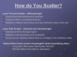
2. What is Flow Cytometry?
Flow Cytometry is a laser-based,
biophysical technology, that measures
cell properties while in a fluid stream.
FLOW = Cells in motion
CYTO = Cell
METRY = Measure
Blood Chromosomes Bone-Marrow Bacteria
3. How Does It Work?
Technology is used to
analyze the physical and
chemical characteristics
of particles in a fluid, that
pass through a laser, one
cell at a time.
Cell components are
fluorescently labeled and
then excited by lasers to
emit light at varying
wavelengths.
LASER = Light Amplification by Stimulated Emission of Radiation
4. What Makes It Work?
Fluidics:
Cells are moved through system by fluidics. Cells are taken up through
the sample injection port. Cells are then focused into single file and
carried to flow cell at the core, in the center of the sheath fluid.
Hydrodynamic focusing produces a single file stream of particles.
Optics:
Excitation ~ Laser and lenses, which shape and focus the laser beam.
Collection ~ Collection lens (fibers) and a system of optical mirrors and
filters that route specified wavelengths of collected light to optical
detectors.
Electronics:
Convert the optical signals to proportionate electronic signals and
digitize them for computer analysis.
Monitor and control the operation of the cytometer.
5. How do You Scatter?
Laser Forward Scatter - diffracted light
Lasers illuminate fluorescence particles
Creates scatter in a forward direction
Related to relative cell surface area and refractive index of the cell
Laser Side Scatter - reflected and refracted light
Detected at 90°to the laser beam
Related to cell granularity and complexity
Occurs at any interface where there is a change in the refractive index
Optical filters block certain wavelengths while transmitting others
Long pass, Short pass, Band pass, Dichroic
All these filters block light by absorption
7. Why is Flow Cytometry a Powerful
Research Tool?
Areas of Cells Analyzed
Immunology ~ Immunophenotyping, Infectious Diseases ,
Transplantation, Autoimmunity
Molecular ~ DNA, RNA, Chromosomes, Genes
Pathology ~ Biopsy, Surgical Specimens, Autopsy, Neoplasm,
Genetic Defects
Marine Science ~ Plankton (photosynthetic picoplankton)
Diagnostics ~ Leukemia, Oncology, HIV/A
Application of Methodology
Analyze many cells rapidly, Accurately quantitate rare cell subtypes,
correlate multiple parameters measured simultaneously on each cell,
no radioisotopes, time saving.
8. What are other applications?
Purpose of Flow Cytometry in the Laboratory is to analyze the physical and
chemical characteristics one cell at a time by exciting fluorescently labeled
cells by a laser to emit light at varying wavelengths.
Monocyte phagocytosis
Multicolor phenotyping cell
surface
Neutrophil oxidative burst
Microbiological analysis
Neutrophil phagocytosis
Multicolor phenotyping
intracellular
Cell sorting
Intracellular Calcium fluxes
Cell Cycle Analysis
Apoptosis
Protein engineering
DNA/Cell analysis
Detection of biomarkers
Cell proliferation assays
9. Current Application of Methodology
Pediatric Leukemia common type of
cancer in children
Flow cytometry is most commonly
used technology in diagnosis of
childhood leukemia
Leukemia cells can be recognized
by:
Unique cell marker
combinations
Visualized monoclonal
antibodies
DNA index
Immunophenotyping
10. References
Applications of Flow Cytometry. (n.d.). Seattle Children's Hospital. Retrieved September 15, 2014, from
https://www.seattlechildrens.org/research/cores/flow-cytometry/applications-of-flow-cytometry/
Brown, M., & Wittwer, C. (2000, April 10). Flow Cytometry: Principles and Clinical Applications in Hematology. Flow
Cytometry: Principles and Clinical Applications in Hematology. Retrieved September 15, 2014, from
http://www.clinchem.org/content/46/8/1221.full.pdf+html
Flow Cytometry. (n.d.). Flow Cytometry Analysis from Applied Cytometry. Retrieved September 9, 2014, from
http://www.appliedcytometry.com/flow_cytometry.php#work AppliedCytometry 2010
Mandal, A. (2014, July 2). Flow Cytometry - What is Flow Cytometry?. News-Medical.net. Retrieved September 15,
2014, from http://www.news-medical.net/health/Flow-Cytometry-What-is-Flow-Cytometry.aspx
Moore, J. (Director) (2014, February 4). Flow Cytometry 101. Goldenson 113. Lecture conducted from HMS, Boston,
MA 02115. introflowcytometry.pdf. (n.d.). University of Minnesota Duluth. Retrieved September 15, 2014, from
http://www.d.umn.edu
11. References continued
Rahman, M. (n.d.). Introduction to Flow Cytometry. Introduction to Flow Cytometry. Retrieved September 15, 2014,
from http://www.abdserotec.com/introduction-to-flow-cytometry.html
University of Minnesota Duluth. (n.d.). University of Minnesota Duluth. Retrieved September 15, 2014, from
http://www.d.umn.edu/
Uses of flow cytometry, Flow Cytometry Core Facility. (2014, June 12). Uses of flow cytometry, Flow Cytometry Core
Facility. Retrieved September 15, 2014, from http://www.icms.qmul.ac.uk/flowcytometry/uses/
Wang, X. M. (2014). Advances and issues in flow cytometric detection. Translational Pediatrics, 3(2). Retrieved
September 15, 2014, from http://www.thetp.org/article/view/3551/4426Wang, X. M. (2014). Advances and issues in
flow cytometric detection. Translational Pediatrics, 3(2). Retrieved September 15, 2014, from
http://www.thetp.org/article/view/3551/4426
Zankl, Janine. "INTRODUCTION TO FLOW CYTOMETRY."
http://www.biozentrum.unibas.ch/fileadmin/redaktion/Forschung/Research_Groups/FACS/Introduction_to_Flow_Cyto
metry-2012.pdf. FACS Core Facility, 17 Oct. 2012. Web. 15 Sept. 2014.
Editor's Notes
Measure molecular material for diagnostics purposes in the lab
Reference: http://en.wikipedia.org/wiki/Flow_cytometry
Three to six properties or components are quantitated in a single sample. 10,000 cells in less than one minute.
Reference: Mandal, A. (2014, July 2). Flow Cytometry - What is Flow Cytometry?. News-Medical.net. Retrieved September 15, 2014, from http://www.news-medical.net/health/Flow-Cytometry-What-is-Flow-Cytometry.aspx
Photo: Compliments of http://www.biozentrum.unibas.chf
Hydrodynamic focusing produces a single file stream of particles
Without the nozzle, the instrument would become blocked and it would not be possible to analyze one cell at a time.
Reference: Rahman, M. (n.d.). Introduction to Flow Cytometry. Introduction to Flow Cytometry. Retrieved September 15, 2014, from http://www.abdserotec.com/introduction-to-flow-cytometry.html.
Fluidics; Introduction to Flow Cytometry By AbD Serotec. Chapter 1 page 4
Introduction to Flow Cytometry: A Learning Guide, BD BioSciences
The Flow Cytometer: How does it work? www.abcam.com
Optics; Introduction to Flow Cytometry By AbD Serotec. Chapter 1 page 5
Laser beam is processed by several filters to generate excitation wavelengths for several fluorochromes
This beam strikes each individual cell as the cell passes through the flow cell in single file.
Upon striking the cell the laser beam can generate two kinds of signals.
• For all cells, the deflection of incident light is read as forward scatter (FSC) and side scatter (SSC).
• If the cell is labeled with a fluorophore, the laser beam results in excitation and emission of photons.
Reference: Rahman, M. (n.d.). Introduction to Flow Cytometry. Introduction to Flow Cytometry. Retrieved September 15, 2014, from http://www.abdserotec.com/introduction-to-flow-cytometry.html
Photo: Courtesy of http://www.assay-protocol.com/cell-biology/flow-cytometry
Sheath Flow Pr • Refers to the process whereby the sample stream is injected into a surrounding sheath stream under conditions of laminar flow.
• Properties of laminar flow through a narrowing orifice at the neck of the flow cell facilitate hydrodynamic focusing and coaxial flow of particles/cells through the interrogation point of the flow chamber.
Introduce and focus the cells for interrogation. Sample is injected into the central channel (core). Enclosed by an outer sheath that contains faster flowing fluid, the sample then pass in front of an illumination source. Hydrodynamic focusing produces a single file stream of particles, without it the nozzle of the instrument would become blocked and it would not be possible to analyze one cell at a time.
Reference: Rahman, M. (n.d.). Introduction to Flow Cytometry. Introduction to Flow Cytometry. Retrieved September 15, 2014, from http://www.abdserotec.com/introduction-to-flow-cytometry.html. Picture 1 Chapter 1 page 4 and Picture 2 Chapter 1 page 7.
Most common use for Flow Cytometry is for total DNA per cell in a biopsy specimen from tumors, for clinical cancer diagnosis and prognosis.
Quantiation of CD+4 and CD+8 T lymphcocytes in blood to determine when an HIV infection has resulted in AIDS .
Chromosomes and Blood 1000 cells per second and bone marrow and bacteria 100 cells per second.
Reference: Mandal, A. (2014, July 2). Flow Cytometry - What is Flow Cytometry?. News-Medical.net. Retrieved September 15, 2014, from http://www.news-medical.net/health/Flow-Cytometry-What-is-Flow-Cytometry.aspx
Reference: Applications of Flow Cytometry. (n.d.). Seattle Children's Hospital. Retrieved September 15, 2014, from https://www.seattlechildrens.org/research/cores/flow-cytometry/applications-of-flow-cytometry/
Photo: Courtesy of www.harvard.edu
Propidium iodide is an intercalating agent and a fluorescent molecule with a molecular mass of 668.4 Da that can be used to stain cells
Flow cytometry is now a widely used method for analyzing expression of cell surface and intracellular molecules, characterizing and defining different cell types in heterogeneous cell populations, assessing the purity of isolated subpopulations, and analyzing cell size and volume. It allows simultaneous multi-parameter analysis of single cells. It is predominantly used to measure fluorescence intensity produced by fluorescent-labeled antibodies detecting proteins or ligands that bind to specific cell-associated molecules, such as DNA binding by Propidium iodide.
Reference: Wang, X. M. (2014). Advances and issues in flow cytometric detection. Translational Pediatrics, 3(2). Retrieved September 15, 2014, from http://www.thetp.org/article/view/3551/4426