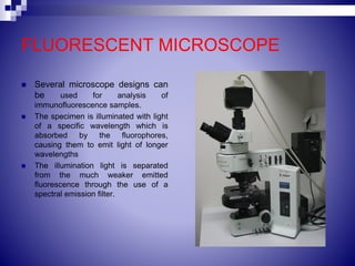
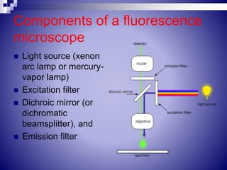
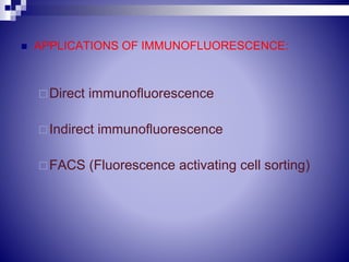
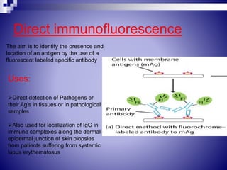
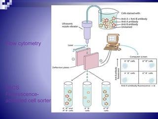
Immunofluorescence is a technique developed in 1944 that labels antibodies with fluorescent dyes to visualize antigens under a fluorescence microscope. The process employs light of specific wavelengths to excite fluorochromes like fluorescein and rhodamine, which emit light of longer wavelengths. Applications include direct and indirect immunofluorescence for disease diagnosis and flow cytometry (FACS) for cell sorting and analysis.