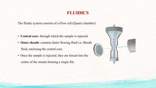
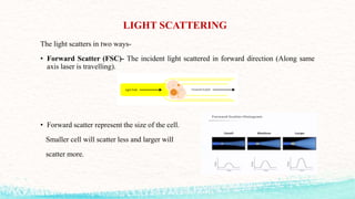
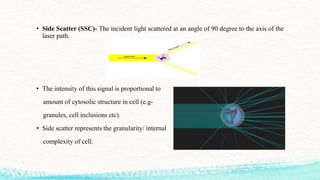
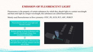
SlideShare a Scribd company logo
FLOW CYTOMETRY
Subject- Pharmaceutical Biotechnology (P)
DEPARTMENT OF PHARMACEUTICAL SCIENCES
Dr. Hari Singh Gour Vishwavidyalaya
Sagar(M.P)-470003, India
(A Central University)
Submitted By:
DEBASIS SEN
(Y22254008)
Submitted To:
Prof. UMESH K. PATIL
(Professor, DOPS)
Dr. UDITAAGRAWAL
(Guest Faculty, DOPS)
Ms. POOJA DAS BIDLA
(Research Scholar, DOPS)
CONTENT
• INTRODUCTION
• PRINCIPLE
• BASIC MECHANISM
• SAMPLE PREPARATION
• INSTRUMENTATION
• DATAANALYSIS
• GATING
• COMPENSATION
• WORKING
• APPLICATION
• LIMITATIONS
• CONCLUSION
• REFERENCES
INTRODUCTION
Flow- the motion characteristics of fluids
Cyto- combining form of cells
Metry- measurement
• Hence, flow cytometry is the measurement of cell properties as cell move in a single
hydrodynamic flow and interrupt a beam of light.
• Flow cytometry is a popular cell biology technique that utilizes laser-based technology to
count, sort, and profile cells in a heterogeneous fluid mixture.
• The technique was first described by Wallace Coulter in the 1950s.
• The present flow cytometers are capable of analyzing upto 13 parametrs (forward scatter,
side scatter, 11 colours of immunofluorescence).
PRINCIPLE
Flow
Cytometry
Principle of Laminar
Flow
Electrostatics
Optics and Light
Scattering
Coulter Principle
BASIC MECHANISM
Biological sample
Label it with a fluorescent marker
Cells move in a linear stream through a focused light source(laser
beam)
Fluorescent molecule gets activated and emits light that is filtered
and detected by sensitive light detectors(usually a photomultiplier
tube)
Conversion of analogue fluorescent signals to digital signal
SAMPLE PREPARATION
The exact steps for sample preparation can vary depending on the specific experimental design
and research question. Here is the general steps which are followed for sample preparation in
flow cytometry-
Collect the
cells or
particles of
interest
Prepare the
cells or
particles by
washing them
in a buffer
solution to
remove any
debris or
contaminants
Stain the cells
or particles
with
fluorescent
dyes or
antibodies
Fixation: In
order to stop
any further
degradation
Filtering: to
eliminate any
cellular
aggregates and
debris
Final
resuspension:
involves
resuspending
the cells or
particles in a
buffer solution
INSTRUMENTATION
COMPONENTS
Flow Cytometry has three main components-
Fluidics
• Transports cells in a
stream to the laser
beam for
interrogation.
Optics
• It consists of lasers
to illuminate the
particles in the
sample stream and
optical filters to
direct the resulting
light signals to the
appropriate
detectors
Electronics
• The electronics
system converts the
detected light
signals into
electronic signals
that can processed
by the computer
FLUIDICS
The fluidic system consists of a Flow cell (Quartz chamber)
• Central core- through which the sample is injected.
• Outer sheath- contains faster flowing fluid i.e; Sheath
fluid, enclosing the central core.
• Once the sample is injected, they are forced into the
centre of the stream forming a single file.
OPTICS
Single wavelength, coherent light
Light Source
• Provide energy that excite the fluorochrome of interest, helps to
create stable and reliable signal.
Focus the beam
Lenses
• Keep the beam perpendicular to sample stream flow
• Make beam small enough to illuminate only one cell at a time
Filters
• Ensures that each photodetector receives light bands of various
wavelenghts
When a light intersects a laser beam at the so called ‘interrogation point’ two events occur-
A. Light scattering
B. Emission of fluorescent light
LIGHT SCATTERING
The light scatters in two ways-
• Forward Scatter (FSC)- The incident light scattered in forward direction (Along same
axis laser is travelling).
• Forward scatter represent the size of the cell.
Smaller cell will scatter less and larger will
scatter more.
• Side Scatter (SSC)- The incident light scattered at an angle of 90 degree to the axis of the
laser path.
• The intensity of this signal is proportional to
amount of cytosolic structure in cell (e.g-
granules, cell inclusions etc).
• Side scatter represents the granularity/ internal
complexity of cell.
EMISSION OF FLUORESCENT LIGHT
Fluorescence is the property of certain substances by which they absorb light at a certain wavelength
and then emit light at a longer wavelength, this substances are called fluorochromes.
Mainly used fluorochromes in flow cytometry- FITC, PE, ECD, PC5, APC, PERCP
Fluorochrome absorb some light and
become excited
Release energy in form of photons with
specific wavelength (larger than
excitation wavelength)
Photons pass through the collection lens
and splits down specific channel with the
use of filter
OPTICAL FILTERS
Many wavelengths of light will be scattered from a cell.
But how to split the light into its specific wavelengths to detect them independently?
Optical filters are designed such that they absorb or reflect some wavelengths of light, while
transmitting others.
Optical
Filters
Short pass
filter
Long pass
filter
Band pass
filter
Dichroic
filter
DETECTOR
There are mainly two types of detectors used in flow cytometry-
• Photodiode
• Photomultiplier tube (PMT)
Photodiode
• Used for strong signals
• When saturation is a potential
problem
• e.g- Forward scatter
Photomultiplier tube
(PMT)
• More sensitive and used to
detect small amounts of
fluorescence
• Band pass filter in front to
allow only specific band width
of light reach it
ELECTRONICS
• Detectors basically collects photos of light and convert them to current.
• The electronics process that light signal and convert the current to a digitalized value.
• This digitalized value is represented as a graph by the computer.
• Softwares-
1. Kaluza
2. CellQuest
3. FlowJo
4. WinMDI
5. FCS Express
DATA DISPLAY
The data are usually presented in the form of single parameter histograms or as plots or
correlated parameters, which are referred to as cytograms.
Cytograms may display data in the form of-
• Dot plot
• Density plot
• Contour plot
• Histogram
GATING
Gating is used to isolate a subset of cells on a plot and set a region on a histogram or
cytogram.
It allows the ability to look at parameters only on that subset.
COMPENSATION
Why do we need to compensate?
Fluorochromes typically give fluorescence over a large part of the spectrums (100nm or
more).
• Detectors detect fluorescence based on wavelengths
• Thus detects fluorescence from more than one fluorochrome which is called ‘Bleed Over’
• You need to compensate this bleed over so that one detector reports signals from only one
fluorochrome.
WORKING OF FLOW CYTOMETRY
APPLICATIONS
Erythropoiesis
and red cell
disorder
Platelet
function
analysis
Phagocytosis
analysis
Detection of
apoptosis
Cell
counting
and cell
sorting
Cell cycle
analysis
Staging of
Lymphomas
LIMITATIONS
• Flow cytometry is limited by its requirement that analyzed cells be in suspension, making
information on tissue architecture and cell-cell interactions unavailable.
• Cell subpopulations with similar marker expression and difficult to differentiate and
analyses that employ more fluorophores are subject to signal slipover.
• Flow cytometry may generate massive amounts of data, making analyses complicated.
CONCLUSION
Flow cytometry is a versatile and powerful technique that has revolutionized our
understanding of cells and their functions. Flow cytometry has a wide range of applications,
including immunology, hematology, oncology, microbiology, and stem cell research. It can
be used to analyze complex mixtures of cells, such as blood or bone marrow samples, to
identify and quantify different cell types. Flow cytometry can also be used to study the cell
cycle, apoptosis, and cell signaling pathways. So it has widespread use in research and
clinical settings has led to numerous discoveries and advancements in medicine and biology.
REFERENCES
1. Büscher M. Flow cytometry instrumentation–an overview. Current Protocols in
Cytometry. 2019 Jan;87(1):e52.
2. Nunez R. Flow cytometry: principles and instrumentation. Current Issues in Molecular
Biology. 2001 Apr;3(2):39-45.
3. Hoffman RA. Flow cytometry: instrumentation, applications, future trends and
limitations. Standardization and Quality Assurance in Fluorescence Measurements II:
Bioanalytical and Biomedical Applications. 2008:307-42.
4. Chapman GV. Instrumentation for flow cytometry. Journal of immunological methods.
2000 Sep 21;243(1-2):3-12.
THANK YOU

More Related Content

Similar to Flowcytometry.pptx

Flow cytometry
Flow cytometryFlow cytometry
Flow cytometry
Saranya Ganesh
 
flowcytometry-basic principle of flowcyto
flowcytometry-basic principle of flowcytoflowcytometry-basic principle of flowcyto
flowcytometry-basic principle of flowcyto
Dr. Ajit Surya Singh
 
flowcytometry-150403122734-conversion-gate01.pdf
flowcytometry-150403122734-conversion-gate01.pdfflowcytometry-150403122734-conversion-gate01.pdf
flowcytometry-150403122734-conversion-gate01.pdf
dmfrmicro
 
Flowcytometry
FlowcytometryFlowcytometry
Flowcytometry
rameshkandimalla1723
 
Flow cytometry
Flow cytometryFlow cytometry
Flow cytometry
tashagarwal
 
Flow Cytometry (FCM)
Flow Cytometry (FCM) Flow Cytometry (FCM)
Flow Cytometry (FCM)
Dr. Amer Ali Khaleel /HMU
 
Flow cytometry
Flow cytometryFlow cytometry
Flow cytometry
RajanPaudel9
 
Flowcytometry by asif
Flowcytometry by asifFlowcytometry by asif
Flowcytometry by asif
Asif Zeb
 
Flow cytometry
Flow cytometryFlow cytometry
Flow cytometry
Anushi Jain
 
Flow cytometry in cell biology
Flow cytometry in cell biologyFlow cytometry in cell biology
Flow cytometry in cell biology
Amani Riyadh
 
Flow cytometry definition, principle, parts, steps, types, uses
Flow cytometry definition, principle, parts, steps, types, usesFlow cytometry definition, principle, parts, steps, types, uses
Flow cytometry definition, principle, parts, steps, types, uses
Gayathri Devi S
 
Flow cytometry
Flow cytometryFlow cytometry
Flow cytometry
Pradeep Singh Narwat
 
Flow cytometry
Flow cytometryFlow cytometry
Flow cytometry
Nijas Mohamed
 
flow cytometry.pptx
flow cytometry.pptxflow cytometry.pptx
flow cytometry.pptx
jasveer15
 
flow cytometry presentation
flow cytometry  presentationflow cytometry  presentation
flow cytometry presentation
Jamile Saberzade
 
Flow cytometry
Flow cytometryFlow cytometry
Flow cytometry
prachann
 
An introduction to flow cytometry- Ashwini.R
An introduction to flow cytometry- Ashwini.RAn introduction to flow cytometry- Ashwini.R
An introduction to flow cytometry- Ashwini.R
Ashwini R
 
Flow cytometry
Flow cytometryFlow cytometry
Flow cytometry
Ankita Rungta Kapoor
 
FACS and MACS with their applications in biological research.
FACS and MACS with their applications in biological research.FACS and MACS with their applications in biological research.
FACS and MACS with their applications in biological research.
Deepak Agarwal
 
Flow Cytometry Principle
Flow Cytometry PrincipleFlow Cytometry Principle
Flow Cytometry Principle
Boster Biological Technology
 

Similar to Flowcytometry.pptx (20)

Flow cytometry
Flow cytometryFlow cytometry
Flow cytometry
 
flowcytometry-basic principle of flowcyto
flowcytometry-basic principle of flowcytoflowcytometry-basic principle of flowcyto
flowcytometry-basic principle of flowcyto
 
flowcytometry-150403122734-conversion-gate01.pdf
flowcytometry-150403122734-conversion-gate01.pdfflowcytometry-150403122734-conversion-gate01.pdf
flowcytometry-150403122734-conversion-gate01.pdf
 
Flowcytometry
FlowcytometryFlowcytometry
Flowcytometry
 
Flow cytometry
Flow cytometryFlow cytometry
Flow cytometry
 
Flow Cytometry (FCM)
Flow Cytometry (FCM) Flow Cytometry (FCM)
Flow Cytometry (FCM)
 
Flow cytometry
Flow cytometryFlow cytometry
Flow cytometry
 
Flowcytometry by asif
Flowcytometry by asifFlowcytometry by asif
Flowcytometry by asif
 
Flow cytometry
Flow cytometryFlow cytometry
Flow cytometry
 
Flow cytometry in cell biology
Flow cytometry in cell biologyFlow cytometry in cell biology
Flow cytometry in cell biology
 
Flow cytometry definition, principle, parts, steps, types, uses
Flow cytometry definition, principle, parts, steps, types, usesFlow cytometry definition, principle, parts, steps, types, uses
Flow cytometry definition, principle, parts, steps, types, uses
 
Flow cytometry
Flow cytometryFlow cytometry
Flow cytometry
 
Flow cytometry
Flow cytometryFlow cytometry
Flow cytometry
 
flow cytometry.pptx
flow cytometry.pptxflow cytometry.pptx
flow cytometry.pptx
 
flow cytometry presentation
flow cytometry  presentationflow cytometry  presentation
flow cytometry presentation
 
Flow cytometry
Flow cytometryFlow cytometry
Flow cytometry
 
An introduction to flow cytometry- Ashwini.R
An introduction to flow cytometry- Ashwini.RAn introduction to flow cytometry- Ashwini.R
An introduction to flow cytometry- Ashwini.R
 
Flow cytometry
Flow cytometryFlow cytometry
Flow cytometry
 
FACS and MACS with their applications in biological research.
FACS and MACS with their applications in biological research.FACS and MACS with their applications in biological research.
FACS and MACS with their applications in biological research.
 
Flow Cytometry Principle
Flow Cytometry PrincipleFlow Cytometry Principle
Flow Cytometry Principle
 

Recently uploaded

Ophthalmology Clinical Tests for OSCE exam
Ophthalmology Clinical Tests for OSCE examOphthalmology Clinical Tests for OSCE exam
Ophthalmology Clinical Tests for OSCE exam
KafrELShiekh University
 
Maxilla, Mandible & Hyoid Bone & Clinical Correlations by Dr. RIG.pptx
Maxilla, Mandible & Hyoid Bone & Clinical Correlations by Dr. RIG.pptxMaxilla, Mandible & Hyoid Bone & Clinical Correlations by Dr. RIG.pptx
Maxilla, Mandible & Hyoid Bone & Clinical Correlations by Dr. RIG.pptx
Dr. Rabia Inam Gandapore
 
Novas diretrizes da OMS para os cuidados perinatais de mais qualidade
Novas diretrizes da OMS para os cuidados perinatais de mais qualidadeNovas diretrizes da OMS para os cuidados perinatais de mais qualidade
Novas diretrizes da OMS para os cuidados perinatais de mais qualidade
Prof. Marcus Renato de Carvalho
 
How STIs Influence the Development of Pelvic Inflammatory Disease.pptx
How STIs Influence the Development of Pelvic Inflammatory Disease.pptxHow STIs Influence the Development of Pelvic Inflammatory Disease.pptx
How STIs Influence the Development of Pelvic Inflammatory Disease.pptx
FFragrant
 
Physiology of Chemical Sensation of smell.pdf
Physiology of Chemical Sensation of smell.pdfPhysiology of Chemical Sensation of smell.pdf
Physiology of Chemical Sensation of smell.pdf
MedicoseAcademics
 
Colonic and anorectal physiology with surgical implications
Colonic and anorectal physiology with surgical implicationsColonic and anorectal physiology with surgical implications
Colonic and anorectal physiology with surgical implications
Dr Maria Tamanna
 
ARTHROLOGY PPT NCISM SYLLABUS AYURVEDA STUDENTS
ARTHROLOGY PPT NCISM SYLLABUS AYURVEDA STUDENTSARTHROLOGY PPT NCISM SYLLABUS AYURVEDA STUDENTS
ARTHROLOGY PPT NCISM SYLLABUS AYURVEDA STUDENTS
Dr. Vinay Pareek
 
Hemodialysis: Chapter 4, Dialysate Circuit - Dr.Gawad
Hemodialysis: Chapter 4, Dialysate Circuit - Dr.GawadHemodialysis: Chapter 4, Dialysate Circuit - Dr.Gawad
Hemodialysis: Chapter 4, Dialysate Circuit - Dr.Gawad
NephroTube - Dr.Gawad
 
The Electrocardiogram - Physiologic Principles
The Electrocardiogram - Physiologic PrinciplesThe Electrocardiogram - Physiologic Principles
The Electrocardiogram - Physiologic Principles
MedicoseAcademics
 
Journal Article Review on Rasamanikya
Journal Article Review on RasamanikyaJournal Article Review on Rasamanikya
Journal Article Review on Rasamanikya
Dr. Jyothirmai Paindla
 
Pharynx and Clinical Correlations BY Dr.Rabia Inam Gandapore.pptx
Pharynx and Clinical Correlations BY Dr.Rabia Inam Gandapore.pptxPharynx and Clinical Correlations BY Dr.Rabia Inam Gandapore.pptx
Pharynx and Clinical Correlations BY Dr.Rabia Inam Gandapore.pptx
Dr. Rabia Inam Gandapore
 
Adv. biopharm. APPLICATION OF PHARMACOKINETICS : TARGETED DRUG DELIVERY SYSTEMS
Adv. biopharm. APPLICATION OF PHARMACOKINETICS : TARGETED DRUG DELIVERY SYSTEMSAdv. biopharm. APPLICATION OF PHARMACOKINETICS : TARGETED DRUG DELIVERY SYSTEMS
Adv. biopharm. APPLICATION OF PHARMACOKINETICS : TARGETED DRUG DELIVERY SYSTEMS
AkankshaAshtankar
 
Local Advanced Lung Cancer: Artificial Intelligence, Synergetics, Complex Sys...
Local Advanced Lung Cancer: Artificial Intelligence, Synergetics, Complex Sys...Local Advanced Lung Cancer: Artificial Intelligence, Synergetics, Complex Sys...
Local Advanced Lung Cancer: Artificial Intelligence, Synergetics, Complex Sys...
Oleg Kshivets
 
Thyroid Gland- Gross Anatomy by Dr. Rabia Inam Gandapore.pptx
Thyroid Gland- Gross Anatomy by Dr. Rabia Inam Gandapore.pptxThyroid Gland- Gross Anatomy by Dr. Rabia Inam Gandapore.pptx
Thyroid Gland- Gross Anatomy by Dr. Rabia Inam Gandapore.pptx
Dr. Rabia Inam Gandapore
 
Integrating Ayurveda into Parkinson’s Management: A Holistic Approach
Integrating Ayurveda into Parkinson’s Management: A Holistic ApproachIntegrating Ayurveda into Parkinson’s Management: A Holistic Approach
Integrating Ayurveda into Parkinson’s Management: A Holistic Approach
Ayurveda ForAll
 
basicmodesofventilation2022-220313203758.pdf
basicmodesofventilation2022-220313203758.pdfbasicmodesofventilation2022-220313203758.pdf
basicmodesofventilation2022-220313203758.pdf
aljamhori teaching hospital
 
Netter's Atlas of Human Anatomy 7.ed.pdf
Netter's Atlas of Human Anatomy 7.ed.pdfNetter's Atlas of Human Anatomy 7.ed.pdf
Netter's Atlas of Human Anatomy 7.ed.pdf
BrissaOrtiz3
 
Pictures of Superficial & Deep Fascia.ppt.pdf
Pictures of Superficial & Deep Fascia.ppt.pdfPictures of Superficial & Deep Fascia.ppt.pdf
Pictures of Superficial & Deep Fascia.ppt.pdf
Dr. Rabia Inam Gandapore
 
Best Ayurvedic medicine for Gas and Indigestion
Best Ayurvedic medicine for Gas and IndigestionBest Ayurvedic medicine for Gas and Indigestion
Best Ayurvedic medicine for Gas and Indigestion
Swastik Ayurveda
 
Top-Vitamin-Supplement-Brands-in-India List
Top-Vitamin-Supplement-Brands-in-India ListTop-Vitamin-Supplement-Brands-in-India List
Top-Vitamin-Supplement-Brands-in-India List
SwisschemDerma
 

Recently uploaded (20)

Ophthalmology Clinical Tests for OSCE exam
Ophthalmology Clinical Tests for OSCE examOphthalmology Clinical Tests for OSCE exam
Ophthalmology Clinical Tests for OSCE exam
 
Maxilla, Mandible & Hyoid Bone & Clinical Correlations by Dr. RIG.pptx
Maxilla, Mandible & Hyoid Bone & Clinical Correlations by Dr. RIG.pptxMaxilla, Mandible & Hyoid Bone & Clinical Correlations by Dr. RIG.pptx
Maxilla, Mandible & Hyoid Bone & Clinical Correlations by Dr. RIG.pptx
 
Novas diretrizes da OMS para os cuidados perinatais de mais qualidade
Novas diretrizes da OMS para os cuidados perinatais de mais qualidadeNovas diretrizes da OMS para os cuidados perinatais de mais qualidade
Novas diretrizes da OMS para os cuidados perinatais de mais qualidade
 
How STIs Influence the Development of Pelvic Inflammatory Disease.pptx
How STIs Influence the Development of Pelvic Inflammatory Disease.pptxHow STIs Influence the Development of Pelvic Inflammatory Disease.pptx
How STIs Influence the Development of Pelvic Inflammatory Disease.pptx
 
Physiology of Chemical Sensation of smell.pdf
Physiology of Chemical Sensation of smell.pdfPhysiology of Chemical Sensation of smell.pdf
Physiology of Chemical Sensation of smell.pdf
 
Colonic and anorectal physiology with surgical implications
Colonic and anorectal physiology with surgical implicationsColonic and anorectal physiology with surgical implications
Colonic and anorectal physiology with surgical implications
 
ARTHROLOGY PPT NCISM SYLLABUS AYURVEDA STUDENTS
ARTHROLOGY PPT NCISM SYLLABUS AYURVEDA STUDENTSARTHROLOGY PPT NCISM SYLLABUS AYURVEDA STUDENTS
ARTHROLOGY PPT NCISM SYLLABUS AYURVEDA STUDENTS
 
Hemodialysis: Chapter 4, Dialysate Circuit - Dr.Gawad
Hemodialysis: Chapter 4, Dialysate Circuit - Dr.GawadHemodialysis: Chapter 4, Dialysate Circuit - Dr.Gawad
Hemodialysis: Chapter 4, Dialysate Circuit - Dr.Gawad
 
The Electrocardiogram - Physiologic Principles
The Electrocardiogram - Physiologic PrinciplesThe Electrocardiogram - Physiologic Principles
The Electrocardiogram - Physiologic Principles
 
Journal Article Review on Rasamanikya
Journal Article Review on RasamanikyaJournal Article Review on Rasamanikya
Journal Article Review on Rasamanikya
 
Pharynx and Clinical Correlations BY Dr.Rabia Inam Gandapore.pptx
Pharynx and Clinical Correlations BY Dr.Rabia Inam Gandapore.pptxPharynx and Clinical Correlations BY Dr.Rabia Inam Gandapore.pptx
Pharynx and Clinical Correlations BY Dr.Rabia Inam Gandapore.pptx
 
Adv. biopharm. APPLICATION OF PHARMACOKINETICS : TARGETED DRUG DELIVERY SYSTEMS
Adv. biopharm. APPLICATION OF PHARMACOKINETICS : TARGETED DRUG DELIVERY SYSTEMSAdv. biopharm. APPLICATION OF PHARMACOKINETICS : TARGETED DRUG DELIVERY SYSTEMS
Adv. biopharm. APPLICATION OF PHARMACOKINETICS : TARGETED DRUG DELIVERY SYSTEMS
 
Local Advanced Lung Cancer: Artificial Intelligence, Synergetics, Complex Sys...
Local Advanced Lung Cancer: Artificial Intelligence, Synergetics, Complex Sys...Local Advanced Lung Cancer: Artificial Intelligence, Synergetics, Complex Sys...
Local Advanced Lung Cancer: Artificial Intelligence, Synergetics, Complex Sys...
 
Thyroid Gland- Gross Anatomy by Dr. Rabia Inam Gandapore.pptx
Thyroid Gland- Gross Anatomy by Dr. Rabia Inam Gandapore.pptxThyroid Gland- Gross Anatomy by Dr. Rabia Inam Gandapore.pptx
Thyroid Gland- Gross Anatomy by Dr. Rabia Inam Gandapore.pptx
 
Integrating Ayurveda into Parkinson’s Management: A Holistic Approach
Integrating Ayurveda into Parkinson’s Management: A Holistic ApproachIntegrating Ayurveda into Parkinson’s Management: A Holistic Approach
Integrating Ayurveda into Parkinson’s Management: A Holistic Approach
 
basicmodesofventilation2022-220313203758.pdf
basicmodesofventilation2022-220313203758.pdfbasicmodesofventilation2022-220313203758.pdf
basicmodesofventilation2022-220313203758.pdf
 
Netter's Atlas of Human Anatomy 7.ed.pdf
Netter's Atlas of Human Anatomy 7.ed.pdfNetter's Atlas of Human Anatomy 7.ed.pdf
Netter's Atlas of Human Anatomy 7.ed.pdf
 
Pictures of Superficial & Deep Fascia.ppt.pdf
Pictures of Superficial & Deep Fascia.ppt.pdfPictures of Superficial & Deep Fascia.ppt.pdf
Pictures of Superficial & Deep Fascia.ppt.pdf
 
Best Ayurvedic medicine for Gas and Indigestion
Best Ayurvedic medicine for Gas and IndigestionBest Ayurvedic medicine for Gas and Indigestion
Best Ayurvedic medicine for Gas and Indigestion
 
Top-Vitamin-Supplement-Brands-in-India List
Top-Vitamin-Supplement-Brands-in-India ListTop-Vitamin-Supplement-Brands-in-India List
Top-Vitamin-Supplement-Brands-in-India List
 

Flowcytometry.pptx

  • 1. FLOW CYTOMETRY Subject- Pharmaceutical Biotechnology (P) DEPARTMENT OF PHARMACEUTICAL SCIENCES Dr. Hari Singh Gour Vishwavidyalaya Sagar(M.P)-470003, India (A Central University) Submitted By: DEBASIS SEN (Y22254008) Submitted To: Prof. UMESH K. PATIL (Professor, DOPS) Dr. UDITAAGRAWAL (Guest Faculty, DOPS) Ms. POOJA DAS BIDLA (Research Scholar, DOPS)
  • 2. CONTENT • INTRODUCTION • PRINCIPLE • BASIC MECHANISM • SAMPLE PREPARATION • INSTRUMENTATION • DATAANALYSIS • GATING • COMPENSATION • WORKING • APPLICATION • LIMITATIONS • CONCLUSION • REFERENCES
  • 3. INTRODUCTION Flow- the motion characteristics of fluids Cyto- combining form of cells Metry- measurement • Hence, flow cytometry is the measurement of cell properties as cell move in a single hydrodynamic flow and interrupt a beam of light. • Flow cytometry is a popular cell biology technique that utilizes laser-based technology to count, sort, and profile cells in a heterogeneous fluid mixture. • The technique was first described by Wallace Coulter in the 1950s. • The present flow cytometers are capable of analyzing upto 13 parametrs (forward scatter, side scatter, 11 colours of immunofluorescence).
  • 5. BASIC MECHANISM Biological sample Label it with a fluorescent marker Cells move in a linear stream through a focused light source(laser beam) Fluorescent molecule gets activated and emits light that is filtered and detected by sensitive light detectors(usually a photomultiplier tube) Conversion of analogue fluorescent signals to digital signal
  • 6. SAMPLE PREPARATION The exact steps for sample preparation can vary depending on the specific experimental design and research question. Here is the general steps which are followed for sample preparation in flow cytometry- Collect the cells or particles of interest Prepare the cells or particles by washing them in a buffer solution to remove any debris or contaminants Stain the cells or particles with fluorescent dyes or antibodies Fixation: In order to stop any further degradation Filtering: to eliminate any cellular aggregates and debris Final resuspension: involves resuspending the cells or particles in a buffer solution
  • 7. INSTRUMENTATION COMPONENTS Flow Cytometry has three main components- Fluidics • Transports cells in a stream to the laser beam for interrogation. Optics • It consists of lasers to illuminate the particles in the sample stream and optical filters to direct the resulting light signals to the appropriate detectors Electronics • The electronics system converts the detected light signals into electronic signals that can processed by the computer
  • 8.
  • 9. FLUIDICS The fluidic system consists of a Flow cell (Quartz chamber) • Central core- through which the sample is injected. • Outer sheath- contains faster flowing fluid i.e; Sheath fluid, enclosing the central core. • Once the sample is injected, they are forced into the centre of the stream forming a single file.
  • 10. OPTICS Single wavelength, coherent light Light Source • Provide energy that excite the fluorochrome of interest, helps to create stable and reliable signal. Focus the beam Lenses • Keep the beam perpendicular to sample stream flow • Make beam small enough to illuminate only one cell at a time Filters • Ensures that each photodetector receives light bands of various wavelenghts
  • 11. When a light intersects a laser beam at the so called ‘interrogation point’ two events occur- A. Light scattering B. Emission of fluorescent light
  • 12. LIGHT SCATTERING The light scatters in two ways- • Forward Scatter (FSC)- The incident light scattered in forward direction (Along same axis laser is travelling). • Forward scatter represent the size of the cell. Smaller cell will scatter less and larger will scatter more.
  • 13. • Side Scatter (SSC)- The incident light scattered at an angle of 90 degree to the axis of the laser path. • The intensity of this signal is proportional to amount of cytosolic structure in cell (e.g- granules, cell inclusions etc). • Side scatter represents the granularity/ internal complexity of cell.
  • 14. EMISSION OF FLUORESCENT LIGHT Fluorescence is the property of certain substances by which they absorb light at a certain wavelength and then emit light at a longer wavelength, this substances are called fluorochromes. Mainly used fluorochromes in flow cytometry- FITC, PE, ECD, PC5, APC, PERCP Fluorochrome absorb some light and become excited Release energy in form of photons with specific wavelength (larger than excitation wavelength) Photons pass through the collection lens and splits down specific channel with the use of filter
  • 15. OPTICAL FILTERS Many wavelengths of light will be scattered from a cell. But how to split the light into its specific wavelengths to detect them independently? Optical filters are designed such that they absorb or reflect some wavelengths of light, while transmitting others. Optical Filters Short pass filter Long pass filter Band pass filter Dichroic filter
  • 16. DETECTOR There are mainly two types of detectors used in flow cytometry- • Photodiode • Photomultiplier tube (PMT) Photodiode • Used for strong signals • When saturation is a potential problem • e.g- Forward scatter Photomultiplier tube (PMT) • More sensitive and used to detect small amounts of fluorescence • Band pass filter in front to allow only specific band width of light reach it
  • 17. ELECTRONICS • Detectors basically collects photos of light and convert them to current. • The electronics process that light signal and convert the current to a digitalized value. • This digitalized value is represented as a graph by the computer. • Softwares- 1. Kaluza 2. CellQuest 3. FlowJo 4. WinMDI 5. FCS Express
  • 18. DATA DISPLAY The data are usually presented in the form of single parameter histograms or as plots or correlated parameters, which are referred to as cytograms. Cytograms may display data in the form of- • Dot plot • Density plot • Contour plot • Histogram
  • 19. GATING Gating is used to isolate a subset of cells on a plot and set a region on a histogram or cytogram. It allows the ability to look at parameters only on that subset.
  • 20. COMPENSATION Why do we need to compensate? Fluorochromes typically give fluorescence over a large part of the spectrums (100nm or more). • Detectors detect fluorescence based on wavelengths • Thus detects fluorescence from more than one fluorochrome which is called ‘Bleed Over’ • You need to compensate this bleed over so that one detector reports signals from only one fluorochrome.
  • 21. WORKING OF FLOW CYTOMETRY
  • 22. APPLICATIONS Erythropoiesis and red cell disorder Platelet function analysis Phagocytosis analysis Detection of apoptosis Cell counting and cell sorting Cell cycle analysis Staging of Lymphomas
  • 23. LIMITATIONS • Flow cytometry is limited by its requirement that analyzed cells be in suspension, making information on tissue architecture and cell-cell interactions unavailable. • Cell subpopulations with similar marker expression and difficult to differentiate and analyses that employ more fluorophores are subject to signal slipover. • Flow cytometry may generate massive amounts of data, making analyses complicated.
  • 24. CONCLUSION Flow cytometry is a versatile and powerful technique that has revolutionized our understanding of cells and their functions. Flow cytometry has a wide range of applications, including immunology, hematology, oncology, microbiology, and stem cell research. It can be used to analyze complex mixtures of cells, such as blood or bone marrow samples, to identify and quantify different cell types. Flow cytometry can also be used to study the cell cycle, apoptosis, and cell signaling pathways. So it has widespread use in research and clinical settings has led to numerous discoveries and advancements in medicine and biology.
  • 25. REFERENCES 1. Büscher M. Flow cytometry instrumentation–an overview. Current Protocols in Cytometry. 2019 Jan;87(1):e52. 2. Nunez R. Flow cytometry: principles and instrumentation. Current Issues in Molecular Biology. 2001 Apr;3(2):39-45. 3. Hoffman RA. Flow cytometry: instrumentation, applications, future trends and limitations. Standardization and Quality Assurance in Fluorescence Measurements II: Bioanalytical and Biomedical Applications. 2008:307-42. 4. Chapman GV. Instrumentation for flow cytometry. Journal of immunological methods. 2000 Sep 21;243(1-2):3-12.