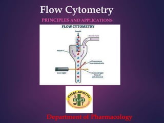
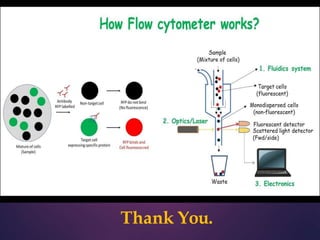
Flow cytometry is a technique that allows for the measurement and analysis of physical and chemical characteristics of cells and particles as they flow in a fluid stream through a beam of light. It works by using laser beams to interrogate cells as they flow through the instrument. Light scattering and fluorescent signals are detected and analyzed to provide information about properties like cell size, granularity, and expression of cell surface markers or intracellular proteins. Key applications of flow cytometry include immunophenotyping, cell sorting, cell cycle analysis, and detection of DNA content. It is a powerful tool widely used in research, clinical diagnostics, and other fields.
Equipments used , types of culture and media, subculturing, secondary culture, finite & continuous cell lines, cryopreservation and applications of cell culture
Nucleic Acid Quantification Methods - DNA / RNA Quantificationajithnandanam
Nucleic acids are quantified to check the concentration and purity of DNA/RNA present in the solution mixture.it is important to know the concentration and purity of the nucleic acid for the use in further applications like PCR, restriction digestion etc. Spectrophotometric analysis is the most commonly used method of quantifying DNA, agarose gel electrophoresis can also be used to analyse the DNA sample for purity.
The present document provide the information about Methods of extraction of drugs from the biological matrix (protein precipitation method, liquid extraction)
The Main Advantage
The main advantages of flow cytometry over histology and IHC is the possibility to precisely measure the quantities of antigens and the possibility to stain each cell with multiple antibodies-fluorophores, in current laboratories around 10 antibodies can be bound to each cell. This is much less than mass cytometer where up to 40 can be currently measured, but at a higher and slower pace.
Aquatic research
In aquatic systems, flow cytometry is used for the analysis of autofluorescing cells or cells that are fluorescently-labeled with added stains.
This research started in 1981 when Clarice Yentsch used flow cytometry to measure the fluorescence in a red tide producing dinoflagellates
Marine scientists use the sorting ability of flow cytometers to make discrete measurements of cellular activity and diversity, to conduct investigations into the mutualistic relationships between microorganisms that live in close proximity,and to measure biogeochemical rates of multiple processes in the ocean
Cell Proliferation assay
Cell proliferation is the major function in the immune system. Often it is required to analyse the proliferative nature of the cells in order to make some conclusions. One such assay to determine the cell proliferation is the tracking dye carboxyfluorescein diacetate succinimidyl ester (CFSE). It helps to monitor proliferative cells. This assay gives quantitative as well as qualitative data during time-series experiments
Cell counting
Cell sorting
Determining cell characteristics and function
Detecting microorganisms
Biomarker detection
Protein engineering detection
Diagnosis of health disorders such as blood cancers
Flow cytometry can be used for cell cycle analysis to estimate the percentages of a cell population in the different phases of the cell cycle, or it can be used with other reagents to analyze just the S phase.
Why flow cytometry is ideal for cell cycle analysis
Live-cell cycle analysis stains—Vybrant DyeCycle stains
Classic DNA cell cycle stains such as Hoechst 33342 and DRAQ5 for cell cycle analysis, but most of these have limitations that have to be considered when using them in an experiment which is why the Invitrogen Vybrant DyeCycle stains for live-cell cycle analysis were developed.
Fixed-cell cycle analysis stains FxCycle reagents
We offer classic DNA cell cycle stains such as DAPI, PI, and 7-AAD for fixed cell cycle analysis, but these reagents do not cover the full spectrum of laser excitation available.
The FxCycle reagents offer options for the 405 nm (violet) and 633 nm (red) laser thereby increasing the ability to multiplex by freeing up the 488 nm and 633 nm lasers for other cellular analyses such as immunophenotyping, apoptosis analysis, and dead cell discrimination.
Precise—Accurate cell cycle analysis in living cells
Safe—Low cytotoxicity for combining with additional live cell experiments
Cell sort compatible—Easily sort cells based on phase of the cell cycle
The technique of flow cytometry is used to evaluate cells for a number of functions, such as cell counting, phenotyping, cell cycle analysis, and viability.
diagnosis of cancer, bioluminescent detection, diagnosis of cancer, haplotype mapping, imaging gene expression in vivo, types of cancer diagnosis method, ultrasound imaging
Fluorescence- Activated Cell Sorter is a powerful technique used in cell sorting, cell-cycle analysis etc.
The presentation gives a basic understanding of the principle of FACS, instrumentation, interpretation of results, applications, how to do cell-cycle analysis using FACS and various troubleshooting tips.
Couples presenting to the infertility clinic- Do they really have infertility...Sujoy Dasgupta
Dr Sujoy Dasgupta presented the study on "Couples presenting to the infertility clinic- Do they really have infertility? – The unexplored stories of non-consummation" in the 13th Congress of the Asia Pacific Initiative on Reproduction (ASPIRE 2024) at Manila on 24 May, 2024.
These simplified slides by Dr. Sidra Arshad present an overview of the non-respiratory functions of the respiratory tract.
Learning objectives:
1. Enlist the non-respiratory functions of the respiratory tract
2. Briefly explain how these functions are carried out
3. Discuss the significance of dead space
4. Differentiate between minute ventilation and alveolar ventilation
5. Describe the cough and sneeze reflexes
Study Resources:
1. Chapter 39, Guyton and Hall Textbook of Medical Physiology, 14th edition
2. Chapter 34, Ganong’s Review of Medical Physiology, 26th edition
3. Chapter 17, Human Physiology by Lauralee Sherwood, 9th edition
4. Non-respiratory functions of the lungs https://academic.oup.com/bjaed/article/13/3/98/278874
micro teaching on communication m.sc nursing.pdfAnurag Sharma
Microteaching is a unique model of practice teaching. It is a viable instrument for the. desired change in the teaching behavior or the behavior potential which, in specified types of real. classroom situations, tends to facilitate the achievement of specified types of objectives.
- Video recording of this lecture in English language: https://youtu.be/lK81BzxMqdo
- Video recording of this lecture in Arabic language: https://youtu.be/Ve4P0COk9OI
- Link to download the book free: https://nephrotube.blogspot.com/p/nephrotube-nephrology-books.html
- Link to NephroTube website: www.NephroTube.com
- Link to NephroTube social media accounts: https://nephrotube.blogspot.com/p/join-nephrotube-on-social-media.html
Flu Vaccine Alert in Bangalore Karnatakaaddon Scans
As flu season approaches, health officials in Bangalore, Karnataka, are urging residents to get their flu vaccinations. The seasonal flu, while common, can lead to severe health complications, particularly for vulnerable populations such as young children, the elderly, and those with underlying health conditions.
Dr. Vidisha Kumari, a leading epidemiologist in Bangalore, emphasizes the importance of getting vaccinated. "The flu vaccine is our best defense against the influenza virus. It not only protects individuals but also helps prevent the spread of the virus in our communities," he says.
This year, the flu season is expected to coincide with a potential increase in other respiratory illnesses. The Karnataka Health Department has launched an awareness campaign highlighting the significance of flu vaccinations. They have set up multiple vaccination centers across Bangalore, making it convenient for residents to receive their shots.
To encourage widespread vaccination, the government is also collaborating with local schools, workplaces, and community centers to facilitate vaccination drives. Special attention is being given to ensuring that the vaccine is accessible to all, including marginalized communities who may have limited access to healthcare.
Residents are reminded that the flu vaccine is safe and effective. Common side effects are mild and may include soreness at the injection site, mild fever, or muscle aches. These side effects are generally short-lived and far less severe than the flu itself.
Healthcare providers are also stressing the importance of continuing COVID-19 precautions. Wearing masks, practicing good hand hygiene, and maintaining social distancing are still crucial, especially in crowded places.
Protect yourself and your loved ones by getting vaccinated. Together, we can help keep Bangalore healthy and safe this flu season. For more information on vaccination centers and schedules, residents can visit the Karnataka Health Department’s official website or follow their social media pages.
Stay informed, stay safe, and get your flu shot today!
Title: Sense of Taste
Presenter: Dr. Faiza, Assistant Professor of Physiology
Qualifications:
MBBS (Best Graduate, AIMC Lahore)
FCPS Physiology
ICMT, CHPE, DHPE (STMU)
MPH (GC University, Faisalabad)
MBA (Virtual University of Pakistan)
Learning Objectives:
Describe the structure and function of taste buds.
Describe the relationship between the taste threshold and taste index of common substances.
Explain the chemical basis and signal transduction of taste perception for each type of primary taste sensation.
Recognize different abnormalities of taste perception and their causes.
Key Topics:
Significance of Taste Sensation:
Differentiation between pleasant and harmful food
Influence on behavior
Selection of food based on metabolic needs
Receptors of Taste:
Taste buds on the tongue
Influence of sense of smell, texture of food, and pain stimulation (e.g., by pepper)
Primary and Secondary Taste Sensations:
Primary taste sensations: Sweet, Sour, Salty, Bitter, Umami
Chemical basis and signal transduction mechanisms for each taste
Taste Threshold and Index:
Taste threshold values for Sweet (sucrose), Salty (NaCl), Sour (HCl), and Bitter (Quinine)
Taste index relationship: Inversely proportional to taste threshold
Taste Blindness:
Inability to taste certain substances, particularly thiourea compounds
Example: Phenylthiocarbamide
Structure and Function of Taste Buds:
Composition: Epithelial cells, Sustentacular/Supporting cells, Taste cells, Basal cells
Features: Taste pores, Taste hairs/microvilli, and Taste nerve fibers
Location of Taste Buds:
Found in papillae of the tongue (Fungiform, Circumvallate, Foliate)
Also present on the palate, tonsillar pillars, epiglottis, and proximal esophagus
Mechanism of Taste Stimulation:
Interaction of taste substances with receptors on microvilli
Signal transduction pathways for Umami, Sweet, Bitter, Sour, and Salty tastes
Taste Sensitivity and Adaptation:
Decrease in sensitivity with age
Rapid adaptation of taste sensation
Role of Saliva in Taste:
Dissolution of tastants to reach receptors
Washing away the stimulus
Taste Preferences and Aversions:
Mechanisms behind taste preference and aversion
Influence of receptors and neural pathways
Impact of Sensory Nerve Damage:
Degeneration of taste buds if the sensory nerve fiber is cut
Abnormalities of Taste Detection:
Conditions: Ageusia, Hypogeusia, Dysgeusia (parageusia)
Causes: Nerve damage, neurological disorders, infections, poor oral hygiene, adverse drug effects, deficiencies, aging, tobacco use, altered neurotransmitter levels
Neurotransmitters and Taste Threshold:
Effects of serotonin (5-HT) and norepinephrine (NE) on taste sensitivity
Supertasters:
25% of the population with heightened sensitivity to taste, especially bitterness
Increased number of fungiform papillae
Ethanol (CH3CH2OH), or beverage alcohol, is a two-carbon alcohol
that is rapidly distributed in the body and brain. Ethanol alters many
neurochemical systems and has rewarding and addictive properties. It
is the oldest recreational drug and likely contributes to more morbidity,
mortality, and public health costs than all illicit drugs combined. The
5th edition of the Diagnostic and Statistical Manual of Mental Disorders
(DSM-5) integrates alcohol abuse and alcohol dependence into a single
disorder called alcohol use disorder (AUD), with mild, moderate,
and severe subclassifications (American Psychiatric Association, 2013).
In the DSM-5, all types of substance abuse and dependence have been
combined into a single substance use disorder (SUD) on a continuum
from mild to severe. A diagnosis of AUD requires that at least two of
the 11 DSM-5 behaviors be present within a 12-month period (mild
AUD: 2–3 criteria; moderate AUD: 4–5 criteria; severe AUD: 6–11 criteria).
The four main behavioral effects of AUD are impaired control over
drinking, negative social consequences, risky use, and altered physiological
effects (tolerance, withdrawal). This chapter presents an overview
of the prevalence and harmful consequences of AUD in the U.S.,
the systemic nature of the disease, neurocircuitry and stages of AUD,
comorbidities, fetal alcohol spectrum disorders, genetic risk factors, and
pharmacotherapies for AUD.
Explore natural remedies for syphilis treatment in Singapore. Discover alternative therapies, herbal remedies, and lifestyle changes that may complement conventional treatments. Learn about holistic approaches to managing syphilis symptoms and supporting overall health.
ARTIFICIAL INTELLIGENCE IN HEALTHCARE.pdfAnujkumaranit
Artificial intelligence (AI) refers to the simulation of human intelligence processes by machines, especially computer systems. It encompasses tasks such as learning, reasoning, problem-solving, perception, and language understanding. AI technologies are revolutionizing various fields, from healthcare to finance, by enabling machines to perform tasks that typically require human intelligence.
2. What is Flow Cytometry?
• ‘Flow Cytometry’ as the name suggests is a technique for cell
counting and measurement of different properties of the cell
(‘cyto’= cell; ‘metry’=count/measurement).
• It is a laser based technology that measures and analyses
different physical and chemical properties of the cells/particles
flowing in a stream of fluid through a beam of light.
4. Historical Perspective -
Evolution of Flow Cytometry
17th
Century
1934
1947 to
1949
1953 1965
1879 1968
1970s
onwards…
Development of
light microscope
by
Leeuwenhoek.
Principles of Droplet
formation by Lord
Rayleigh.
Counting of RBCs
by Moldavan by
forcing a
suspension of cells
through capillary
tube.
Development of
Coulter Principle by
Wallace Coulter and
counting of RBCs
using the first Coulter
Counter.
Optical counting of
RBCs by Crosland-
Taylor by use of
laminar flow
principles
Development of
electrostatic
inkjet droplet
deflection by
Richard Sweet
Application of
Sweet’s principle
and Coulter principle
to develop the first
cell sorter by M.
Fulwyler
Development of
fluorescence
based cell sorter
by Wolfgang
Gohde
Development of
FACS and other
advances.
5. Principles of working of
Flow Cytometer
Coulter
Principle
Principle s
of Laminar
Flow
Electro
statics
Optics
& Light
Scattering
Flow
Cytometry
6. Components of a Flow Cytometer
• A flow cytometer is made up of three main systems: fluidics, optics
and electronics.
The fluidics system transports particles in a stream to the laser
beam for interrogation.
The optics system consists of lasers to illuminate the particles in
the sample stream and optical filters to direct the resulting light
signals to the appropriate detectors.
The electronics system converts the detected light signals into
electronic signals that can be processed by the computer.
For some instruments equipped with a sorting feature, the
electronics system is also capable of initiating sorting decisions to
charge and deflect particles.
7. Working of a Flow Cytometer
• In the flow cytometer, particles are carried to the laser intercept in a
fluid stream. Any suspended particle or cell from 0.2–150
micrometers in size is suitable for analysis.
• The portion of the fluid stream where particles are located is called the
sample core. When particles pass through the laser intercept, they
scatter laser light.
• Any fluorescent molecules present on the particle fluoresce. The
scattered and fluorescent light is collected by appropriately positioned
lenses.
• A combination of beam splitters and filters steers the scattered and
fluorescent light to the appropriate detectors. The detectors produce
electronic signals proportional to the optical signals striking them.
8. Applications of Flow Cytometry
• Flow cytometry is the sine qua non (without which, nothing)of the
modern researcher’s toolbox.
• Flow cytometry measures multiple characteristics of individual
particles flowing in single file in a stream of fluid.
• Light scattering at different angles can distinguish differences in size
and internal complexity, whereas light emitted from fluorescently
labeled antibodies can identify a wide array of cell surface and
cytoplasmic antigens.
• This approach makes flow cytometry a powerful tool for detailed
analysis of complex populations in a short period of time.
9. Applications
Immunophenotyping
• Cell subsets are measured by labeling population-specific proteins
with a fluorescent tag on the cell surface.
• In clinical labs, immunophenotyping is useful in diagnosing
hematological malignancies such as lymphomas and leukemia.
Cell Sorting
• The cell sorter is a specialized flow cytometer with the ability to
physically isolate cells of interest into separate collection tubes.
• The sorter uses sophisticated electronics and fluidics to identify
and "kick" the cells of interest out of the fluidic stream into a test
tube.
10. Cell Cycle Analysis
• Flow cytometry can analyze replication states using fluorescent
dyes to measure the four distinct phases of the cell cycle.
• Along with determining cell cycle replication states, the assay can
measure cell aneuploidy associated with chromosomal
abnormalities.
Apoptosis
• The two distinct types of cell death, apoptosis and necrosis, can be
distinguished by flow cytometry on the basis of differences in
morphological, biochemical and molecular changes occurring in
the dying cells.
11. Cell Proliferation Assays
• The flow cytometer can measure proliferation by labeling resting
cells with a cell membrane fluorescent dye, carboxyfluorescein
succinimidyl ester (CFSE).
• When the cells are activated, they begin to proliferate and undergo
mitosis. As the cells divide, half of the original dye is passed on to
each daughter cell.
• By measuring the reduction of the fluorescence signal, researchers
can calculate cellular activation and proliferation.
12. DNA Content Analysis
• The measurement of cellular DNA content by flow cytometry uses
fluorescent dyes, such as propidium iodide, that intercalate into the
DNA helical structure.
• The fluorescent signal is directly proportional to the amount of
DNA in the nucleus and can identify gross gains or losses in DNA.
13. Fluorescence Activated Cell Sorting (FACS)
• Consider a group of lymphocytes from a mouse that have been stained
with green fluorescent antibodies specific for CD4 (e.g., fluorescein
isothiocyanate, or FITC anti-CD4) and red fluorescent antibodies specific
for CD8 (e.g., phycoerythrin, or PE anti-CD8).
• Both the labeled cells generate SSC and FSC as they pass through the
laser beam creating voltage pulses that are recorded by the computer.
• However, each labeled cell will also emit light of specific wavelength as
a result of the fluorescent label. For instance, CD4 cells will emit green
fluorescent light of wavelength 525-530 nm while CD8 cells emit orange
light of wavelength 560 nm.
• These fluorescent signals pass through the Photomultiplier tubes and
generate voltage pulses. The software integrates all the information for
a particular cell allowing characterization of individual cells.
14.
15. Clinical Applications – DNA Content Analysis
• Investigators are currently using techniques of DNA flow cytometry
to measure ploidy status (DNA content) and proliferative potential
(S phase fraction) in a wide variety of solid tumors.
• These measurements have shown relevance for diagnosis, prognosis,
and treatment for patients with cancer.
• The measurement of cellular DNA content by flow cytometry uses
fluorescent dyes, such as propidium iodide, that intercalate into the
DNA helical structure.
• The fluorescent signal is directly proportional to the amount of DNA
in the nucleus and can identify gross gains or losses in DNA.
• Abnormal DNA content, also known as “DNA content aneuploidy”,
can be determined in a tumor cell population.
16. • DNA aneuploidy generally is associated with malignancy;
however, certain benign conditions may appear aneuploid.
Cell Cycle Analysis
• This technique is based on the premise that cells in G0 or G1
phases of the cell cycle possess a normal diploid
chromosomal, and hence DNA content (2n) whereas cells in
G2 and just prior to mitosis (M) contain exactly twice this
amount (4n).
• As DNA is synthesized during S-phase, cells are found with a
DNA content ranging between 2n and 4n.
• A histogram plot of DNA content against cell numbers gives
the classical DNA profile for a proliferating cell culture.
17. Flow Cytometry and Ecology
• Assessments of diversity, abundance, and activity of water column
microorganisms are fundamental to studies in aquatic microbiology.
• Currently, most applications of flow cytometry to environmental
samples make use of various morphological and physiological
characteristics of the cells (e.g., size and pigment content of
photosynthetic organisms).
• These criteria generally are not sufficient for identification at the genus
or species level. Staining with DNA-specific fluorochromes offers
information about numbers of bacterial cells but not about their identity.
• The combined use of dyes that bind preferentially to G- C or A. T base
pairs has been used to distinguish organisms of different G+C content
18. Flow Cytometry and Cancer Research
• The prognosis of patients with cancer is largely determined by the specific
histological diagnosis, tumor mass stage, and host performance status.
• Quantitative cytology in the form of flow cytometry has greatly advanced
the objective elucidation of tumor cell heterogeneity by using probes that
discriminate tumor and normal cells and assess differentiate as well as
proliferative tumor cell properties.
• Both DNA content analysis and FACS can be utilised in cancer research.
• Abnormal nuclear DMA content is a conclusive marker of malignancy and
is found with increasing frequency in leukemia (23% among 793 patients),
in lymphoma (53% among 360 patients), and in myeloma (76% among 177
patients), as well as in solid tumors (75% among 3611 patients), for an
overall incidence of 67% in 4941 patients.
19. • Flow cytometric immunophenotyping (FCI) aids in the differentiation
of chronic lymphocytic leukemia (CLL) from mantle cell lymphoma
(MCL); however, overlapping phenotypes may occur.
• CD11c expression has been reported in up to 90% of CLL cases but
has rarely been reported in MCL.
• Whether CD11c can be used to exclude MCL has not been directly
addressed.
• FCI reports were reviewed for 90 MCL cases (44 patients) and 355
CLL/small lymphocytic lymphoma (SLL) cases (158 patients).