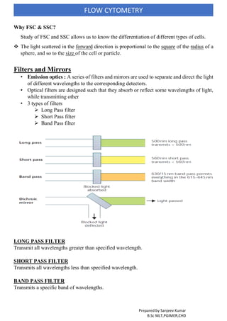
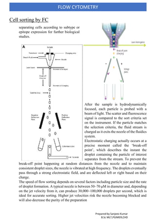
FLOW CYTOMETRY, PRINCIPLE, APPLICATION, USE IN HAEMATOLOGY, COMPONENT OF FLOW CYTOMETRY, DATA INTERPRETATION, DATA ANALYSIS, CELL SHORTING ADVANTAGES AND DISADVANTAGES, IMMUNOLOGICAL CLASSIFICATION OF ACUTE
LEUKEMIA
This slide show forms part of the Introduction to Flow Cytometry seminar help by The Garvan MLC Flow Cytometry Facility. The Garvan MLC Flow Cytometry Facility is part of the Garvan Institute of Medical Research and is located in Sydney NSW.
It is a laser based technology that measures and analyses different physical and chemical properties of the cells/particles flowing in a stream of fluid through a beam of light
Flow cytometry is a standard laser-based technology that is used in the detection and measurement of physical and chemical characteristics of cells or particles in a heterogeneous fluid mixture.
Fluorescence- Activated Cell Sorter is a powerful technique used in cell sorting, cell-cycle analysis etc.
The presentation gives a basic understanding of the principle of FACS, instrumentation, interpretation of results, applications, how to do cell-cycle analysis using FACS and various troubleshooting tips.
Introduction
Definition
Basic mechanism
Prerequisite of flow cytometer
Components of flow cytometry
Flow system
Optics system
Concept of scattering
Advantage
Limitation
Application
Conclusion
References
CHAPTER 1 SEMESTER V - ROLE OF PEADIATRIC NURSE.pdfSachin Sharma
Pediatric nurses play a vital role in the health and well-being of children. Their responsibilities are wide-ranging, and their objectives can be categorized into several key areas:
1. Direct Patient Care:
Objective: Provide comprehensive and compassionate care to infants, children, and adolescents in various healthcare settings (hospitals, clinics, etc.).
This includes tasks like:
Monitoring vital signs and physical condition.
Administering medications and treatments.
Performing procedures as directed by doctors.
Assisting with daily living activities (bathing, feeding).
Providing emotional support and pain management.
2. Health Promotion and Education:
Objective: Promote healthy behaviors and educate children, families, and communities about preventive healthcare.
This includes tasks like:
Administering vaccinations.
Providing education on nutrition, hygiene, and development.
Offering breastfeeding and childbirth support.
Counseling families on safety and injury prevention.
3. Collaboration and Advocacy:
Objective: Collaborate effectively with doctors, social workers, therapists, and other healthcare professionals to ensure coordinated care for children.
Objective: Advocate for the rights and best interests of their patients, especially when children cannot speak for themselves.
This includes tasks like:
Communicating effectively with healthcare teams.
Identifying and addressing potential risks to child welfare.
Educating families about their child's condition and treatment options.
4. Professional Development and Research:
Objective: Stay up-to-date on the latest advancements in pediatric healthcare through continuing education and research.
Objective: Contribute to improving the quality of care for children by participating in research initiatives.
This includes tasks like:
Attending workshops and conferences on pediatric nursing.
Participating in clinical trials related to child health.
Implementing evidence-based practices into their daily routines.
By fulfilling these objectives, pediatric nurses play a crucial role in ensuring the optimal health and well-being of children throughout all stages of their development.
How many patients does case series should have In comparison to case reports.pdfpubrica101
Pubrica’s team of researchers and writers create scientific and medical research articles, which may be important resources for authors and practitioners. Pubrica medical writers assist you in creating and revising the introduction by alerting the reader to gaps in the chosen study subject. Our professionals understand the order in which the hypothesis topic is followed by the broad subject, the issue, and the backdrop.
https://pubrica.com/academy/case-study-or-series/how-many-patients-does-case-series-should-have-in-comparison-to-case-reports/
The dimensions of healthcare quality refer to various attributes or aspects that define the standard of healthcare services. These dimensions are used to evaluate, measure, and improve the quality of care provided to patients. A comprehensive understanding of these dimensions ensures that healthcare systems can address various aspects of patient care effectively and holistically. Dimensions of Healthcare Quality and Performance of care include the following; Appropriateness, Availability, Competence, Continuity, Effectiveness, Efficiency, Efficacy, Prevention, Respect and Care, Safety as well as Timeliness.
Trauma Outpatient Center is a comprehensive facility dedicated to addressing mental health challenges and providing medication-assisted treatment. We offer a diverse range of services aimed at assisting individuals in overcoming addiction, mental health disorders, and related obstacles. Our team consists of seasoned professionals who are both experienced and compassionate, committed to delivering the highest standard of care to our clients. By utilizing evidence-based treatment methods, we strive to help our clients achieve their goals and lead healthier, more fulfilling lives.
Our mission is to provide a safe and supportive environment where our clients can receive the highest quality of care. We are dedicated to assisting our clients in reaching their objectives and improving their overall well-being. We prioritize our clients' needs and individualize treatment plans to ensure they receive tailored care. Our approach is rooted in evidence-based practices proven effective in treating addiction and mental health disorders.
COVID-19 PCR tests remain a critical component of safe and responsible travel in 2024. They ensure compliance with international travel regulations, help detect and control the spread of new variants, protect vulnerable populations, and provide peace of mind. As we continue to navigate the complexities of global travel during the pandemic, PCR testing stands as a key measure to keep everyone safe and healthy. Whether you are planning a business trip, a family vacation, or an international adventure, incorporating PCR testing into your travel plans is a prudent and necessary step. Visit us at https://www.globaltravelclinics.com/
Health Education on prevention of hypertensionRadhika kulvi
Hypertension is a chronic condition of concern due to its role in the causation of coronary heart diseases. Hypertension is a worldwide epidemic and important risk factor for coronary artery disease, stroke and renal diseases. Blood pressure is the force exerted by the blood against the walls of the blood vessels and is sufficient to maintain tissue perfusion during activity and rest. Hypertension is sustained elevation of BP. In adults, HTN exists when systolic blood pressure is equal to or greater than 140mmHg or diastolic BP is equal to or greater than 90mmHg. The
India Clinical Trials Market: Industry Size and Growth Trends [2030] Analyzed...Kumar Satyam
According to TechSci Research report, "India Clinical Trials Market- By Region, Competition, Forecast & Opportunities, 2030F," the India Clinical Trials Market was valued at USD 2.05 billion in 2024 and is projected to grow at a compound annual growth rate (CAGR) of 8.64% through 2030. The market is driven by a variety of factors, making India an attractive destination for pharmaceutical companies and researchers. India's vast and diverse patient population, cost-effective operational environment, and a large pool of skilled medical professionals contribute significantly to the market's growth. Additionally, increasing government support in streamlining regulations and the growing prevalence of lifestyle diseases further propel the clinical trials market.
Growing Prevalence of Lifestyle Diseases
The rising incidence of lifestyle diseases such as diabetes, cardiovascular diseases, and cancer is a major trend driving the clinical trials market in India. These conditions necessitate the development and testing of new treatment methods, creating a robust demand for clinical trials. The increasing burden of these diseases highlights the need for innovative therapies and underscores the importance of India as a key player in global clinical research.
Empowering ACOs: Leveraging Quality Management Tools for MIPS and BeyondHealth Catalyst
Join us as we delve into the crucial realm of quality reporting for MSSP (Medicare Shared Savings Program) Accountable Care Organizations (ACOs).
In this session, we will explore how a robust quality management solution can empower your organization to meet regulatory requirements and improve processes for MIPS reporting and internal quality programs. Learn how our MeasureAble application enables compliance and fosters continuous improvement.
For those battling kidney disease and exploring treatment options, understanding when to consider a kidney transplant is crucial. This guide aims to provide valuable insights into the circumstances under which a kidney transplant at the renowned Hiranandani Hospital may be the most appropriate course of action. By addressing the key indicators and factors involved, we hope to empower patients and their families to make informed decisions about their kidney care journey.
1. Prepared by Sanjeev Kumar
B.Sc MLT,PGIMER,CHD
FLOW CYTOMETRY
Definition:
Measuring properties of cell as they flow in a fluid suspension across an illuminated
light path.
Flow - fluid
Cyto - cells
Metry - measure
• Flow cytometry is a laser based, biophysical technology employed in cell counting,
sorting and biomarker detection by suspending cells in a stream of fluid and passing
them by an electronic detection apparatus.
• Measurement of cellular properties as they are moving in a fluids stream.
• Flow cytometry: Flow: cell move in single file & Cytometer: measurement of
numerous cell properties
• Unique ability to demonstrate protein expressed by cell at the rate of 5,000-10,000 cells
per second.
• This method allows the quantitative and qualitative analysis of several properties of cell
populations from virtually any type of fresh unfixed tissue or body fluid.
Most commonly analyzed materials are:
blood,
bone marrow aspirate and
lymph node suspensions.
INTRODUCTION
The concept of flow cytometry has been in existence for more than five decades.
Flow cytometric immunophenotyping (FCI) first appeared in clinical laboratories in the
1980s, in the wake of the AIDS epidemic.
Initially utilized to assess CD4 T-cells, the technique was soon applied to lymphoid and
eventually myeloid neoplasms.
Current flow cytometers have the capability of simultaneously measuring multiple
parameters of individual cells in a cell suspension.
Thus, a large number of cell specimens can be processed with a quick turnaround time.
In addition, flow cytometry is also highly sensitive and can detect immunophenotype of
cells in a specimen with thousands of cells.
The parameters analyzed by flow cytometry include
physical properties of cells; the size, cytoplasmic granularity, and amount of DNA
contents; and
cell antigens/markers (surface, cytoplasmic, and nuclear) that can be recognized by
specific antibodies.
By using appropriate antibody panels, flow cytometry can reveal
the cell type (hematopoietic, lymphoid, or nonhematopoietic),
cell lineage (B- and T cells, natural killer cells, myeloid/ monocytic cells,
neuro/neuroendocrine cells, and epithelial cells),
cell maturation stage (precursors vs. matured cells)
2. Prepared by Sanjeev Kumar
B.Sc MLT,PGIMER,CHD
FLOW CYTOMETRY
Basic mechanism
Biological sample
Label it with a fluorescent marker
Cells move in a linear stream through a focused light source (laser beam)
Fluorescent molecule gets activated and emits light that is filtered and detected by sensitive
light detectors (usually a photomultiplier tube)
Conversion of analog fluorescent signals to digital signals
Basic principle of FC
• Cell or particle incubated with fluorescent dye labeled antibodies in suspension is hydro
dynamically focused in a single file using sheath fluid under optimized pressure and when
a laser beam strikes a cell provide information about properties of cell.
It includes
• Relative size of cell
• Relative granularity or internal complexity, an
• Relative fluorescence intensity
• Based in these properties FC identifies cell population of interest
3. Prepared by Sanjeev Kumar
B.Sc MLT,PGIMER,CHD
FLOW CYTOMETRY
Component of FC
1. Fluidics-Flow chamber and sheath Fluid
2. Optics-laser, Light Sensing (FSC and SSC), Photomultiplier diode(PMT) and Filters-
Mirrors
3. Electronics-Software, hardware and Digital
Light
source
Flow
Chamber
Optical
system
Light
Detector
ElectronicComputer
4. Prepared by Sanjeev Kumar
B.Sc MLT,PGIMER,CHD
FLOW CYTOMETRY
Fluidics-Flow chamber
One of the fundamentals of flow cytometry is
the ability to measure the properties of
individual particles, which is managed by the
fluidics system.
Heart of instrument.
It is designed to deliver cells in single file at
point of measurement.
Sample is injected into the center of sheath fluid.
Flow chamber wall made up of quartz cuvette.
Once the sample is injected into a stream of
sheath fluid within the flow chamber, they are
forced into the center of the stream forming a
single file by the PRINCIPLE OF
HYDRODYNAMIC FOCUSING.
'Only one cell or particle can pass through the laser beam at a given moment.'
As the sheath fluid moves, it creates a massive drag effect on the narrowing central
chamber. This alters the velocity of the central fluid whose flow front becomes parabolic
with greatest velocity at its centre and zero velocity at the wall. The effect creates a single
file of particles and is called hydrodynamic focusing.
Closed camber -used for analysis.
Open chamber -ample stream emerges into open chamber- used for analyzing and sorting
cell
Optical- Light Sensing
• Laser or an arc lamp or LED
• Commonly used laser is air cooled argon ion laser producing blue light at 448 nm
• Laser produce a high intensity beam of monochromatic light
• Occur at the point of interception of stationary light and cell.
• Occur light scattering and emission of fluorescence light.
• Factors that affect light scattering are the cell's membrane, nucleus, and any granular
material inside the cell.
• Light that is scattered in the forward direction (along the same axis the laser is traveling)
is detected in the Forward Scatter Channel.
• Laser light that is scattered at 90° to the axis of the laser path is detected in the Side
Scatter Channel.
• Why Lasers are more common?
• They are highly coherent and uniform. They can be easily focused on a very small
area (like a sample stream). They are monochromatic, emitting single
wavelengths of light.
5. Prepared by Sanjeev Kumar
B.Sc MLT,PGIMER,CHD
FLOW CYTOMETRY
When a light intersects a laser beam at the so called 'interrogation point' two events occur:
a) light scattering
b) emission of light (fluorescence)
Fluorescence is light emitted during decay of excited electron to its basal state.
When light from a laser interrogates a cell, that cell scatters light in all directions.
The scattered light can travel from the interrogation point down a path to a detector.
FORWARD SCATTER (FSC)
• Light that is scattered in the forward direction (along the same axis the laser is traveling)
is detected in the Forward Scatter Channel.
• The intensity of this signal has been attributed to cell size, refractive index (membrane
permeability)
SIDE SCATTER (SSC)
Laser light that is scattered at 90 degrees
to the axis of the laser path is detected in
the Side Scatter Channel.
The intensity of this signal is proportional
to the amount of cytosolic structure in the
cell (e.g. granules, cell inclusions, etc.)
Side scatter detector
Measuring cell
granularity
FSC
Detector
Collection Lens
SSC Detector
Laser
Beam
6. Prepared by Sanjeev Kumar
B.Sc MLT,PGIMER,CHD
FLOW CYTOMETRY
Why FSC & SSC?
Study of FSC and SSC allows us to know the differentiation of different types of cells.
The light scattered in the forward direction is proportional to the square of the radius of a
sphere, and so to the size of the cell or particle.
Filters and Mirrors
• Emission optics : A series of filters and mirrors are used to separate and direct the light
of different wavelengths to the corresponding detectors.
• Optical filters are designed such that they absorb or reflect some wavelengths of light,
while transmitting other
• 3 types of filters
Long Pass filter
Short Pass filter
Band Pass filter
LONG PASS FILTER
Transmit all wavelengths greater than specified wavelength.
SHORT PASS FILTER
Transmits all wavelengths less than specified wavelength.
BAND PASS FILTER
Transmits a specific band of wavelengths.
7. Prepared by Sanjeev Kumar
B.Sc MLT,PGIMER,CHD
FLOW CYTOMETRY
Dichroic Filters
Can be a long pass or short pass filter
Filter is placed at a 45º angle to the incident light
Part of the light is reflected at 90º to the incident light, and part of the light is transmitted
and continues on.
Detectors
There are two main types of photo detectors used in flow cytometer
Photodiodes
Used for strong signals, when saturation is a potential problem (e.g. FSC detector)
Photo Detectors usually have a band pass filter in front of them to only allow a
specific band width of light to reach it.
These are sensitive instrument and ideal for scatter and fluorescence detection.
The amount of light or the number of photons are proportional to the amount of
fluorochrome present in the cell.
Photo multiplier tubes (PMT)
More sensitive than a Photodiode, but can be destroyed by exposure to too much
light.
PMT is used for detecting small amounts of fluorescence emitted from
fluorochromes (e.g. SSC & fluorescent signals)
PMT Convert the incident light into electronic pulses.
Once PMT generates a pulse, the signal needs to be amplified & ultimately digitized
for computer analysis by use of amplifiers & analogue-to- digital converters.
(ADCs). Silicon photodiodes are usually enough to measure forward scatter.
Electronic of FC
• When light hits a photo detector a small
current (a few microamperes) is
generated. Its associated voltage has an
amplitude proportional to the total
number of light photons received by the
detector.
• This voltage is then amplified by a
series of linear or logarithmic
amplifiers, and by analogue to digital
convertors (ADCs), into electrical
signals large enough (5–10 volts) to be
plotted graphically.
• Computer system is used for analysis,
presentation & storage of cytometric
data.This raw data in computer permits
repeated analysis, often with different
gating or compensation.
8. Prepared by Sanjeev Kumar
B.Sc MLT,PGIMER,CHD
FLOW CYTOMETRY
Data Analysis
In a Flow- cytometer data is analyses as-
Single parameter
Multiple parameter
In a single parameter, we analyses only one
parameter on each cell & prepared a scatter gram
FSC-SSC
In multiple parameter, we analyses more than
one parameter on each cell & prepared a scatter gram
FL1-FL2
Data Interpretation
The digitized data, stored in a computer software & interpret as-
Histogram
Dot-plot analysis
Histogram relies on the
measurement of pulses of a
given value & their
assignment to channels
which represent diff.
Voltage levels. The
process of counting each
pulse in the appropriate
channel is known as ADC.
Software present in the flow
cytometer, interpret the signals in
dot plot analysis as-
FSC
SSC
Single Parameter Graph
Multiple parameter
FSC
SSC
9. Prepared by Sanjeev Kumar
B.Sc MLT,PGIMER,CHD
FLOW CYTOMETRY
Cell sorting by FC
separating cells according to subtype or
epitope expression for further biological
studies.
After the sample is hydrodynamically
focused, each particle is probed with a
beam of light. The scatter and fluorescence
signal is compared to the sort criteria set
on the instrument. If the particle matches
the selection criteria, the fluid stream is
charged as it exits the nozzle of the fluidics
system.
Electrostatic charging actually occurs at a
precise moment called the ‘break-off
point’, which describes the instant the
droplet containing the particle of interest
separates from the stream. To prevent the
break-off point happening at random distances from the nozzle and to maintain
consistent droplet sizes, the nozzle is vibrated at high frequency. The droplets eventually
pass through a strong electrostatic field, and are deflected left or right based on their
charge.
The speed of flow sorting depends on several factors including particle size and the rate
of droplet formation. A typical nozzle is between 50–70 μM in diameter and, depending
on the jet velocity from it, can produce 30,000–100,000 droplets per second, which is
ideal for accurate sorting. Higher jet velocities risk the nozzle becoming blocked and
will also decrease the purity of the preparation
10. Prepared by Sanjeev Kumar
B.Sc MLT,PGIMER,CHD
FLOW CYTOMETRY
Clinical Applications in Hematology
Today, flow cytometer has become a principal tool for the diagnosis of various disorders
in clinical practice. Some applications of flow cytometer are as follow-
For the diagnosis of Acute Leukemia.
For the diagnosis of CLPD.
For the diagnosis of PNH.
For the detection of MRD.
For the diagnosis of Platelet abnormality.
Detection of fetal-cells in mother-circulation.
For reticulocyte count.
IMMUNOLOGICAL CLASSIFICATION OF ACUTE
LEUKEMIA
Acute Lymphoblastic Leukemia (ALL) Tdt+ve-
B-cell lineage (CD19+ve, CD79a+ve &CD22+ve)-
Pro B-ALL( CD10-, CD15+)
Common ALL (CD10+, cIgM- )
Pre B ALL ( cIg M+ )
Mature B ALL (sIg +)
T-Cell lineage ( cCD3+, CD7+)-
Pro T-ALL( CD3+, CD7+)
Pre T-ALL(CD2+, CD5+)
Corticol T-ALL( CD1a+)
Mature T-ALL (CD3+)
Acute Myeloid Leukemia (AML)-
AML(M0-M5) (anti MPO+ ,CD13+, CD33+, CD117+ )
Pure erythroid leukaemia (antiglycophorin A+, antiblood group antigen+, CD36+ )
Megakaryoblastic leukaemia ( CD41+, CD42+, CD61+)
11. Prepared by Sanjeev Kumar
B.Sc MLT,PGIMER,CHD
FLOW CYTOMETRY
Panel of monoclonal Abs for the Diagnosis of Acute leukemia
ALL AML
B-lineage T-lineage
First Line CD19,CD22,
CD79a,
CD10
CD7,
CD2,
cyCD3
CD13, CD33,
CD117, anti-
MPO
TdT, HLA-DR, CD34
Second
Line
cymu, SmIg CD1a,
CD5,
CD8,
anti-TCR
CD41, CD42,
CD61,
anti-
glycophorin A
Chronic Lymphoproliferative Disorder (CLPD)
Chronic Lymphocytic Leukemia(CLL).
Prolymphocytic Leukemia(PLL).
Hairy cell Leukemia(HCL).
Splenic lymphoma with villous lymphocyte (SLVL).
Adult T-cell Leukemia /Lymphoma.
Panel of monoclonal Abs for the diagnosis of lymphoid
disorders
B-Cell T-Cell
First Line SmIg (kappa /lambda)
CD19, CD23, FMC7
mCD79b, mCD22, CD5
CD2,
CD5,
Second
Line
CD11c, CD25, CD103
CD123, CD38, CD138, CyIg
CD3, CD4, CD7, CD8
12. Prepared by Sanjeev Kumar
B.Sc MLT,PGIMER,CHD
FLOW CYTOMETRY
PNH
Acquired hematological disorder characterized by anemia, intravascular hemolysis
specially at night while patient is sleeping as well as tendency to thrombosis and
infection.
Results from the mutation in the PGPA gene in the haemopoietic stem cells, which leads
to reduced / absent synthesis of GPI proteins.
Increased sensitivity of erythrocytes to complement-mediated cell lysis due to
deficiency of membrane-bound GPI anchored proteins which normally function as the
inhibitors of reactive hemolysis. GPI-anchored proteins include: complement regulatory
proteins like DAF (decay accelerating factor, CD55), MIRL (membrane inhibitor of
reactive lysis, CD59),
Used to detect the deficiency of GPI anchored proteins on RBC & WBC (neutrophils).
Reticulocyte enumeration
This method provides excellent discrimination between reticulocytes and mature RBCs,
with greater precision, sensitivity, and reproducibility than the traditional method.
The flow cytometric enumeration of reticulocytes uses fluorescent dyes that bind the
residual RNA, such as thiazole orange
The fluorescence intensity is directly proportional to the amount of RNA and related to
the immaturity of the RBC, a reticulocyte maturity index can be given.
Fetomaternal Hemorrhage
The use of flow cytometry for the detection of fetal cells is much more objective,
reproducible, and sensitive than the other traditional method.
Fluorescent labeled antibodies to the rhesus (D) antigen can be used, or more recently,
antibodies directed against hemoglobin F.
This method has the ability to distinguish fetal cells from Adult-cells (adult red cells
with small amounts of hemoglobin F).
This intracellular approach, which uses permeabilization of the red cell membrane and
an antibody to the g chain of human hemoglobin, is precise and sensitive.
These are the few applications of flow cytometer, but now days a flow cytometer is
broadly used in DNA analysis, cell cycle analysis, chromosome analysis, cell-culture
studies, detection of blood-parasites & bacteria in blood and body fluids etc. The main
principle is the same, only the change in the nature of sample & monoclonal Ab’s
tagged to cell surface.
13. Prepared by Sanjeev Kumar
B.Sc MLT,PGIMER,CHD
FLOW CYTOMETRY
Advantages of flow cytometry
• Rapid assessment of large no. of cells
• Multi parameter analysis
• High accuracy & reproducibility
• Objective analysis
• Ability to analyze many samples quickly
• Capable of data reduction
• Permanent data storage
• Ability to reanalyze data
• Requires relatively small sample
Disadvantage
• Expensive
• Need trained Flow cytometrist.
• Difficult to obtain the antibodies according to panel list.
• Chance of auto fluorescence and false negative result in case of deteriorate reagent