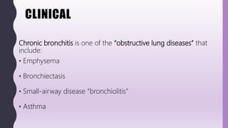
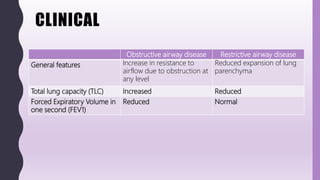
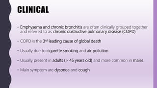
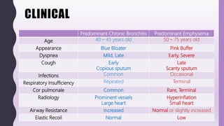
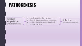
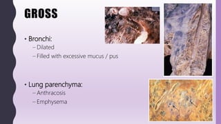
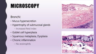
FLASHPATH
H a z e m A l i
CHRONIC
BRONCHITIS
H a z e m A l i
CLINICAL
Chronic bronchitis is one of the “obstructive lung diseases” that
include:
• Emphysema
• Bronchiectasis
• Small-airway disease “bronchiolitis”
• Asthma
CLINICAL
Obstructive airway disease Restrictive airway disease
General features Increase in resistance to
airflow due to obstruction at
any level
Reduced expansion of lung
parenchyma
Total lung capacity (TLC) Increased Reduced
Forced Expiratory Volume in
one second (FEV1)
Reduced Normal
CLINICAL
• Emphysema and chronic bronchitis are often clinically grouped together
and referred to as chronic obstructive pulmonary disease (COPD)
• COPD is the 3rd leading cause of global death
• Usually due to cigarette smoking and air pollution
• Usually present in adults (> 45 years old) and more common in males
• Main symptom are dyspnea and cough
CLINICAL
Predominant Chronic Bronchitis Predominant Emphysema
Age 40 – 45 years old 50 – 75 years old
Appearance Blue Bloater Pink Buffer
Dyspnea Mild, Late Early, Severe
Cough Early
Copious sputum
Late
Scanty sputum
Infections Common Occasional
Respiratory Insufficiency Repeated Terminal
Cor pulmonale Common Rare, Terminal
Radiology Prominent vessels
Large heart
Hyperinflation
Small heart
Airway Resistance Increased Normal or slightly increased
Elastic Recoil Normal Low
CLINICAL
• Chronic bronchitis is:
– Chronic disease of large airways
– Persistent “productive” cough
– For at least 3 months
– In at least 2 consecutive years
– Without other apparent explanation
CLINICAL
Lines of treatment:
• Smoking cessation
• Medications (Bronchodilators, Steroids)
• Oxygen therapy and Ventilatory support
• Antibiotics (for 2ry infections)
• Lung transplantation (for end-stage diseases)
Main causes of death:
• Coronary artery disease
• Respiratory failure
• Right-sided heart failure
PATHOGENESIS
Smoking
Air pollution
(initiate bronchitis)
• Interferes with ciliary action
• Directly damages airway epithelium
• Inhibits ability of white blood cells
to clear bacteria
Infection
(maintain bronchitis)
GROSS
• Bronchi:
– Dilated
– Filled with excessive mucus / pus
• Lung parenchyma:
– Anthracosis
– Emphysema
MICROSCOPY
Bronchi:
• Mucus hypersecretion
• Hypertrophy of submucosal glands
– Increased Reid index
• Goblet cell hyperplasia
• Squamous metaplasia, Dysplasia
• Chronic inflammation
– No eosinophils
MICROSCOPY
Reid index
• Ratio of
Thickness of mucus gland layer
"superficial surface of glandular layer to superficial perichondrium"
Thickness of bronchial wall
"surface epithelial basement membrane to superficial perichondrium"
REMEMBER:
• Measured at main or lobar bronchi
• The epithelium should be parallel to the cartilage
• Normal index  up to 0.4
• Chronic bronchitis  > 0.4 (average 0.6)
MICROSCOPY
• Other Smoking-related conditions:
– Anthracosis
– Emphysema
– Desquamative interstitial pneumonia
– Respiratory bronchiolitis
– Pulmonary eosinophilic granuloma
• “Langerhans cell histiocytosis”
– Usual interstitial pneumonia
• Pulmonary hypertension
CYTOLOGY
Benign bronchial cell changes
1. Reactive changes
– Columnar, ciliated bronchial cells
– Nuclear enlargement
– Coarse chromatin
– Prominent nucleoli
2. Creola bodies
– Spherical 3D Clusters
– Columnar, ciliated bronchial cells
– Also seen in Asthma
• Charcot-leyden crystals
• Curschmann's spirals
• Eosinophils
Retainedciliaisanevidenceofbenign
natureofthebronchialcells
CYTOLOGY
3. Goblet cell hyperplasia
– Large sheets or round clusters
– Composed almost exclusively of goblet cells
• Abundant mucin-filled cytoplasm
– Benign columnar, ciliated bronchial cells are also seen
• The bronchi are lined with ciliated or columnar epithelium with scattered goblet
cells. Goblet cell hyperplasia is an indication of irritation, such as in bronchitis or asthma
Benign bronchial cells changes can mimic Adenocarcinoma:
– LOSS OF CILIA
– Do not rush for calling adenocarcinoma on just few small group of cells
• Especially with inflammatory background OR history of COPD, Asthma
– Reactive changes resolve within 1 month  wait and repeat cytology
CYTOLOGY
Other injury-associated findings
1. Bronchial reserve cell hyperplasia
– Tightly packed small cells
– Scant cytoplasm
– Smudged dark chromatin
– Nuclear molding may be seen
– No mitoses or necrosis
Bronchial reserve cell hyperplasia can mimic Small cell carcinoma:
– LESS COHESIVE
– MARKED MITOSIS / NECROSIS
– CLINICAL HISTORY
CYTOLOGY
Other injury-associated findings
2. Reparative “re-epithelialization” of respiratory tract
– Flat, cohesive sheets
– Abundant cytoplasm
– Enlarged nuclei
– prominent nucleoli
– Mitoses
Reparative epithelium can mimic Non-Small cell carcinoma:
– LESS COHESIVE
– MARKED MITOSIS / NECROSIS
– CLINICAL HISTORY
DIFFERENTIAL DIAGNOSIS
Chronic
bronchitis
Bronchiectasis Asthma
Small-airway
disease
“bronchiolitis”
Emphysema
Site B r o n c h u s Bronchioles Alveoli
Major
pathology
• Mucous gland
hyperplasia
• Excess mucus
• Inflammation
• Airway
dilation &
scarring
• Thickened
basement
membrane
• Smooth
muscle
hyperplasia
• Excess mucus
• Inflammation
(eosinophils)
• Inflammatory
scarring &
obliteration
• Airspace
enlargement
• Wall
destruction
• No fibrosis
Other obstructive lung diseases:
DIFFERENTIAL DIAGNOSIS
Chronic Bronchitis Bronchial Asthma
Age Usually adults Any age
Smoking history Almost invariable Possible
Cough Persistent
Productive
Intermittent
Non-productive
Breathlessness Persistent Intermittent
Nocturnal symptoms Uncommon Common
Family history Uncommon
“unless family members smoke”
Common
Other allergic diseases Uncommon Common
“eczema or allergic rhinitis”
Airflow obstruction Irreversible Reversible
Sputum Macrophages
Neutrophils
Creola bodies
Eosinophils
Charcot–Leyden crystals
Curschmann’s spirals
Creola bodies
DIFFERENTIAL DIAGNOSIS
Chronic Bronchitis Bronchial Asthma
Gross Excess mucus
Bronchial dilatation
Associated emphysema
Mucous plugs
Hyperinflation but no emphysema
Airway inflammation CD8+ T cells
Neutrophils periodically
CD4+ T cells
Eosinophils
Mast cells
Airway epithelium Intact
Goblet cell hyperplasia
Squamous metaplasia
Fragile with stripping
Goblet cell hyperplasia
Squamous metaplasia
Basement membrane thickening Mild to moderate Marked
Bronchial glands enlargement Marked Moderate
Airway muscle hypertrophy May be seen Marked
Major complications Cor pulmonale Allergic bronchopulmonary
aspergillosis
DIFFERENTIAL DIAGNOSIS
Other causes of chronic cough
• Lung carcinoma
• Bronchiectasis
• Cystic fibrosis
• Congestive heart failure
• Tuberculosis
WWW.
DO NOT FORGET TO SEARCH FOR MORE PICS
AND VIRTUAL SLIDES
THANK YOU
H a z e m A l i

FlashPath - Lung - Chronic Bronchitis

  • 1.
    FLASHPATH H a ze m A l i
  • 2.
  • 3.
    CLINICAL Chronic bronchitis isone of the “obstructive lung diseases” that include: • Emphysema • Bronchiectasis • Small-airway disease “bronchiolitis” • Asthma
  • 4.
    CLINICAL Obstructive airway diseaseRestrictive airway disease General features Increase in resistance to airflow due to obstruction at any level Reduced expansion of lung parenchyma Total lung capacity (TLC) Increased Reduced Forced Expiratory Volume in one second (FEV1) Reduced Normal
  • 5.
    CLINICAL • Emphysema andchronic bronchitis are often clinically grouped together and referred to as chronic obstructive pulmonary disease (COPD) • COPD is the 3rd leading cause of global death • Usually due to cigarette smoking and air pollution • Usually present in adults (> 45 years old) and more common in males • Main symptom are dyspnea and cough
  • 6.
    CLINICAL Predominant Chronic BronchitisPredominant Emphysema Age 40 – 45 years old 50 – 75 years old Appearance Blue Bloater Pink Buffer Dyspnea Mild, Late Early, Severe Cough Early Copious sputum Late Scanty sputum Infections Common Occasional Respiratory Insufficiency Repeated Terminal Cor pulmonale Common Rare, Terminal Radiology Prominent vessels Large heart Hyperinflation Small heart Airway Resistance Increased Normal or slightly increased Elastic Recoil Normal Low
  • 7.
    CLINICAL • Chronic bronchitisis: – Chronic disease of large airways – Persistent “productive” cough – For at least 3 months – In at least 2 consecutive years – Without other apparent explanation
  • 8.
    CLINICAL Lines of treatment: •Smoking cessation • Medications (Bronchodilators, Steroids) • Oxygen therapy and Ventilatory support • Antibiotics (for 2ry infections) • Lung transplantation (for end-stage diseases) Main causes of death: • Coronary artery disease • Respiratory failure • Right-sided heart failure
  • 9.
    PATHOGENESIS Smoking Air pollution (initiate bronchitis) •Interferes with ciliary action • Directly damages airway epithelium • Inhibits ability of white blood cells to clear bacteria Infection (maintain bronchitis)
  • 10.
    GROSS • Bronchi: – Dilated –Filled with excessive mucus / pus • Lung parenchyma: – Anthracosis – Emphysema
  • 11.
    MICROSCOPY Bronchi: • Mucus hypersecretion •Hypertrophy of submucosal glands – Increased Reid index • Goblet cell hyperplasia • Squamous metaplasia, Dysplasia • Chronic inflammation – No eosinophils
  • 12.
    MICROSCOPY Reid index • Ratioof Thickness of mucus gland layer "superficial surface of glandular layer to superficial perichondrium" Thickness of bronchial wall "surface epithelial basement membrane to superficial perichondrium" REMEMBER: • Measured at main or lobar bronchi • The epithelium should be parallel to the cartilage • Normal index  up to 0.4 • Chronic bronchitis  > 0.4 (average 0.6)
  • 13.
    MICROSCOPY • Other Smoking-relatedconditions: – Anthracosis – Emphysema – Desquamative interstitial pneumonia – Respiratory bronchiolitis – Pulmonary eosinophilic granuloma • “Langerhans cell histiocytosis” – Usual interstitial pneumonia • Pulmonary hypertension
  • 14.
    CYTOLOGY Benign bronchial cellchanges 1. Reactive changes – Columnar, ciliated bronchial cells – Nuclear enlargement – Coarse chromatin – Prominent nucleoli 2. Creola bodies – Spherical 3D Clusters – Columnar, ciliated bronchial cells – Also seen in Asthma • Charcot-leyden crystals • Curschmann's spirals • Eosinophils Retainedciliaisanevidenceofbenign natureofthebronchialcells
  • 15.
    CYTOLOGY 3. Goblet cellhyperplasia – Large sheets or round clusters – Composed almost exclusively of goblet cells • Abundant mucin-filled cytoplasm – Benign columnar, ciliated bronchial cells are also seen • The bronchi are lined with ciliated or columnar epithelium with scattered goblet cells. Goblet cell hyperplasia is an indication of irritation, such as in bronchitis or asthma Benign bronchial cells changes can mimic Adenocarcinoma: – LOSS OF CILIA – Do not rush for calling adenocarcinoma on just few small group of cells • Especially with inflammatory background OR history of COPD, Asthma – Reactive changes resolve within 1 month  wait and repeat cytology
  • 16.
    CYTOLOGY Other injury-associated findings 1.Bronchial reserve cell hyperplasia – Tightly packed small cells – Scant cytoplasm – Smudged dark chromatin – Nuclear molding may be seen – No mitoses or necrosis Bronchial reserve cell hyperplasia can mimic Small cell carcinoma: – LESS COHESIVE – MARKED MITOSIS / NECROSIS – CLINICAL HISTORY
  • 17.
    CYTOLOGY Other injury-associated findings 2.Reparative “re-epithelialization” of respiratory tract – Flat, cohesive sheets – Abundant cytoplasm – Enlarged nuclei – prominent nucleoli – Mitoses Reparative epithelium can mimic Non-Small cell carcinoma: – LESS COHESIVE – MARKED MITOSIS / NECROSIS – CLINICAL HISTORY
  • 18.
    DIFFERENTIAL DIAGNOSIS Chronic bronchitis Bronchiectasis Asthma Small-airway disease “bronchiolitis” Emphysema SiteB r o n c h u s Bronchioles Alveoli Major pathology • Mucous gland hyperplasia • Excess mucus • Inflammation • Airway dilation & scarring • Thickened basement membrane • Smooth muscle hyperplasia • Excess mucus • Inflammation (eosinophils) • Inflammatory scarring & obliteration • Airspace enlargement • Wall destruction • No fibrosis Other obstructive lung diseases:
  • 19.
    DIFFERENTIAL DIAGNOSIS Chronic BronchitisBronchial Asthma Age Usually adults Any age Smoking history Almost invariable Possible Cough Persistent Productive Intermittent Non-productive Breathlessness Persistent Intermittent Nocturnal symptoms Uncommon Common Family history Uncommon “unless family members smoke” Common Other allergic diseases Uncommon Common “eczema or allergic rhinitis” Airflow obstruction Irreversible Reversible Sputum Macrophages Neutrophils Creola bodies Eosinophils Charcot–Leyden crystals Curschmann’s spirals Creola bodies
  • 20.
    DIFFERENTIAL DIAGNOSIS Chronic BronchitisBronchial Asthma Gross Excess mucus Bronchial dilatation Associated emphysema Mucous plugs Hyperinflation but no emphysema Airway inflammation CD8+ T cells Neutrophils periodically CD4+ T cells Eosinophils Mast cells Airway epithelium Intact Goblet cell hyperplasia Squamous metaplasia Fragile with stripping Goblet cell hyperplasia Squamous metaplasia Basement membrane thickening Mild to moderate Marked Bronchial glands enlargement Marked Moderate Airway muscle hypertrophy May be seen Marked Major complications Cor pulmonale Allergic bronchopulmonary aspergillosis
  • 21.
    DIFFERENTIAL DIAGNOSIS Other causesof chronic cough • Lung carcinoma • Bronchiectasis • Cystic fibrosis • Congestive heart failure • Tuberculosis
  • 22.
    WWW. DO NOT FORGETTO SEARCH FOR MORE PICS AND VIRTUAL SLIDES
  • 23.
    THANK YOU H az e m A l i