SlideShare a Scribd company logo
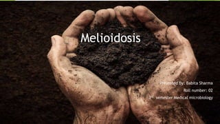
Melioidosis
Presented by: Babita Sharma
Roll number: 02
3rd semester Medical microbiology
Introduction
 Melioidosis is an infectious disease caused by bacterium Burkholderia
pseudomallei( previously known as Pseudomonas pseudomallei) that can infect
humans or animals.
 Also called Whitmore’s disease, pseudoglanders, night cliff disease, paddy-field
disease.
 It has several forms like, the formation of skin abscess, sepsis, septic shock,
abscess formation in several internal organ and pulmonary disease.
 Human acquired infection by inhalation of infected aerosol, inoculation of
contaminated material or excreta of infected animals and direct contact with soil.
 Melioidosis is primarily a disease of rats, but also occurs in guinea pig, rabbits,
goats and dog.
 Melioidosis is clinically and pathologically similar to glanders disease.
History
 This disease, now termed melioidosis, was named from Greek word “ Melis ”(
distemper of asses) and “eidos”(resemblance) by Stanton and Fletcher in 1932.
 Melioidosis was first discovered in Burma( now Myanmar) by Whitemore and
Krishmaswami in 1911.
 Also reported in Malaysia and Singapore in 1913 and then Vietnam in 1925.
 First diagnosed case in nepal was in 2005.
 Potential use of B. pseudomallei as a biological Weapon.
 Glanders together with Anthrax was implicated in the first modern era as
biological warfare (1915-1918) to infect horses in the united state, Romania,
Spain, Norway and Argentina.
Reason to use in Bioterrorism
 High mortality rate
 Easy transmitability
 Short incubation period
 Difficulty is diagnosis and treatment
 Ability to survive outside its natural environment
 Intrinsic antibiotic resistance
Epidemiology
 Melioidosis is predominantly disease of tropical climates. occurs worldwide but
most common in Southeast Asia ,Australia , Thailand Africa, India and china.
 A modelling study estimate that there are 165,000 cases of melioidosis in human
per year worldwide, of which 89,000 (54%) are estimated to be fatal.
 Melioidosis is prevalent in the northern Australia and northeast Thailand, where
the annual incidence is up to 50 cases per100,000 individual.
 Bacteria occurs as an environmental saprophyte found in the soil, rice paddies
and muddy water particularly common in moist clay soil.
 Polluted and contaminated atmosphere contribute to spread.
Bacteriology
Morphology
 Burkholderia pseudomallei is a Gram -negative, Motile, obligate aerobic, non
spore forming bacillus that is straight or slightly curved.
 Measure about 2-5µm in length and 0.3-0.8 µm in diameter.
 Typical bipolar safety pin like appearance in the Methylene blue stain.
 Facultative intracellular pathogen.
Agent and Reservoir
 Agent: Burkholderia pseudomallei
 Reservoir:
 Soil, rice paddies and muddy water.
 Unchlorinated water supply and drinking water in rural area.
 B. pseudomallei can survive in extreme conditions, such as in distilled (without
nutrients) water (for ≥16 years), 40% soil moisture content for longer than 2
years and 0% moisture content around 30 days survive.
 No animal reservoir.
Mode of transmission
 Inhalation
inhalation of Dust, contaminated soil and aerosol containing bacteria.
 Ingestion
by consuming contaminated water, Unchlorinated domestic water , ingestion
of food and water contaminated with infected animal excreta and soil.
 Inoculation
inoculation during work on a soil through injuries and puncture of skin of
farmer.
 Breast milk
Transmitted to infants through breast milk from infected mothers.
 Direct contact with contaminated soil and water.
 Vector born transmission via mosquito (Aedes aegypti) and rat flea (Xenopsylla
cheopsis)
 Vertical, sexual, Human to human and animal to human transmission is rare.
Risk factor
Diabetes mellitus
Alcohol use
Chronic lung disease
Chronic renal disease
Immunocompromised patients
Cancer patients
Virulence Factor and pathogenesis
Virulence factor function
Flagella host cell attachment
Pilli host cell attachment
Cps Antiphagocytic, Biofilm production
T3ss Intracellular invasion and escape phagocytosis
T6ss1 Intracellular invasion and intracellular spread
BipB and BipD Intracellular invasion
BipC Adhesion, invasion, actin formation
INOS Intracellular survival
BIMA Actin polymerization
Pathogenesis
 Bacteria first enter at a break in the skin or mucous membrane and replicate in
epithelial cell.
 Use flagellar motility to spread and infect various cell types.
 T3ss transport the protein across the cell membrane which causes invasion.
 Attachment via adhesion protein, including the type IV pilus protein pilA and
adhesion protein BoaA and BoaB.
 Enters host cell by endocytosis.
 Bacteria replicates in both phagocyte and non-phagocyte cell intracellularly
which causes the lysis and infect the adjacent cell.
 Bacteria trigger the autophagy by the activation of NOD2 ie intracellular
pathogen recognition receptor protein that causes bacteria killing.
 T3SS inject effector proteins( BopA )which help to escape and protection by
host autophagy. macrophage lysis is mechanism for bacterial survival.
 Replication occur in host cytoplasm.
 Inside the host cell, polymerization of host actin propel bacteria to move
forward until reaches to CNS.
 Then, profusion occur which fuses the neighbouring cells forming
multinucleated giant cell(MNGC) that results in plague and the damage of host
cell.
 Plaque (a central clear area with a ring of fused cells) that provide shelter for
bacteria for further replication or latent infection.
 Direct cell to cell spread occurs in blood stream causing sepsis and infection in
antigen presenting cell that transport bacteria to the lymphatic
system(secondary spread).
 Bacteria can remain dormant for 19-29 years until imunosupression or other
host stress reactivates bacteria proliferation.
 Toxin anti-toxin system host immune response and selective pressure of
antibiotics are the factors contributing to latency.
Clinical manifestation
The disease shows different stage such as acute, local and chronic infections. The
incubation period is variable. It ranges from 2 day to as high as to many years.
Acute melioidosis
 Development of nodule, pain or swelling ,ulceration, abscess at the site of
infection of bacteria in the skin.
 Bacteria subsequently spread, causing secondary lymphangitis, reginal
lymphangitis, fever and myalgia.
 Progress rapidly to acute septicaemia with high mortality rate.
 Acute blood stream infection is most commonly seen in patients with HIV,
diabetes, renal failure etc. the condition results in septic shock.
Pulmonary infection
 Manifests as mild bronchitis to severe pneumonia.
 The condition is associated with high fever, headache, chest pain, anorexia and
general myalgia.
 Productive and non productive cough with normal sputum is typical
manifestation.
Chronic suppurative infection
 It is associated with multiple caseous or suppurative foci of infection in several
organs including joints, skin, lymph nodes, spleen, lungs, liver and brain.
 Bacteria remains as intracellular pathogens of the reticuloendothelial system,
which contributes to long latency and reactivation of the infection. Hence this
disease is called as Vietnam time bomb disease.
Clinical sign in animal
 Widely vary within a species, depending on the site of infection ; range from
acute to chronic.
 Subclinical infection is common.
 Single or multiple suppurative or caseous nodules/abscesses formation.
 Mastitis in goats
 The respiratory infection in sheep.
 Fatality occur when vital organs are infected.
Laboratory diagnosis
 Sample specimen
 Sputum
 BAL
 Blood or bone marrow
 Urine
 Throat swab
 Pus and wound swab
 Skin lesions
 Rectal swab
 Microscopy :
Gram stain : gram negative
Methylene blue stain :bipolar safety pin appearance
 Culture(gold standard)
 Not fastidious and grow on a large variety of culture media( BA,CA,MA etc).
 Ability to grow on 42°c. culture typically positive in 24 to 48 hours.
 Ashdown’s medium, Brukholderia pseudomallei selective media, francis media
and modified Ashdown’s medium are used for selective isolation.
 Ashdown’s medium contains crystal violet and gentamicin as selective agent. It
usually produce irregular-edge, rough, flat wrinkled purple colonies.
 Required BSL 3 laboratory for handling of organism.
 Resistance pattern
 Gentamicin and colistin resistance
 Amoxicillin-clavulanate sensitivity
 Biochemical reaction
 motility : positive
 Oxidase : positive
 Nitrate reduction: positive
 Indole : negative
 Methyl Red : negative
 Voges-Proskauer : Negative
 H2S : negative
 TSI : A/K
 Lactose and maltose fermenter
 Serological test
Rapid antigen detection
 Latex agglutination : Target LPS( sensitivity-94% and specificity—83%)
:Target exopolysaccharide (sensitivity-98.7% and specificity-97.2%)
 Immunofluorescence assay(IFA): Target exopolysaccharide
blood: sensitivity 100% and specificity 99.6%
Nonblood sample: Sensitivities 32.7% (respiratory sample ) to 50%(pus)
 Lateral flow assay: Target capsular polysaccharide
Sensitivity-98.7% and a specificity-97.2%
 Indirect hemagglutination assay(IHA): titer≥ 1:160 in the absence of a positive
culture is therefore regarded as supportive rather than definitive.
Poorly defined antigens from strain of B. pseudomellei absorbed to red blood cell.
 ELISA: commercial kit for melioidosis appears to perform well.
 Molecular technique
 PCR: Detection of pathogenic B. pseudomallei DNA from a clinical specimen.
 DNA Sequencing
Radiological test
 CXR: for diagnosis of pulmonary melioidosis.
 CTscan : used to diagnosis abscesses in the liver and spleen.
Treatment
Antimicrobial therapy is separated into the initial intensive phase and the subsequent
eradication phase. No validated vaccine are available till now.
Intensive phase
 Ceftazime, 2g( child, 50mg/kg upto 2gm), every 6 hours.
 Meropenem, 1g( child, 25mg/kg upto 1 gm), every 8 hours.
Eradication phase
 Trimethoprime- sulfamethoxazole ( child 6/30mg/kg upto 240/1,200mg orally,
every 12 hours.
 Folic acid, 5 mg (child, 0.1mg/kg upto 5 mg) orally daily.
Prevention and control
 Wearing personal protective equipment such as gloves and suitable clothing for
high risk group like agricultural worker.
 Disinfection (chlorination and chloramination) of the drinking water supply.
 Mechanization of agricultural activities in disease endemic area.
 Awareness raiser among veterinary and human health authorities.
 Avoid contact with animal urine, infected animals or an infected environment
e.g. waterlogged places where infected animals may have urinated.
 Drink only safe or boiled water during the rainy season, especially in flood-
prone areas.
 Practice good personal hygiene, washing hands before eating and after
defecating.
 Consult a physician for prophylactic use of antibiotics during flooding times.
 Protect the water supply from animal contamination.
 Use of disinfectant such as 70% ethanol, benzalkonium chloride, iodine etc.
Melioidosis in Nepal
 Shrestha N.K et al (2005) reported first case of melioidosis in a patient of 34 years
old male who had returned to Nepal from Malaysia.
 According to Shrestha N et al(2019) there was 2 cases reported at Tribhuvan
University Teaching Hospital. Both of them were diabetic male patients.
 According to Chaudhary R et al (2019) reported the case of cerebral melioidosis in
22years old male at Tertiary Care Hospitals . Who was also an soldier from
commando Training Academy.
Bibliography
 Tille, Patricia M. (2014). Bailey and Scott’s Diagnostic Microbiology. 14th edition. St.
Louis, Missouri: Elsevier.
 Parija SC (2012).Textbook of Microbiology and Immunology. 2nd edition. Noida, India:
Elsevier.
 WHO (2014). A Brief Guide to Emerging Infectious Diseases and Zoonoses.
 Shrestha NK, Sharma SK, Khanal B, Dhakal SS (2005). Melioidosis imported into
Nepal. Scandinavian Journal of infectious disease 37(1):64-80.
 Mukhopadhyay C, Shaw T, Varghese GM, Dance DAB(2018). Melioidosis in South Asia
(India, Nepal, Pakistan, Bhutan and Afghanistan). Tropical Medicine and Infectious
Disease.3(2):51
 Wiersinga WJ, Virk HS, Torres AG et al (2018). Melioidosis. Nat Rev Dis Primers ;
4:17107.
 https://www.uptodate.com/contents/melioidosis-epidemiology-clinical-manifestations-
and-diagnosis.
 https://wwwnc.cdc.gov/eid/syn/en/article/21/2/14-1045.
 Shrestha P, Adhikari M, Pant V, Baral S, Shrestha A, Bashyal B and Sherchand
JB(2019). Melioidosis misdiagnosed in Nepal. BMC infectious disease,19:176.
 Chaudhary R, Singh A, Pradhan M, Karki R, and Bhandari PB(2019). A fatal case
of cerebral Melioidosis. MJSB, 18:64.
Thank you

More Related Content

What's hot

Tick borne infections
Tick borne infectionsTick borne infections
Tick borne infections
thojo23
 
Yersenia
YerseniaYersenia
Yersenia
Arooosa
 

What's hot (20)

Dec provocation test (lymphatic filiarisis)
Dec provocation test (lymphatic filiarisis)Dec provocation test (lymphatic filiarisis)
Dec provocation test (lymphatic filiarisis)
 
Tularemia
TularemiaTularemia
Tularemia
 
Non typhoid salmonellosis
Non typhoid salmonellosisNon typhoid salmonellosis
Non typhoid salmonellosis
 
Lecture brucellosis 5
Lecture   brucellosis 5Lecture   brucellosis 5
Lecture brucellosis 5
 
MRSA
MRSAMRSA
MRSA
 
Actinomyces + nocardia
Actinomyces + nocardiaActinomyces + nocardia
Actinomyces + nocardia
 
Pseudomonas aeruginosa an epitome of a drug resistance in bacteria
Pseudomonas aeruginosa an epitome of a drug resistance in bacteriaPseudomonas aeruginosa an epitome of a drug resistance in bacteria
Pseudomonas aeruginosa an epitome of a drug resistance in bacteria
 
Pseudomonas aeruginosa mini-presentation
Pseudomonas aeruginosa mini-presentationPseudomonas aeruginosa mini-presentation
Pseudomonas aeruginosa mini-presentation
 
Pneumocystis jirovecii infection
Pneumocystis jirovecii infectionPneumocystis jirovecii infection
Pneumocystis jirovecii infection
 
Pathogenesis,pathology and diagnosis of Cryptococcosis,Histoplasmosis, Epizoo...
Pathogenesis,pathology and diagnosis of Cryptococcosis,Histoplasmosis, Epizoo...Pathogenesis,pathology and diagnosis of Cryptococcosis,Histoplasmosis, Epizoo...
Pathogenesis,pathology and diagnosis of Cryptococcosis,Histoplasmosis, Epizoo...
 
Brucellosis
BrucellosisBrucellosis
Brucellosis
 
Tick borne infections
Tick borne infectionsTick borne infections
Tick borne infections
 
Brucellosis ppt
Brucellosis pptBrucellosis ppt
Brucellosis ppt
 
Pyrexia of unknown origin
Pyrexia of unknown originPyrexia of unknown origin
Pyrexia of unknown origin
 
Cryptococcal Meningitis SEMINAR
Cryptococcal Meningitis SEMINARCryptococcal Meningitis SEMINAR
Cryptococcal Meningitis SEMINAR
 
Leptospira weil's disease
Leptospira weil's diseaseLeptospira weil's disease
Leptospira weil's disease
 
Neurocysticercosis
NeurocysticercosisNeurocysticercosis
Neurocysticercosis
 
Brucella
BrucellaBrucella
Brucella
 
Yersenia
YerseniaYersenia
Yersenia
 
Burkholderia cepacia
Burkholderia cepaciaBurkholderia cepacia
Burkholderia cepacia
 

Similar to Melioidosis presentation

20 s2. tuberculosis, diptheria
20 s2. tuberculosis, diptheria20 s2. tuberculosis, diptheria
20 s2. tuberculosis, diptheria
Hasan Sultan
 
Organisms Causing Systemic MycosesObjectives -.docx
Organisms Causing Systemic MycosesObjectives   -.docxOrganisms Causing Systemic MycosesObjectives   -.docx
Organisms Causing Systemic MycosesObjectives -.docx
vannagoforth
 
Organisms Causing Systemic MycosesObjectives -.docx
Organisms Causing Systemic MycosesObjectives   -.docxOrganisms Causing Systemic MycosesObjectives   -.docx
Organisms Causing Systemic MycosesObjectives -.docx
amit657720
 

Similar to Melioidosis presentation (20)

CLS Histoplasmosis.pptx
CLS Histoplasmosis.pptxCLS Histoplasmosis.pptx
CLS Histoplasmosis.pptx
 
Corynebacterium Diphtheria. Bordetella Pertusis & Bordetella Parapertusis. Pr...
Corynebacterium Diphtheria. Bordetella Pertusis & Bordetella Parapertusis. Pr...Corynebacterium Diphtheria. Bordetella Pertusis & Bordetella Parapertusis. Pr...
Corynebacterium Diphtheria. Bordetella Pertusis & Bordetella Parapertusis. Pr...
 
Zoonotic tuberculosis
Zoonotic tuberculosisZoonotic tuberculosis
Zoonotic tuberculosis
 
Bohomolets Microbiology Lecture #20
Bohomolets Microbiology Lecture #20Bohomolets Microbiology Lecture #20
Bohomolets Microbiology Lecture #20
 
Zoonotic Infections. Bacillus Anthracis, Brucella. Brucellosis & Anthrax
Zoonotic Infections. Bacillus Anthracis, Brucella. Brucellosis & AnthraxZoonotic Infections. Bacillus Anthracis, Brucella. Brucellosis & Anthrax
Zoonotic Infections. Bacillus Anthracis, Brucella. Brucellosis & Anthrax
 
Dimorphic fungi
Dimorphic fungiDimorphic fungi
Dimorphic fungi
 
Toxoplasmosis and Q-Fever
Toxoplasmosis and Q-FeverToxoplasmosis and Q-Fever
Toxoplasmosis and Q-Fever
 
MLSM 505 Mycobact.pptx
MLSM 505 Mycobact.pptxMLSM 505 Mycobact.pptx
MLSM 505 Mycobact.pptx
 
Mycobacterium spp.ppt
Mycobacterium spp.pptMycobacterium spp.ppt
Mycobacterium spp.ppt
 
MLSM 505 Mycobact.ppt
MLSM 505 Mycobact.pptMLSM 505 Mycobact.ppt
MLSM 505 Mycobact.ppt
 
Mycotic Infections of the Oral cavity . ( Candidiasis )
Mycotic Infections of the Oral cavity . ( Candidiasis )Mycotic Infections of the Oral cavity . ( Candidiasis )
Mycotic Infections of the Oral cavity . ( Candidiasis )
 
20 s2. tuberculosis, diptheria
20 s2. tuberculosis, diptheria20 s2. tuberculosis, diptheria
20 s2. tuberculosis, diptheria
 
Pathogenesis of infection
Pathogenesis of infectionPathogenesis of infection
Pathogenesis of infection
 
Health considerations done
Health considerations doneHealth considerations done
Health considerations done
 
Mycobacterium .pptx
Mycobacterium .pptxMycobacterium .pptx
Mycobacterium .pptx
 
Organisms Causing Systemic MycosesObjectives -.docx
Organisms Causing Systemic MycosesObjectives   -.docxOrganisms Causing Systemic MycosesObjectives   -.docx
Organisms Causing Systemic MycosesObjectives -.docx
 
Organisms Causing Systemic MycosesObjectives -.docx
Organisms Causing Systemic MycosesObjectives   -.docxOrganisms Causing Systemic MycosesObjectives   -.docx
Organisms Causing Systemic MycosesObjectives -.docx
 
Mycobacterium leprae
Mycobacterium lepraeMycobacterium leprae
Mycobacterium leprae
 
Staphylococcus by nissim
Staphylococcus by nissimStaphylococcus by nissim
Staphylococcus by nissim
 
Corynebacterium
CorynebacteriumCorynebacterium
Corynebacterium
 

More from Central Department Of Microbiology, TU

More from Central Department Of Microbiology, TU (20)

Penicillium
PenicilliumPenicillium
Penicillium
 
Chromomycosis
ChromomycosisChromomycosis
Chromomycosis
 
Antifungal agents
Antifungal agentsAntifungal agents
Antifungal agents
 
Aspergillus
AspergillusAspergillus
Aspergillus
 
Scrub typhus
Scrub typhusScrub typhus
Scrub typhus
 
Listeriosis.ppt
Listeriosis.pptListeriosis.ppt
Listeriosis.ppt
 
Rhinosporidiosis
RhinosporidiosisRhinosporidiosis
Rhinosporidiosis
 
Sporotrichosis
SporotrichosisSporotrichosis
Sporotrichosis
 
Chromoblastomycosis
ChromoblastomycosisChromoblastomycosis
Chromoblastomycosis
 
Botulism
BotulismBotulism
Botulism
 
Leptospirosis
LeptospirosisLeptospirosis
Leptospirosis
 
Plague
PlaguePlague
Plague
 
Serological tests-for-the-diagnosis-of-salmonella
Serological tests-for-the-diagnosis-of-salmonellaSerological tests-for-the-diagnosis-of-salmonella
Serological tests-for-the-diagnosis-of-salmonella
 
Salmonella presentation
Salmonella presentationSalmonella presentation
Salmonella presentation
 
Integrated vector management
Integrated vector managementIntegrated vector management
Integrated vector management
 
Tularemia
TularemiaTularemia
Tularemia
 
Antifungal agents
Antifungal agents  Antifungal agents
Antifungal agents
 
Laboratory diagnosis of fungal diseases
Laboratory diagnosis of fungal diseasesLaboratory diagnosis of fungal diseases
Laboratory diagnosis of fungal diseases
 
Selection ,collection, transportation and processing of mycology specimen
Selection ,collection, transportation and processing of mycology specimenSelection ,collection, transportation and processing of mycology specimen
Selection ,collection, transportation and processing of mycology specimen
 
Techniques of diagnostic Mycology- 5 hours
Techniques of diagnostic Mycology- 5 hoursTechniques of diagnostic Mycology- 5 hours
Techniques of diagnostic Mycology- 5 hours
 

Recently uploaded

Aerodynamics. flippatterncn5tm5ttnj6nmnynyppt
Aerodynamics. flippatterncn5tm5ttnj6nmnynypptAerodynamics. flippatterncn5tm5ttnj6nmnynyppt
Aerodynamics. flippatterncn5tm5ttnj6nmnynyppt
sreddyrahul
 
Exomoons & Exorings with the Habitable Worlds Observatory I: On the Detection...
Exomoons & Exorings with the Habitable Worlds Observatory I: On the Detection...Exomoons & Exorings with the Habitable Worlds Observatory I: On the Detection...
Exomoons & Exorings with the Habitable Worlds Observatory I: On the Detection...
Sérgio Sacani
 
Pests of Green Manures_Bionomics_IPM_Dr.UPR.pdf
Pests of Green Manures_Bionomics_IPM_Dr.UPR.pdfPests of Green Manures_Bionomics_IPM_Dr.UPR.pdf
Pests of Green Manures_Bionomics_IPM_Dr.UPR.pdf
PirithiRaju
 
Cancer cell metabolism: special Reference to Lactate Pathway
Cancer cell metabolism: special Reference to Lactate PathwayCancer cell metabolism: special Reference to Lactate Pathway
Cancer cell metabolism: special Reference to Lactate Pathway
AADYARAJPANDEY1
 
The solar dynamo begins near the surface
The solar dynamo begins near the surfaceThe solar dynamo begins near the surface
The solar dynamo begins near the surface
Sérgio Sacani
 
Seminar on Halal AGriculture and Fisheries.pptx
Seminar on Halal AGriculture and Fisheries.pptxSeminar on Halal AGriculture and Fisheries.pptx
Seminar on Halal AGriculture and Fisheries.pptx
RUDYLUMAPINET2
 
Mammalian Pineal Body Structure and Also Functions
Mammalian Pineal Body Structure and Also FunctionsMammalian Pineal Body Structure and Also Functions
Mammalian Pineal Body Structure and Also Functions
YOGESH DOGRA
 
RNA INTERFERENCE: UNRAVELING GENETIC SILENCING
RNA INTERFERENCE: UNRAVELING GENETIC SILENCINGRNA INTERFERENCE: UNRAVELING GENETIC SILENCING
RNA INTERFERENCE: UNRAVELING GENETIC SILENCING
AADYARAJPANDEY1
 

Recently uploaded (20)

Microbial Type Culture Collection (MTCC)
Microbial Type Culture Collection (MTCC)Microbial Type Culture Collection (MTCC)
Microbial Type Culture Collection (MTCC)
 
Aerodynamics. flippatterncn5tm5ttnj6nmnynyppt
Aerodynamics. flippatterncn5tm5ttnj6nmnynypptAerodynamics. flippatterncn5tm5ttnj6nmnynyppt
Aerodynamics. flippatterncn5tm5ttnj6nmnynyppt
 
Hemoglobin metabolism_pathophysiology.pptx
Hemoglobin metabolism_pathophysiology.pptxHemoglobin metabolism_pathophysiology.pptx
Hemoglobin metabolism_pathophysiology.pptx
 
National Biodiversity protection initiatives and Convention on Biological Di...
National Biodiversity protection initiatives and  Convention on Biological Di...National Biodiversity protection initiatives and  Convention on Biological Di...
National Biodiversity protection initiatives and Convention on Biological Di...
 
SCHIZOPHRENIA Disorder/ Brain Disorder.pdf
SCHIZOPHRENIA Disorder/ Brain Disorder.pdfSCHIZOPHRENIA Disorder/ Brain Disorder.pdf
SCHIZOPHRENIA Disorder/ Brain Disorder.pdf
 
NuGOweek 2024 full programme - hosted by Ghent University
NuGOweek 2024 full programme - hosted by Ghent UniversityNuGOweek 2024 full programme - hosted by Ghent University
NuGOweek 2024 full programme - hosted by Ghent University
 
Exomoons & Exorings with the Habitable Worlds Observatory I: On the Detection...
Exomoons & Exorings with the Habitable Worlds Observatory I: On the Detection...Exomoons & Exorings with the Habitable Worlds Observatory I: On the Detection...
Exomoons & Exorings with the Habitable Worlds Observatory I: On the Detection...
 
Pests of Green Manures_Bionomics_IPM_Dr.UPR.pdf
Pests of Green Manures_Bionomics_IPM_Dr.UPR.pdfPests of Green Manures_Bionomics_IPM_Dr.UPR.pdf
Pests of Green Manures_Bionomics_IPM_Dr.UPR.pdf
 
Cancer cell metabolism: special Reference to Lactate Pathway
Cancer cell metabolism: special Reference to Lactate PathwayCancer cell metabolism: special Reference to Lactate Pathway
Cancer cell metabolism: special Reference to Lactate Pathway
 
Topography and sediments of the floor of the Bay of Bengal
Topography and sediments of the floor of the Bay of BengalTopography and sediments of the floor of the Bay of Bengal
Topography and sediments of the floor of the Bay of Bengal
 
The solar dynamo begins near the surface
The solar dynamo begins near the surfaceThe solar dynamo begins near the surface
The solar dynamo begins near the surface
 
mixotrophy in cyanobacteria: a dual nutritional strategy
mixotrophy in cyanobacteria: a dual nutritional strategymixotrophy in cyanobacteria: a dual nutritional strategy
mixotrophy in cyanobacteria: a dual nutritional strategy
 
Erythropoiesis- Dr.E. Muralinath-C Kalyan
Erythropoiesis- Dr.E. Muralinath-C KalyanErythropoiesis- Dr.E. Muralinath-C Kalyan
Erythropoiesis- Dr.E. Muralinath-C Kalyan
 
Structures and textures of metamorphic rocks
Structures and textures of metamorphic rocksStructures and textures of metamorphic rocks
Structures and textures of metamorphic rocks
 
Seminar on Halal AGriculture and Fisheries.pptx
Seminar on Halal AGriculture and Fisheries.pptxSeminar on Halal AGriculture and Fisheries.pptx
Seminar on Halal AGriculture and Fisheries.pptx
 
Mammalian Pineal Body Structure and Also Functions
Mammalian Pineal Body Structure and Also FunctionsMammalian Pineal Body Structure and Also Functions
Mammalian Pineal Body Structure and Also Functions
 
A Giant Impact Origin for the First Subduction on Earth
A Giant Impact Origin for the First Subduction on EarthA Giant Impact Origin for the First Subduction on Earth
A Giant Impact Origin for the First Subduction on Earth
 
Gliese 12 b, a temperate Earth-sized planet at 12 parsecs discovered with TES...
Gliese 12 b, a temperate Earth-sized planet at 12 parsecs discovered with TES...Gliese 12 b, a temperate Earth-sized planet at 12 parsecs discovered with TES...
Gliese 12 b, a temperate Earth-sized planet at 12 parsecs discovered with TES...
 
GBSN - Microbiology (Lab 2) Compound Microscope
GBSN - Microbiology (Lab 2) Compound MicroscopeGBSN - Microbiology (Lab 2) Compound Microscope
GBSN - Microbiology (Lab 2) Compound Microscope
 
RNA INTERFERENCE: UNRAVELING GENETIC SILENCING
RNA INTERFERENCE: UNRAVELING GENETIC SILENCINGRNA INTERFERENCE: UNRAVELING GENETIC SILENCING
RNA INTERFERENCE: UNRAVELING GENETIC SILENCING
 

Melioidosis presentation

  • 1. Melioidosis Presented by: Babita Sharma Roll number: 02 3rd semester Medical microbiology
  • 2. Introduction  Melioidosis is an infectious disease caused by bacterium Burkholderia pseudomallei( previously known as Pseudomonas pseudomallei) that can infect humans or animals.  Also called Whitmore’s disease, pseudoglanders, night cliff disease, paddy-field disease.  It has several forms like, the formation of skin abscess, sepsis, septic shock, abscess formation in several internal organ and pulmonary disease.  Human acquired infection by inhalation of infected aerosol, inoculation of contaminated material or excreta of infected animals and direct contact with soil.  Melioidosis is primarily a disease of rats, but also occurs in guinea pig, rabbits, goats and dog.  Melioidosis is clinically and pathologically similar to glanders disease.
  • 3. History  This disease, now termed melioidosis, was named from Greek word “ Melis ”( distemper of asses) and “eidos”(resemblance) by Stanton and Fletcher in 1932.  Melioidosis was first discovered in Burma( now Myanmar) by Whitemore and Krishmaswami in 1911.  Also reported in Malaysia and Singapore in 1913 and then Vietnam in 1925.  First diagnosed case in nepal was in 2005.  Potential use of B. pseudomallei as a biological Weapon.  Glanders together with Anthrax was implicated in the first modern era as biological warfare (1915-1918) to infect horses in the united state, Romania, Spain, Norway and Argentina.
  • 4. Reason to use in Bioterrorism  High mortality rate  Easy transmitability  Short incubation period  Difficulty is diagnosis and treatment  Ability to survive outside its natural environment  Intrinsic antibiotic resistance
  • 5. Epidemiology  Melioidosis is predominantly disease of tropical climates. occurs worldwide but most common in Southeast Asia ,Australia , Thailand Africa, India and china.  A modelling study estimate that there are 165,000 cases of melioidosis in human per year worldwide, of which 89,000 (54%) are estimated to be fatal.  Melioidosis is prevalent in the northern Australia and northeast Thailand, where the annual incidence is up to 50 cases per100,000 individual.  Bacteria occurs as an environmental saprophyte found in the soil, rice paddies and muddy water particularly common in moist clay soil.  Polluted and contaminated atmosphere contribute to spread.
  • 6. Bacteriology Morphology  Burkholderia pseudomallei is a Gram -negative, Motile, obligate aerobic, non spore forming bacillus that is straight or slightly curved.  Measure about 2-5µm in length and 0.3-0.8 µm in diameter.  Typical bipolar safety pin like appearance in the Methylene blue stain.  Facultative intracellular pathogen.
  • 7. Agent and Reservoir  Agent: Burkholderia pseudomallei  Reservoir:  Soil, rice paddies and muddy water.  Unchlorinated water supply and drinking water in rural area.  B. pseudomallei can survive in extreme conditions, such as in distilled (without nutrients) water (for ≥16 years), 40% soil moisture content for longer than 2 years and 0% moisture content around 30 days survive.  No animal reservoir.
  • 8. Mode of transmission  Inhalation inhalation of Dust, contaminated soil and aerosol containing bacteria.  Ingestion by consuming contaminated water, Unchlorinated domestic water , ingestion of food and water contaminated with infected animal excreta and soil.  Inoculation inoculation during work on a soil through injuries and puncture of skin of farmer.  Breast milk Transmitted to infants through breast milk from infected mothers.  Direct contact with contaminated soil and water.  Vector born transmission via mosquito (Aedes aegypti) and rat flea (Xenopsylla cheopsis)  Vertical, sexual, Human to human and animal to human transmission is rare.
  • 9. Risk factor Diabetes mellitus Alcohol use Chronic lung disease Chronic renal disease Immunocompromised patients Cancer patients
  • 10. Virulence Factor and pathogenesis Virulence factor function Flagella host cell attachment Pilli host cell attachment Cps Antiphagocytic, Biofilm production T3ss Intracellular invasion and escape phagocytosis T6ss1 Intracellular invasion and intracellular spread BipB and BipD Intracellular invasion BipC Adhesion, invasion, actin formation INOS Intracellular survival BIMA Actin polymerization
  • 11. Pathogenesis  Bacteria first enter at a break in the skin or mucous membrane and replicate in epithelial cell.  Use flagellar motility to spread and infect various cell types.  T3ss transport the protein across the cell membrane which causes invasion.  Attachment via adhesion protein, including the type IV pilus protein pilA and adhesion protein BoaA and BoaB.  Enters host cell by endocytosis.  Bacteria replicates in both phagocyte and non-phagocyte cell intracellularly which causes the lysis and infect the adjacent cell.  Bacteria trigger the autophagy by the activation of NOD2 ie intracellular pathogen recognition receptor protein that causes bacteria killing.
  • 12.  T3SS inject effector proteins( BopA )which help to escape and protection by host autophagy. macrophage lysis is mechanism for bacterial survival.  Replication occur in host cytoplasm.  Inside the host cell, polymerization of host actin propel bacteria to move forward until reaches to CNS.  Then, profusion occur which fuses the neighbouring cells forming multinucleated giant cell(MNGC) that results in plague and the damage of host cell.  Plaque (a central clear area with a ring of fused cells) that provide shelter for bacteria for further replication or latent infection.  Direct cell to cell spread occurs in blood stream causing sepsis and infection in antigen presenting cell that transport bacteria to the lymphatic system(secondary spread).
  • 13.  Bacteria can remain dormant for 19-29 years until imunosupression or other host stress reactivates bacteria proliferation.  Toxin anti-toxin system host immune response and selective pressure of antibiotics are the factors contributing to latency.
  • 14. Clinical manifestation The disease shows different stage such as acute, local and chronic infections. The incubation period is variable. It ranges from 2 day to as high as to many years. Acute melioidosis  Development of nodule, pain or swelling ,ulceration, abscess at the site of infection of bacteria in the skin.  Bacteria subsequently spread, causing secondary lymphangitis, reginal lymphangitis, fever and myalgia.  Progress rapidly to acute septicaemia with high mortality rate.  Acute blood stream infection is most commonly seen in patients with HIV, diabetes, renal failure etc. the condition results in septic shock.
  • 15. Pulmonary infection  Manifests as mild bronchitis to severe pneumonia.  The condition is associated with high fever, headache, chest pain, anorexia and general myalgia.  Productive and non productive cough with normal sputum is typical manifestation. Chronic suppurative infection  It is associated with multiple caseous or suppurative foci of infection in several organs including joints, skin, lymph nodes, spleen, lungs, liver and brain.  Bacteria remains as intracellular pathogens of the reticuloendothelial system, which contributes to long latency and reactivation of the infection. Hence this disease is called as Vietnam time bomb disease.
  • 16.
  • 17. Clinical sign in animal  Widely vary within a species, depending on the site of infection ; range from acute to chronic.  Subclinical infection is common.  Single or multiple suppurative or caseous nodules/abscesses formation.  Mastitis in goats  The respiratory infection in sheep.  Fatality occur when vital organs are infected.
  • 18. Laboratory diagnosis  Sample specimen  Sputum  BAL  Blood or bone marrow  Urine  Throat swab  Pus and wound swab  Skin lesions  Rectal swab
  • 19.  Microscopy : Gram stain : gram negative Methylene blue stain :bipolar safety pin appearance  Culture(gold standard)  Not fastidious and grow on a large variety of culture media( BA,CA,MA etc).  Ability to grow on 42°c. culture typically positive in 24 to 48 hours.  Ashdown’s medium, Brukholderia pseudomallei selective media, francis media and modified Ashdown’s medium are used for selective isolation.  Ashdown’s medium contains crystal violet and gentamicin as selective agent. It usually produce irregular-edge, rough, flat wrinkled purple colonies.  Required BSL 3 laboratory for handling of organism.
  • 20.  Resistance pattern  Gentamicin and colistin resistance  Amoxicillin-clavulanate sensitivity  Biochemical reaction  motility : positive  Oxidase : positive  Nitrate reduction: positive  Indole : negative  Methyl Red : negative  Voges-Proskauer : Negative  H2S : negative  TSI : A/K  Lactose and maltose fermenter
  • 21.  Serological test Rapid antigen detection  Latex agglutination : Target LPS( sensitivity-94% and specificity—83%) :Target exopolysaccharide (sensitivity-98.7% and specificity-97.2%)  Immunofluorescence assay(IFA): Target exopolysaccharide blood: sensitivity 100% and specificity 99.6% Nonblood sample: Sensitivities 32.7% (respiratory sample ) to 50%(pus)  Lateral flow assay: Target capsular polysaccharide Sensitivity-98.7% and a specificity-97.2%  Indirect hemagglutination assay(IHA): titer≥ 1:160 in the absence of a positive culture is therefore regarded as supportive rather than definitive. Poorly defined antigens from strain of B. pseudomellei absorbed to red blood cell.  ELISA: commercial kit for melioidosis appears to perform well.
  • 22.  Molecular technique  PCR: Detection of pathogenic B. pseudomallei DNA from a clinical specimen.  DNA Sequencing Radiological test  CXR: for diagnosis of pulmonary melioidosis.  CTscan : used to diagnosis abscesses in the liver and spleen.
  • 23.
  • 24. Treatment Antimicrobial therapy is separated into the initial intensive phase and the subsequent eradication phase. No validated vaccine are available till now. Intensive phase  Ceftazime, 2g( child, 50mg/kg upto 2gm), every 6 hours.  Meropenem, 1g( child, 25mg/kg upto 1 gm), every 8 hours. Eradication phase  Trimethoprime- sulfamethoxazole ( child 6/30mg/kg upto 240/1,200mg orally, every 12 hours.  Folic acid, 5 mg (child, 0.1mg/kg upto 5 mg) orally daily.
  • 25. Prevention and control  Wearing personal protective equipment such as gloves and suitable clothing for high risk group like agricultural worker.  Disinfection (chlorination and chloramination) of the drinking water supply.  Mechanization of agricultural activities in disease endemic area.  Awareness raiser among veterinary and human health authorities.  Avoid contact with animal urine, infected animals or an infected environment e.g. waterlogged places where infected animals may have urinated.
  • 26.  Drink only safe or boiled water during the rainy season, especially in flood- prone areas.  Practice good personal hygiene, washing hands before eating and after defecating.  Consult a physician for prophylactic use of antibiotics during flooding times.  Protect the water supply from animal contamination.  Use of disinfectant such as 70% ethanol, benzalkonium chloride, iodine etc.
  • 27. Melioidosis in Nepal  Shrestha N.K et al (2005) reported first case of melioidosis in a patient of 34 years old male who had returned to Nepal from Malaysia.  According to Shrestha N et al(2019) there was 2 cases reported at Tribhuvan University Teaching Hospital. Both of them were diabetic male patients.  According to Chaudhary R et al (2019) reported the case of cerebral melioidosis in 22years old male at Tertiary Care Hospitals . Who was also an soldier from commando Training Academy.
  • 28. Bibliography  Tille, Patricia M. (2014). Bailey and Scott’s Diagnostic Microbiology. 14th edition. St. Louis, Missouri: Elsevier.  Parija SC (2012).Textbook of Microbiology and Immunology. 2nd edition. Noida, India: Elsevier.  WHO (2014). A Brief Guide to Emerging Infectious Diseases and Zoonoses.  Shrestha NK, Sharma SK, Khanal B, Dhakal SS (2005). Melioidosis imported into Nepal. Scandinavian Journal of infectious disease 37(1):64-80.  Mukhopadhyay C, Shaw T, Varghese GM, Dance DAB(2018). Melioidosis in South Asia (India, Nepal, Pakistan, Bhutan and Afghanistan). Tropical Medicine and Infectious Disease.3(2):51  Wiersinga WJ, Virk HS, Torres AG et al (2018). Melioidosis. Nat Rev Dis Primers ; 4:17107.  https://www.uptodate.com/contents/melioidosis-epidemiology-clinical-manifestations- and-diagnosis.  https://wwwnc.cdc.gov/eid/syn/en/article/21/2/14-1045.
  • 29.  Shrestha P, Adhikari M, Pant V, Baral S, Shrestha A, Bashyal B and Sherchand JB(2019). Melioidosis misdiagnosed in Nepal. BMC infectious disease,19:176.  Chaudhary R, Singh A, Pradhan M, Karki R, and Bhandari PB(2019). A fatal case of cerebral Melioidosis. MJSB, 18:64.