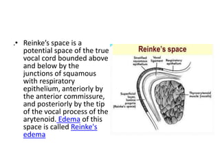
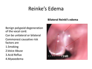
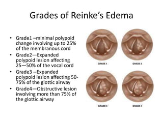
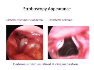
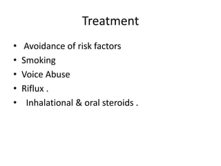
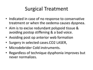
Reinke’s Edema
Dr Shrikant Phatak
Head of ENT Department
Choithram Hospital & Research Center
Indore
.• Reinke’s space is a
potential space of the true
vocal cord bounded above
and below by the
junctions of squamous
with respiratory
epithelium, anteriorly by
the anterior commissure,
and posteriorly by the tip
of the vocal process of the
arytenoid. Edema of this
space is called Reinke's
edema
Reinke’s Edema
Benign polypoid degeneration
of the vocal cord.
Can be unilateral or bilateral
Commonest causative risk
factors are
1.Smoking
2.Voice Abuse
3.Acid Reflux
4.Myxoedema
Bilateral Reinki’s edema
Grades of Reinke’s Edema
• Grade1 –minimal polypoid
change involving up to 25%
of the membranous cord
• Grade2—Expanded
polypoid lesion affecting
25—50% of the vocal cord
• Grade3 --Expanded
polypoid lesion affecting 50-
75% of the glottic airway
• Grade4—Obstructive lesion
involving more than 75% of
the glottic airway
Clinical Features
• Depend on the size of oedema .
• Edema is confined to the membranous vocal
cord the commonest presenting feature is
• Dysphonia
• May or may not be associated Dyspnea & in
rare cases Respiratory Distress
Stroboscopy Appearance
Bilateral asymmetric oedema Unilateral oedema
Oedema is best visualized during inspiration
Treatment
• Avoidance of risk factors
• Smoking
• Voice Abuse
• Riflux .
• Inhalational & oral steroids .
Surgical Treatment
• Indicated in case of no response to conservative
treatment or when the oedema causes dyspnea.
• Aim is to excise redundant polypoid tissue &
avoiding postop stiffening & a bad voice.
• Avoiding post op anterior web formation
• Surgery in selected cases.CO2 LASER,
• Microdebrider Cold instruments.
• Regardless of technique dysphonia improves but
never normalizes.
Thank you
Disclaimer
• The information contained in the presentation is based on the personal
experience and cases collected at Choithram Hospital Indore over the last
20 years.
• It is intended for the use of Medical students ENT post graduates.
• The views expressed are purely on personal opinion. viewers can make
their own opinion. For any confusion please contact sole author.
• Everybody is allowed to copy or download the material best suited to him.
I am not responsible for any controversies arising out of the presentation.
• For any suggestions or corrections you may please contact
phatak_shrikant@yahoo.in

Reinke's oedema

  • 1.
    Reinke’s Edema Dr ShrikantPhatak Head of ENT Department Choithram Hospital & Research Center Indore
  • 2.
    .• Reinke’s spaceis a potential space of the true vocal cord bounded above and below by the junctions of squamous with respiratory epithelium, anteriorly by the anterior commissure, and posteriorly by the tip of the vocal process of the arytenoid. Edema of this space is called Reinke's edema
  • 3.
    Reinke’s Edema Benign polypoiddegeneration of the vocal cord. Can be unilateral or bilateral Commonest causative risk factors are 1.Smoking 2.Voice Abuse 3.Acid Reflux 4.Myxoedema Bilateral Reinki’s edema
  • 4.
    Grades of Reinke’sEdema • Grade1 –minimal polypoid change involving up to 25% of the membranous cord • Grade2—Expanded polypoid lesion affecting 25—50% of the vocal cord • Grade3 --Expanded polypoid lesion affecting 50- 75% of the glottic airway • Grade4—Obstructive lesion involving more than 75% of the glottic airway
  • 5.
    Clinical Features • Dependon the size of oedema . • Edema is confined to the membranous vocal cord the commonest presenting feature is • Dysphonia • May or may not be associated Dyspnea & in rare cases Respiratory Distress
  • 6.
    Stroboscopy Appearance Bilateral asymmetricoedema Unilateral oedema Oedema is best visualized during inspiration
  • 7.
    Treatment • Avoidance ofrisk factors • Smoking • Voice Abuse • Riflux . • Inhalational & oral steroids .
  • 8.
    Surgical Treatment • Indicatedin case of no response to conservative treatment or when the oedema causes dyspnea. • Aim is to excise redundant polypoid tissue & avoiding postop stiffening & a bad voice. • Avoiding post op anterior web formation • Surgery in selected cases.CO2 LASER, • Microdebrider Cold instruments. • Regardless of technique dysphonia improves but never normalizes.
  • 9.
  • 10.
    Disclaimer • The informationcontained in the presentation is based on the personal experience and cases collected at Choithram Hospital Indore over the last 20 years. • It is intended for the use of Medical students ENT post graduates. • The views expressed are purely on personal opinion. viewers can make their own opinion. For any confusion please contact sole author. • Everybody is allowed to copy or download the material best suited to him. I am not responsible for any controversies arising out of the presentation. • For any suggestions or corrections you may please contact phatak_shrikant@yahoo.in