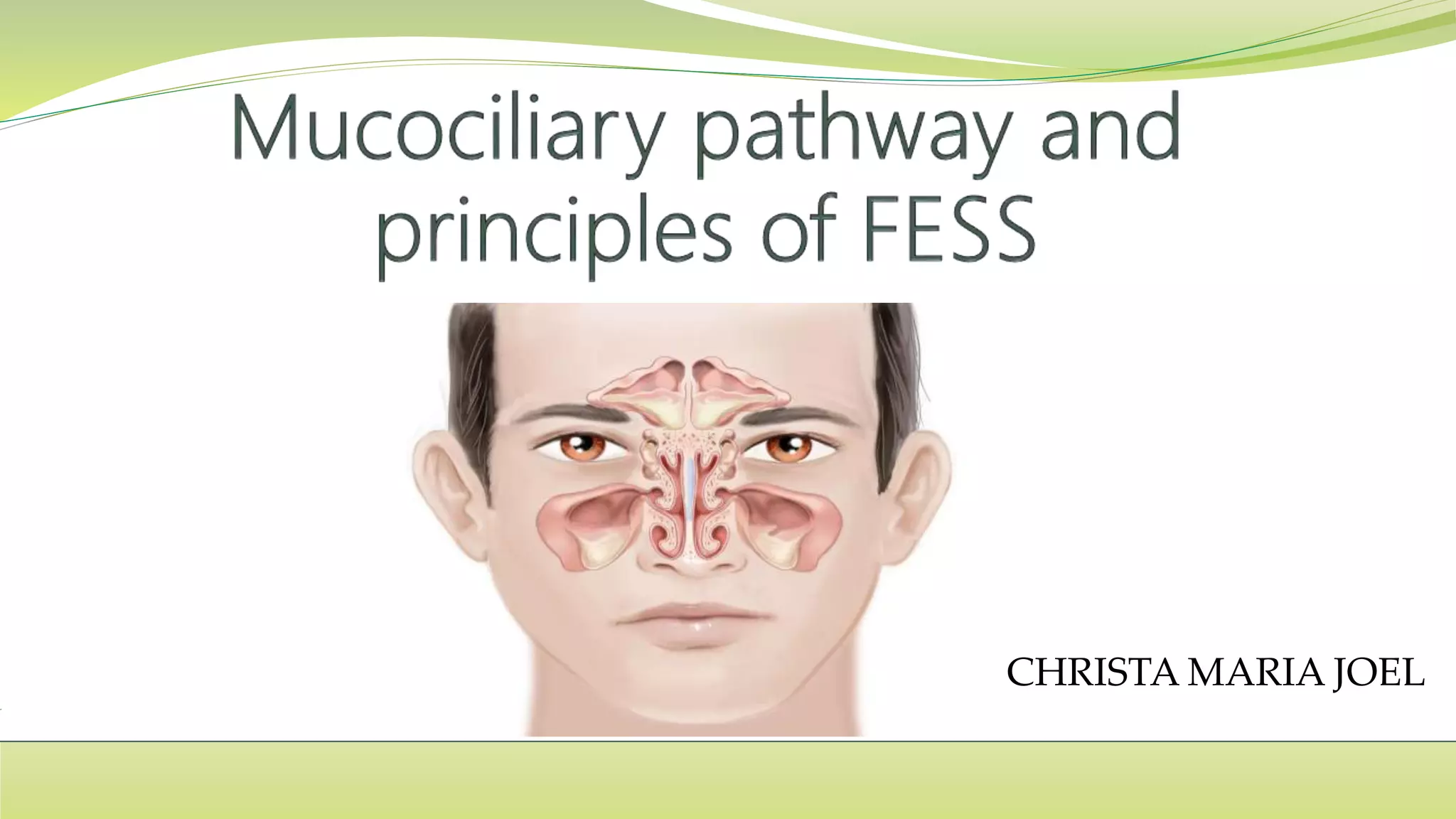
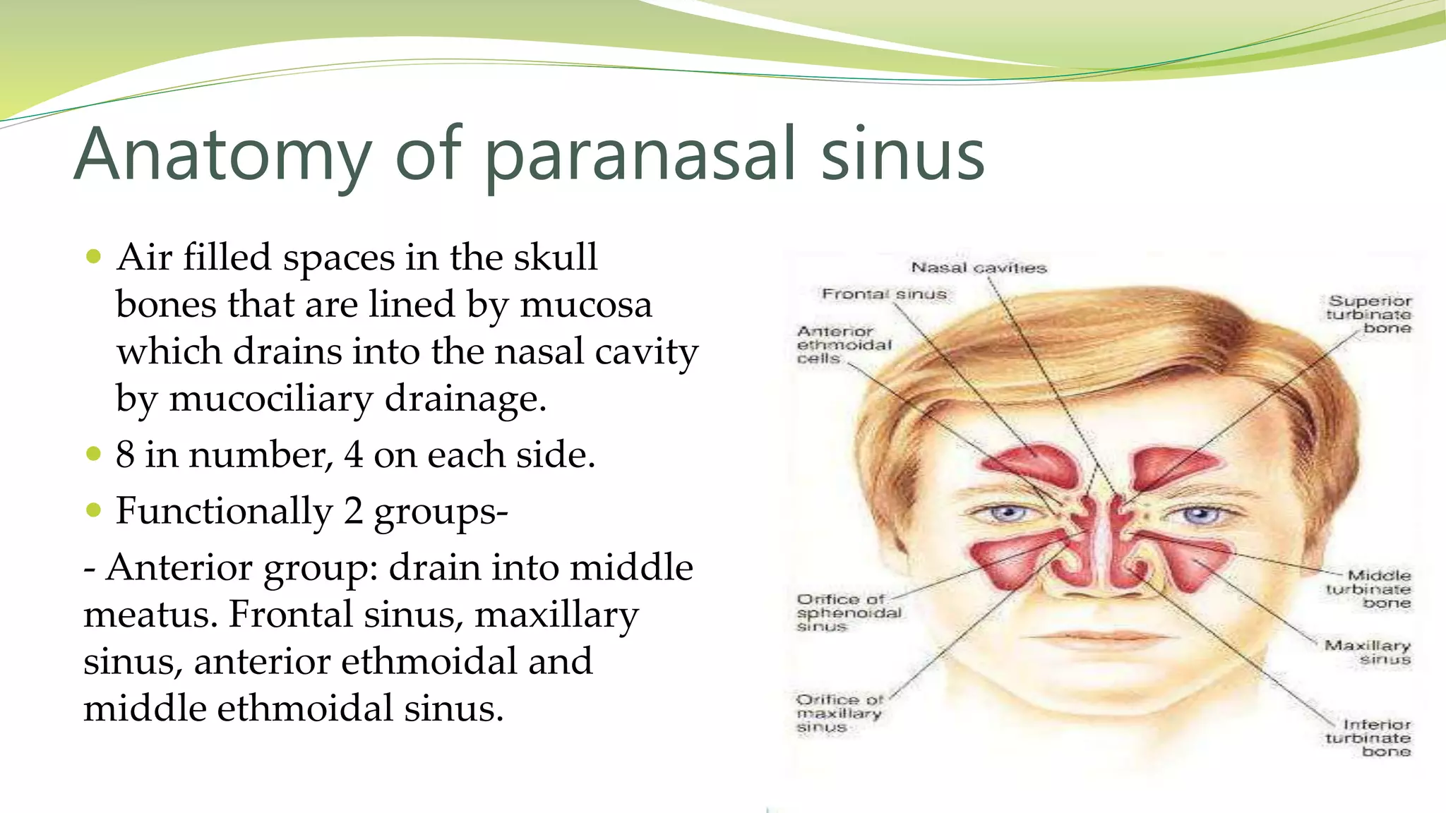
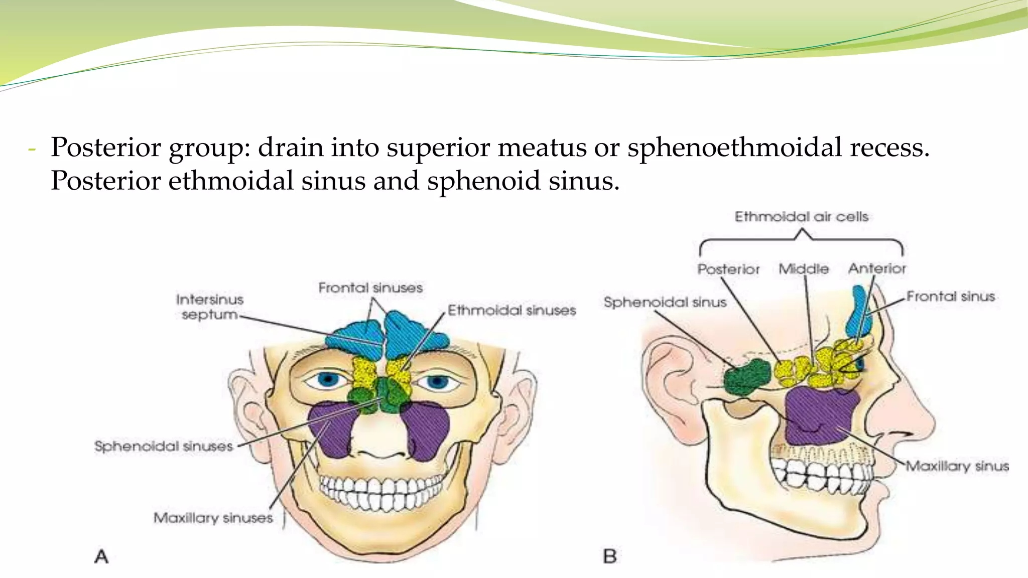
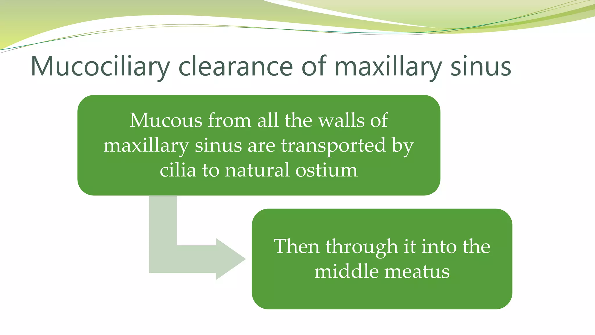
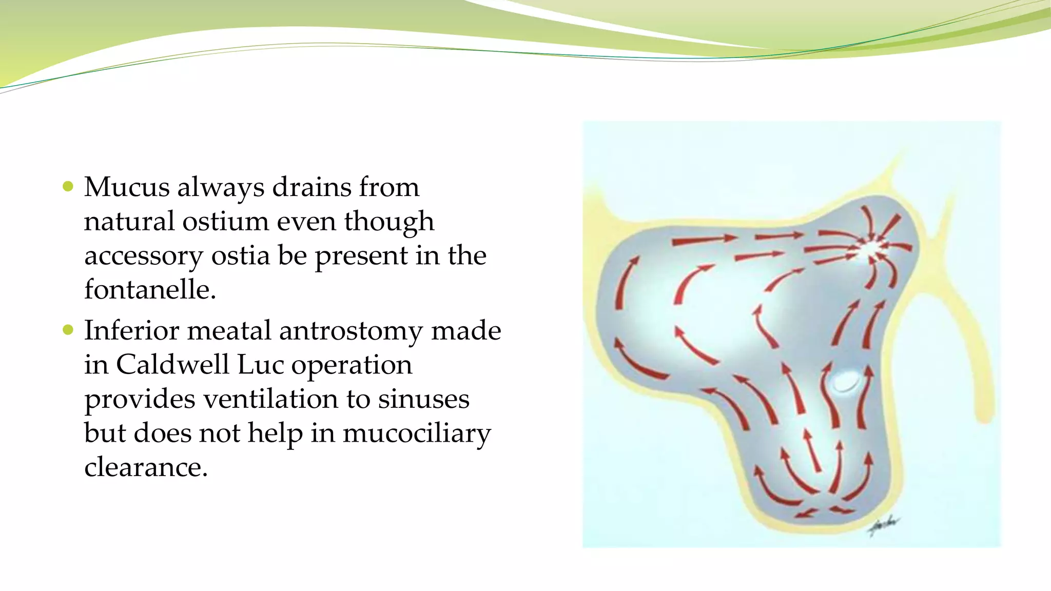
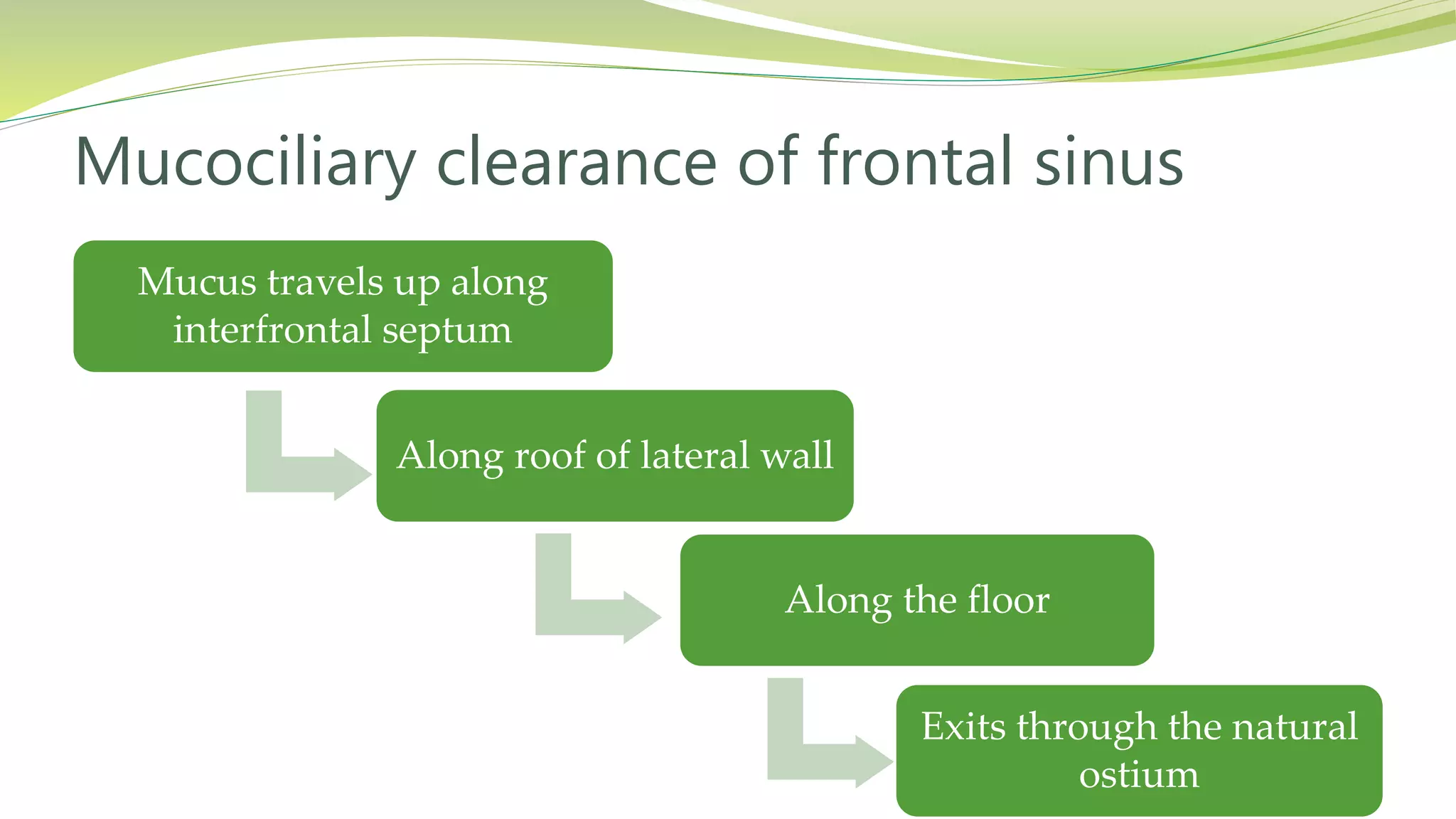
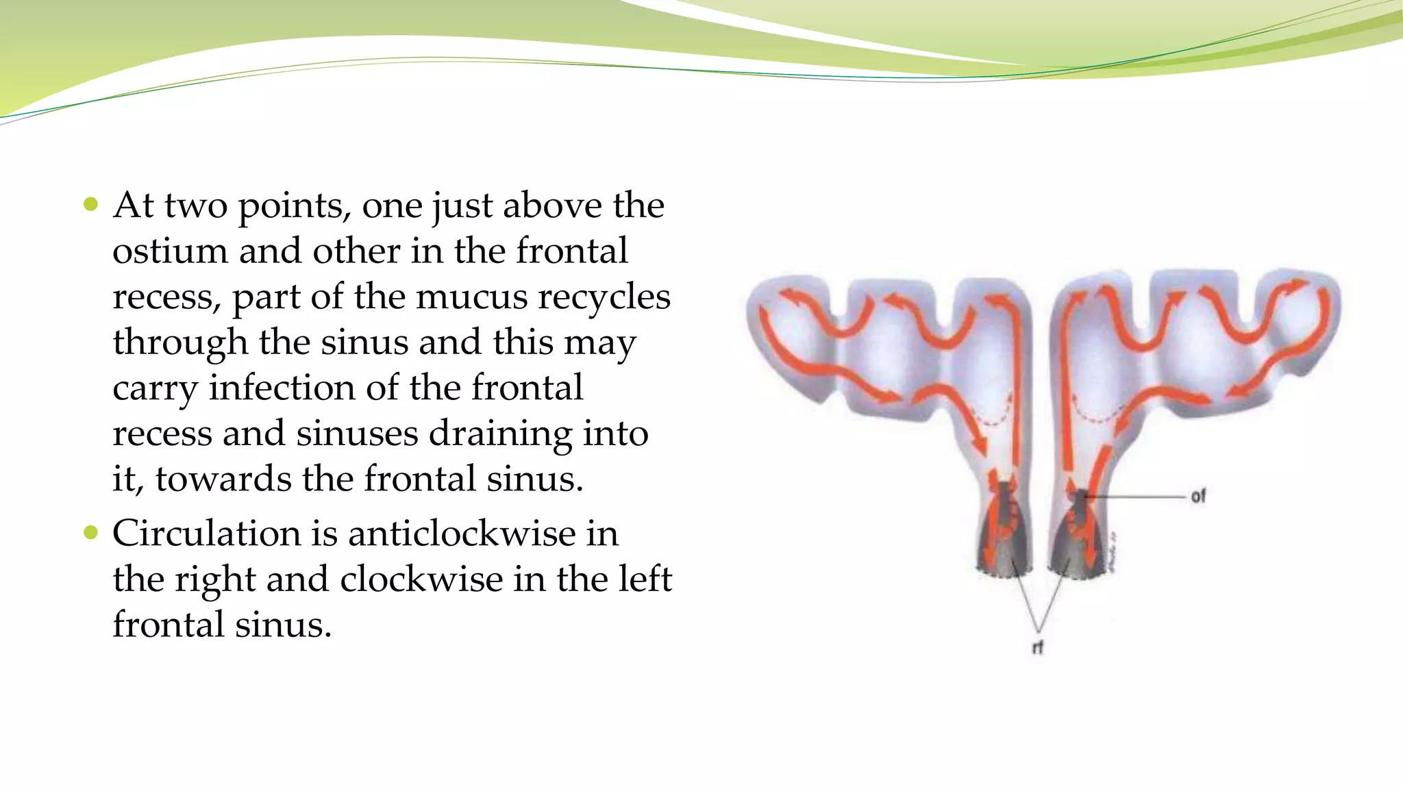
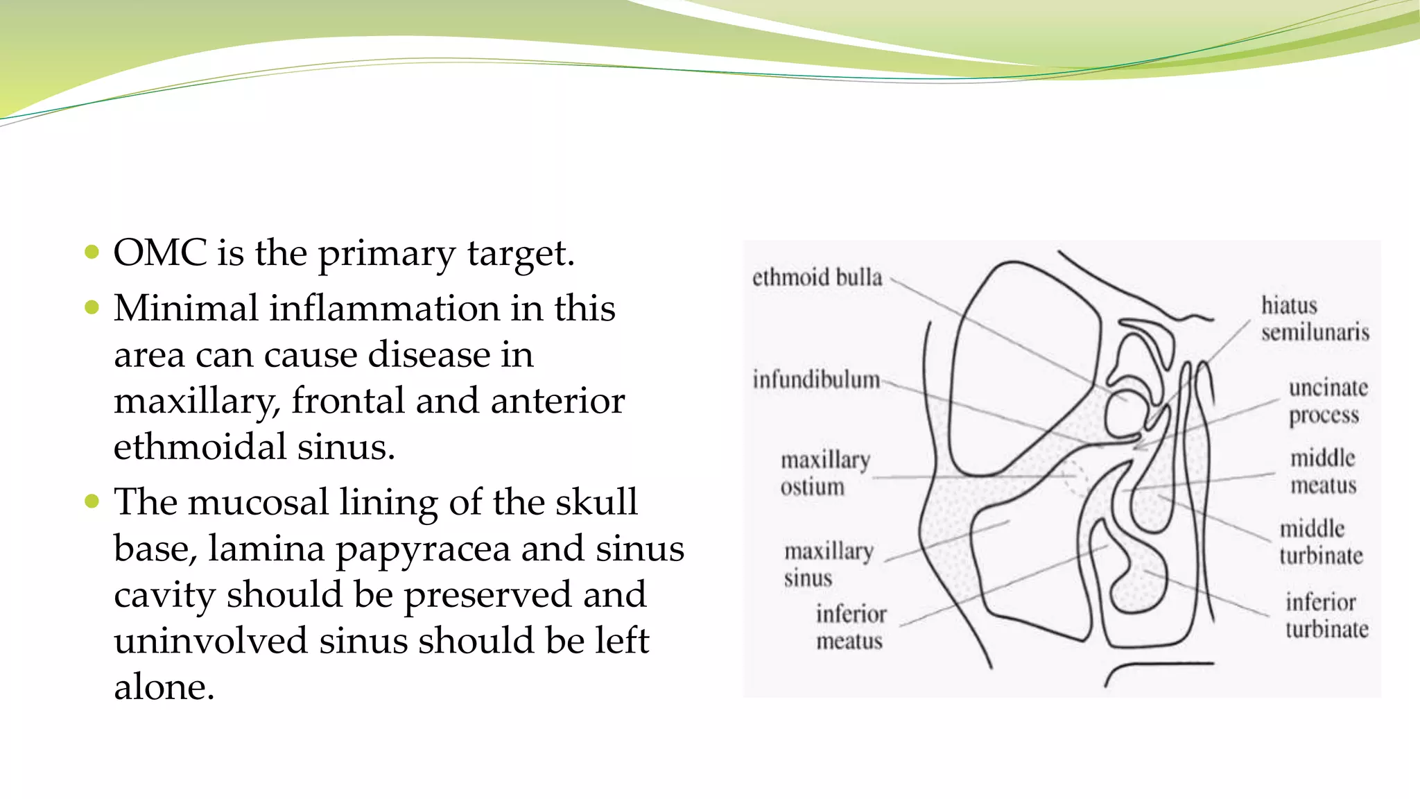
CHRISTA MARIA JOEL
 Air filled spaces in the skull
bones that are lined by mucosa
which drains into the nasal cavity
by mucociliary drainage.
 8 in number, 4 on each side.
 Functionally 2 groups-
- Anterior group: drain into middle
meatus. Frontal sinus, maxillary
sinus, anterior ethmoidal and
middle ethmoidal sinus.
Anatomy of paranasal sinus
- Posterior group: drain into superior meatus or sphenoethmoidal recess.
Posterior ethmoidal sinus and sphenoid sinus.
 Nasal mucosa is rich in goblet cells,
secretory glands both mucous and
serous.
 Their secretion forms a continuous
sheet called mucous blanket spread
over normal mucosa
 Mucous blanket consist of a
superficial mucus layer and deeper
serous layer floating on the top of
cilia which are constantly beating to
carry it like a conveyor belt towards
nasopharynx
Mucociliary mechanism
 In mammals, cilia beat 10-20 times per second at room temperature.
 Rapid effective stroke- extended cilia reach mucus layer.
 Slow recovery stroke- they bend and travel slowly in the reverse
direction in the thin serous layer thus moving the mucous blanket in
only one direction.
 Due to this mechanism the inspired bacteria and virus are trapped on
the viscous membrane and carried to the nasopharynx to get
swallowed.
Ciliary biphasic beat
Mucous from all the walls of
maxillary sinus are transported by
cilia to natural ostium
Then through it into the
middle meatus
Mucociliary clearance of maxillary sinus
 Mucus always drains from
natural ostium even though
accessory ostia be present in the
fontanelle.
 Inferior meatal antrostomy made
in Caldwell Luc operation
provides ventilation to sinuses
but does not help in mucociliary
clearance.
Mucus travels up along
interfrontal septum
Along roof of lateral wall
Along the floor
Exits through the natural
ostium
Mucociliary clearance of frontal sinus
 At two points, one just above the
ostium and other in the frontal
recess, part of the mucus recycles
through the sinus and this may
carry infection of the frontal
recess and sinuses draining into
it, towards the frontal sinus.
 Circulation is anticlockwise in
the right and clockwise in the left
frontal sinus.
Mucociliary
clearance is
towards its ostium
Then into
sphenoethmoidal
recess
Mucociliary clearance of sphenoid sinus
Mucus from anterior group
joins that from frontal and
maxillary sinuses
Travels towards Eustachian tube
Passing in front of torus tubarius into
nasopharynx
Mucociliary clearance of ethmoid sinus
Mucus from posterior
group drains into superior
or supreme meatus
Joins the mucus from sphenoid
sinus in the sphenoethmoidal
recess
Passes above and behind the
torus tubarius into
nasopharynx
 Infected discharge from anterior
group of sinuses passes behind
posterior pillars and cause
hypertrophy of lateral
pharyngeal bands.
 Discharge from posterior group
of sinuses spreads over posterior
pharyngeal wall.
 FESS- functional endoscopic sinus surgery is a minimally invasive
procedure which open the sinus air cells and sinus ostia.
 Primary principle- restore paranasal functions by reestabilishing the
physiological pattern of ventilation and mucociliary clearance.
 Remove irreversibly diseased mucosa and bone.
 Preserve the normal tissue and widen the ostium.
Principles of FESS
 OMC is the primary target.
 Minimal inflammation in this
area can cause disease in
maxillary, frontal and anterior
ethmoidal sinus.
 The mucosal lining of the skull
base, lamina papyracea and sinus
cavity should be preserved and
uninvolved sinus should be left
alone.
THANK YOU

Mucociliary Pathway and Principles of FESS

  • 1.
  • 2.
     Air filledspaces in the skull bones that are lined by mucosa which drains into the nasal cavity by mucociliary drainage.  8 in number, 4 on each side.  Functionally 2 groups- - Anterior group: drain into middle meatus. Frontal sinus, maxillary sinus, anterior ethmoidal and middle ethmoidal sinus. Anatomy of paranasal sinus
  • 3.
    - Posterior group:drain into superior meatus or sphenoethmoidal recess. Posterior ethmoidal sinus and sphenoid sinus.
  • 4.
     Nasal mucosais rich in goblet cells, secretory glands both mucous and serous.  Their secretion forms a continuous sheet called mucous blanket spread over normal mucosa  Mucous blanket consist of a superficial mucus layer and deeper serous layer floating on the top of cilia which are constantly beating to carry it like a conveyor belt towards nasopharynx Mucociliary mechanism
  • 5.
     In mammals,cilia beat 10-20 times per second at room temperature.  Rapid effective stroke- extended cilia reach mucus layer.  Slow recovery stroke- they bend and travel slowly in the reverse direction in the thin serous layer thus moving the mucous blanket in only one direction.  Due to this mechanism the inspired bacteria and virus are trapped on the viscous membrane and carried to the nasopharynx to get swallowed. Ciliary biphasic beat
  • 6.
    Mucous from allthe walls of maxillary sinus are transported by cilia to natural ostium Then through it into the middle meatus Mucociliary clearance of maxillary sinus
  • 7.
     Mucus alwaysdrains from natural ostium even though accessory ostia be present in the fontanelle.  Inferior meatal antrostomy made in Caldwell Luc operation provides ventilation to sinuses but does not help in mucociliary clearance.
  • 8.
    Mucus travels upalong interfrontal septum Along roof of lateral wall Along the floor Exits through the natural ostium Mucociliary clearance of frontal sinus
  • 9.
     At twopoints, one just above the ostium and other in the frontal recess, part of the mucus recycles through the sinus and this may carry infection of the frontal recess and sinuses draining into it, towards the frontal sinus.  Circulation is anticlockwise in the right and clockwise in the left frontal sinus.
  • 10.
    Mucociliary clearance is towards itsostium Then into sphenoethmoidal recess Mucociliary clearance of sphenoid sinus
  • 11.
    Mucus from anteriorgroup joins that from frontal and maxillary sinuses Travels towards Eustachian tube Passing in front of torus tubarius into nasopharynx Mucociliary clearance of ethmoid sinus
  • 12.
    Mucus from posterior groupdrains into superior or supreme meatus Joins the mucus from sphenoid sinus in the sphenoethmoidal recess Passes above and behind the torus tubarius into nasopharynx
  • 13.
     Infected dischargefrom anterior group of sinuses passes behind posterior pillars and cause hypertrophy of lateral pharyngeal bands.  Discharge from posterior group of sinuses spreads over posterior pharyngeal wall.
  • 14.
     FESS- functionalendoscopic sinus surgery is a minimally invasive procedure which open the sinus air cells and sinus ostia.  Primary principle- restore paranasal functions by reestabilishing the physiological pattern of ventilation and mucociliary clearance.  Remove irreversibly diseased mucosa and bone.  Preserve the normal tissue and widen the ostium. Principles of FESS
  • 15.
     OMC isthe primary target.  Minimal inflammation in this area can cause disease in maxillary, frontal and anterior ethmoidal sinus.  The mucosal lining of the skull base, lamina papyracea and sinus cavity should be preserved and uninvolved sinus should be left alone.
  • 16.