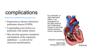
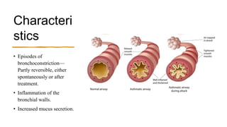
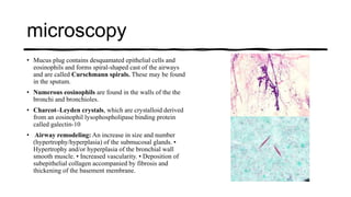
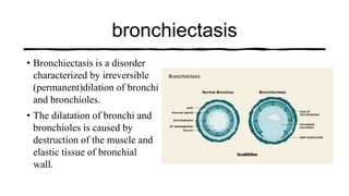
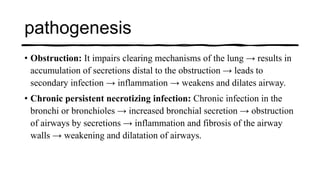
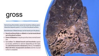
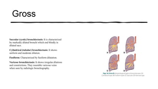
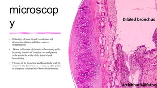
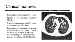
Chronic
Obstructive
Pulmonary
Disease
Dr. Vijay David
Raj R
Types
• Obstructive lung diseases consists of four diseases
namely
• (i) Emphysema,
• (ii) Chronic bronchitis,
• (iii) Asthma and
• (iv) Bronchiectasis.
Chronic Bronchitis
• Chronic bronchitis is defined clinically as
• persistent cough with sputum production (chronic productive cough)
for at least 3 months in at least 2 consecutive years
• in the absence of any other identifiable cause.
Causes
• Cigarette smoking: It is the most important risk factor.
• Air pollutants: Sulfur dioxide, nitrogen dioxide.
• Toxic industrial inhalants: Occupational dust exposure.
• Respiratory tract infection: It may initiate and promote chronic
bronchitis.
pathogenesis
• Irritation by inhaled air pollutants: Irritants cause inflammation → infiltration by CD8+ T lymphocytes, macrophages and
neutrophils.
• Hypersecretion of mucus:
• Hyperplasia/Hypertrophy of Submucosal Glands:
• Occurs in the larger airways such as the trachea and bronchi.
• Triggered by inhaled environmental irritants and proteases released by neutrophils, including elastase, cathepsin, histamine, and IL-13.
• Results in the enlargement and increased activity of submucosal glands.
• Leads to hypersecretion of mucus, contributing to airway obstruction.
• Increase of Goblet Cells in Small Airways:
• Particularly affects small bronchi and bronchioles.
• Goblet cells produce excessive mucus.
• Excessive mucus production can lead to mucus plugging of the bronchial lumen.
• Mucus plugging triggers inflammation and fibrosis of the bronchial wall, further contributing to airway obstruction.
• Chronic inflammation and accompanying fibrosis involving small airways can produce chronic airway obstruction.
• Infection may be secondary rather than primary and probably
important in maintaining and producing acute exacerbations.
• Infection may cause direct damage to airway epithelium and interferes
with ciliary action of the respiratory epithelium → defective clearance
of bacteria by leukocytes.
Morpholog
y
Gross
Airways (large and small)
changes include:
Mucous membrane: It shows
hyperemia, swelling and
edema. It appears dusky red.
Lumen: They are filled with
excessive mucus or
mucopurulent secretion.
microscopy
• Hyperplasia and hypertrophy of mucus-secreting glands in the
submucosa: results in hypersecretion of mucus.
• Reid index: It is the ratio of the thickness of the mucous gland
layer to the thickness of the wall between the base of the surface
epithelium and the inner limit of the cartilage plates. It is useful
for detecting the increase in the size and number of the mucus
glands. Reid index (normally 0.4) is increased in chronic
bronchitis.
• Chronic inflammation of the airways with predominant infiltration
by lymphocytes.
• Marked narrowing of bronchioles:
• In severe cases, there may be obliteration of lumen due to fibrosis
(bronchiolitis obliterans).
Clinical Features
• Usually affects middle-aged men who are heavy smokers.
• Early symptoms: Persistent productive cough (sputum) for many
years.
• Later stage: – Dyspnea on exertion
• Blue bloaters: Patients develop hypercapnia, hypoxemia, and mild
cyanosis. Such patients are called ‘blue bloaters’.
complications
• Progression to chronic obstructive
pulmonary disease (COPD).
• Longstanding cases lead to cor
pulmonale with cardiac failure.
• May develop squamous metaplasia
and dysplasia of the respiratory
epithelium → a rich soil for
cancerous transformation.
asthma
• Asthma is a chronic inflammatory disorder of the bronchial
tree (airways) in which breathing is periodically rendered
difficult by widespread narrowing of the bronchi (reversible
bronchoconstriction).
• It is clinically, characterized by recurrent episodes of
wheezing, breathlessness, tightness of the chest and cough.
Characteri
stics
• Episodes of
bronchoconstriction—
Partly reversible, either
spontaneously or after
treatment.
• Inflammation of the
bronchial walls.
• Increased mucus secretion.
types
Atopic
• Most common type of asthma that usually begins in childhood.
• Family history of asthma or allergic diseases (e.g. eczema, urticaria,
or hay fever) is common.
• Classic example of type I IgE-mediated hypersensitivity reaction.
• Exogenous substances (allergens) causing asthma can be
recognized.
Non atopic
• No causative exogenous factors can be identified and it does not show allergen sensitization.
• Skin tests are usually negative.
• Family history of asthma is less common.
• Respiratory infections due to viruses (e.g. rhinovirus, parainfluenza virus).
• Inhaled air pollutants (e.g. smoke, fumes).
• Drug-induced Asthma Many drugs may trigger asthma (e.g. aspirin-sensitive asthma). Aspirin
inhibits cyclooxygenase pathway of arachidonic acid metabolism → produces leukotrienes
(bronchoconstrictor) → causes asthma.
• Occupational Asthma Triggering occupational agents: • Fumes (epoxy resins, plastics). • Organic
and chemical dusts (wood, cotton, platinum). • Gases (toluene). • Other chemicals (formaldehyde,
penicillin products)
pathogene
sis
• Genetic predisposition: Type
I hypersensitivity (atopy)
reaction and exposure to
environmental trigger. One of
the susceptibility locus is on
the chromosome 5 (5q) →
several genes involved in
regulation of IgE synthesis and
mast cell and eosinophil
growth and differentiation.
• Airway hyper-responsiveness: It is an
abnormality in which there is excessive
tendency for airways to contract
(bronchoconstrictor) too easily in response to
multiple inhaled triggers that usually does
not have any effect on normal individuals.
• Hygiene Hypothesis: proposes that
individuals with lack of infections in early
childhood are more prone to asthma than
children brought up on farms who are
exposed to a high level of endotoxin.
Gross
• Airways (bronchi and
bronchioles) are occluded by
plugs of thick, tenacious
mucus.
• In patients who die due to
status asthmaticus, the lungs
are greatly distended due to
overinflation, and shows small
areas of atelectasis (collapse).
microscopy
• Mucus plug contains desquamated epithelial cells and
eosinophils and forms spiral-shaped cast of the airways
and are called Curschmann spirals. These may be found
in the sputum.
• Numerous eosinophils are found in the walls of the the
bronchi and bronchioles.
• Charcot–Leyden crystals, which are crystalloid derived
from an eosinophil lysophospholipase binding protein
called galectin-10
• Airway remodeling: An increase in size and number
(hypertrophy/hyperplasia) of the submucosal glands. •
Hypertrophy and/or hyperplasia of the bronchial wall
smooth muscle. • Increased vascularity. • Deposition of
subepithelial collagen accompanied by fibrosis and
thickening of the basement membrane.
Clinical
details
• Acute asthmatic attack usually lasts up to several
hours.
• In some mild degree of chest tightness, dyspnea,
wheezing and cough with or without sputum
production, may constantly present.
• Between the asthmatic attacks, patients may be
asymptomatic.
• Status asthmaticus: It is the most severe form of
asthma that persists for days and even weeks. The
bronchoconstriction does not respond to the
drugs. It may cause severe airflow obstruction
leading to severe cyanosis and even death.
bronchiectasis
• Bronchiectasis is a disorder
characterized by irreversible
(permanent)dilation of bronchi
and bronchioles.
• The dilatation of bronchi and
bronchioles is caused by
destruction of the muscle and
elastic tissue of bronchial
wall.
Causes
• Chronic Respiratory Infections: Chronic necrotizing infections. • Bacteria,
Viruses, Fungi
• Cystic Fibrosis: genetic disorder characterized by abnormal transport of chloride
ions, leading to thick, sticky mucus.
• Primary Ciliary Dyskinesia (PCD):
o Kartagener syndrome leading to impaired mucociliary clearance in the respiratory tract,
sinuses, and reproductive organs. Common features include chronic respiratory infections,
sinusitis, bronchiectasis, and situs inversus (reversal of the normal positions of organs).
o Young’s syndrome: Patients develop bronchiectasis, sinusitis and obstructive azoospermia.
The cause is not known.
• Obstructive Lung Diseases like asthma and chronic bronchitis.
• Autoimmune diseases: e.g. rheumatoid
arthritis, Sjögren’s syndrome, systemic lupus
erythematosus, inflammatory bowel disease.
• Immune-mediated disease: e.g. allergic
bronchopulmonary aspergillosis.
• Obstructive causes: Tumour or Foreign
body
Causes
pathogenesis
• Obstruction: It impairs clearing mechanisms of the lung → results in
accumulation of secretions distal to the obstruction → leads to
secondary infection → inflammation → weakens and dilates airway.
• Chronic persistent necrotizing infection: Chronic infection in the
bronchi or bronchioles → increased bronchial secretion → obstruction
of airways by secretions → inflammation and fibrosis of the airway
walls → weakening and dilatation of airways.
gross
Gross
Saccular (cystic) bronchiectasis: It is characterized
by markedly dilated bronchi which end blindly in
dilated sacs.
Cylindrical (tubular) bronchiectasis: It shows
uniform and moderate dilation.
Fusiform: Characterized by fusiform dilatation.
Varicose bronchiectasis: It shows irregular dilations
and constrictions. They resemble varicose veins
when seen by radiologic bronchography.
microscop
y
• Dilatation of bronchi and bronchioles and
destruction of their wall due to severe
inflammation.
• Dense infiltration of chronic inflammatory cells:
It mainly consists of lymphocytes and plasma
cells within the walls of the bronchi and
bronchioles.
• Fibrosis of the bronchial and bronchiolar wall: It
occurs in the chronic cases→ may result in partial
or complete obliteration of bronchiolar lumens.
Clinical features
• Severe persistent productive cough
• Sputum is foul-smelling, sometimes
bloody.
• Symptoms are precipitated by upper
respiratory tract infections.
• Paroxysms of cough occur when the
patient rises in the morning. This is
because, the changes in position
lead to drainage of collections of
pus and secretions into the bronchi.

Chronic Obstructive Lung Disease or Pulmonary Disorder

  • 1.
  • 2.
    Types • Obstructive lungdiseases consists of four diseases namely • (i) Emphysema, • (ii) Chronic bronchitis, • (iii) Asthma and • (iv) Bronchiectasis.
  • 3.
    Chronic Bronchitis • Chronicbronchitis is defined clinically as • persistent cough with sputum production (chronic productive cough) for at least 3 months in at least 2 consecutive years • in the absence of any other identifiable cause.
  • 4.
    Causes • Cigarette smoking:It is the most important risk factor. • Air pollutants: Sulfur dioxide, nitrogen dioxide. • Toxic industrial inhalants: Occupational dust exposure. • Respiratory tract infection: It may initiate and promote chronic bronchitis.
  • 5.
    pathogenesis • Irritation byinhaled air pollutants: Irritants cause inflammation → infiltration by CD8+ T lymphocytes, macrophages and neutrophils. • Hypersecretion of mucus: • Hyperplasia/Hypertrophy of Submucosal Glands: • Occurs in the larger airways such as the trachea and bronchi. • Triggered by inhaled environmental irritants and proteases released by neutrophils, including elastase, cathepsin, histamine, and IL-13. • Results in the enlargement and increased activity of submucosal glands. • Leads to hypersecretion of mucus, contributing to airway obstruction. • Increase of Goblet Cells in Small Airways: • Particularly affects small bronchi and bronchioles. • Goblet cells produce excessive mucus. • Excessive mucus production can lead to mucus plugging of the bronchial lumen. • Mucus plugging triggers inflammation and fibrosis of the bronchial wall, further contributing to airway obstruction. • Chronic inflammation and accompanying fibrosis involving small airways can produce chronic airway obstruction.
  • 6.
    • Infection maybe secondary rather than primary and probably important in maintaining and producing acute exacerbations. • Infection may cause direct damage to airway epithelium and interferes with ciliary action of the respiratory epithelium → defective clearance of bacteria by leukocytes.
  • 7.
    Morpholog y Gross Airways (large andsmall) changes include: Mucous membrane: It shows hyperemia, swelling and edema. It appears dusky red. Lumen: They are filled with excessive mucus or mucopurulent secretion.
  • 8.
    microscopy • Hyperplasia andhypertrophy of mucus-secreting glands in the submucosa: results in hypersecretion of mucus. • Reid index: It is the ratio of the thickness of the mucous gland layer to the thickness of the wall between the base of the surface epithelium and the inner limit of the cartilage plates. It is useful for detecting the increase in the size and number of the mucus glands. Reid index (normally 0.4) is increased in chronic bronchitis. • Chronic inflammation of the airways with predominant infiltration by lymphocytes. • Marked narrowing of bronchioles: • In severe cases, there may be obliteration of lumen due to fibrosis (bronchiolitis obliterans).
  • 9.
    Clinical Features • Usuallyaffects middle-aged men who are heavy smokers. • Early symptoms: Persistent productive cough (sputum) for many years. • Later stage: – Dyspnea on exertion • Blue bloaters: Patients develop hypercapnia, hypoxemia, and mild cyanosis. Such patients are called ‘blue bloaters’.
  • 10.
    complications • Progression tochronic obstructive pulmonary disease (COPD). • Longstanding cases lead to cor pulmonale with cardiac failure. • May develop squamous metaplasia and dysplasia of the respiratory epithelium → a rich soil for cancerous transformation.
  • 11.
    asthma • Asthma isa chronic inflammatory disorder of the bronchial tree (airways) in which breathing is periodically rendered difficult by widespread narrowing of the bronchi (reversible bronchoconstriction). • It is clinically, characterized by recurrent episodes of wheezing, breathlessness, tightness of the chest and cough.
  • 12.
    Characteri stics • Episodes of bronchoconstriction— Partlyreversible, either spontaneously or after treatment. • Inflammation of the bronchial walls. • Increased mucus secretion.
  • 13.
  • 14.
    Atopic • Most commontype of asthma that usually begins in childhood. • Family history of asthma or allergic diseases (e.g. eczema, urticaria, or hay fever) is common. • Classic example of type I IgE-mediated hypersensitivity reaction. • Exogenous substances (allergens) causing asthma can be recognized.
  • 15.
    Non atopic • Nocausative exogenous factors can be identified and it does not show allergen sensitization. • Skin tests are usually negative. • Family history of asthma is less common. • Respiratory infections due to viruses (e.g. rhinovirus, parainfluenza virus). • Inhaled air pollutants (e.g. smoke, fumes). • Drug-induced Asthma Many drugs may trigger asthma (e.g. aspirin-sensitive asthma). Aspirin inhibits cyclooxygenase pathway of arachidonic acid metabolism → produces leukotrienes (bronchoconstrictor) → causes asthma. • Occupational Asthma Triggering occupational agents: • Fumes (epoxy resins, plastics). • Organic and chemical dusts (wood, cotton, platinum). • Gases (toluene). • Other chemicals (formaldehyde, penicillin products)
  • 16.
    pathogene sis • Genetic predisposition:Type I hypersensitivity (atopy) reaction and exposure to environmental trigger. One of the susceptibility locus is on the chromosome 5 (5q) → several genes involved in regulation of IgE synthesis and mast cell and eosinophil growth and differentiation.
  • 17.
    • Airway hyper-responsiveness:It is an abnormality in which there is excessive tendency for airways to contract (bronchoconstrictor) too easily in response to multiple inhaled triggers that usually does not have any effect on normal individuals. • Hygiene Hypothesis: proposes that individuals with lack of infections in early childhood are more prone to asthma than children brought up on farms who are exposed to a high level of endotoxin.
  • 18.
    Gross • Airways (bronchiand bronchioles) are occluded by plugs of thick, tenacious mucus. • In patients who die due to status asthmaticus, the lungs are greatly distended due to overinflation, and shows small areas of atelectasis (collapse).
  • 19.
    microscopy • Mucus plugcontains desquamated epithelial cells and eosinophils and forms spiral-shaped cast of the airways and are called Curschmann spirals. These may be found in the sputum. • Numerous eosinophils are found in the walls of the the bronchi and bronchioles. • Charcot–Leyden crystals, which are crystalloid derived from an eosinophil lysophospholipase binding protein called galectin-10 • Airway remodeling: An increase in size and number (hypertrophy/hyperplasia) of the submucosal glands. • Hypertrophy and/or hyperplasia of the bronchial wall smooth muscle. • Increased vascularity. • Deposition of subepithelial collagen accompanied by fibrosis and thickening of the basement membrane.
  • 20.
    Clinical details • Acute asthmaticattack usually lasts up to several hours. • In some mild degree of chest tightness, dyspnea, wheezing and cough with or without sputum production, may constantly present. • Between the asthmatic attacks, patients may be asymptomatic. • Status asthmaticus: It is the most severe form of asthma that persists for days and even weeks. The bronchoconstriction does not respond to the drugs. It may cause severe airflow obstruction leading to severe cyanosis and even death.
  • 21.
    bronchiectasis • Bronchiectasis isa disorder characterized by irreversible (permanent)dilation of bronchi and bronchioles. • The dilatation of bronchi and bronchioles is caused by destruction of the muscle and elastic tissue of bronchial wall.
  • 22.
    Causes • Chronic RespiratoryInfections: Chronic necrotizing infections. • Bacteria, Viruses, Fungi • Cystic Fibrosis: genetic disorder characterized by abnormal transport of chloride ions, leading to thick, sticky mucus. • Primary Ciliary Dyskinesia (PCD): o Kartagener syndrome leading to impaired mucociliary clearance in the respiratory tract, sinuses, and reproductive organs. Common features include chronic respiratory infections, sinusitis, bronchiectasis, and situs inversus (reversal of the normal positions of organs). o Young’s syndrome: Patients develop bronchiectasis, sinusitis and obstructive azoospermia. The cause is not known. • Obstructive Lung Diseases like asthma and chronic bronchitis.
  • 23.
    • Autoimmune diseases:e.g. rheumatoid arthritis, Sjögren’s syndrome, systemic lupus erythematosus, inflammatory bowel disease. • Immune-mediated disease: e.g. allergic bronchopulmonary aspergillosis. • Obstructive causes: Tumour or Foreign body Causes
  • 24.
    pathogenesis • Obstruction: Itimpairs clearing mechanisms of the lung → results in accumulation of secretions distal to the obstruction → leads to secondary infection → inflammation → weakens and dilates airway. • Chronic persistent necrotizing infection: Chronic infection in the bronchi or bronchioles → increased bronchial secretion → obstruction of airways by secretions → inflammation and fibrosis of the airway walls → weakening and dilatation of airways.
  • 25.
  • 26.
    Gross Saccular (cystic) bronchiectasis:It is characterized by markedly dilated bronchi which end blindly in dilated sacs. Cylindrical (tubular) bronchiectasis: It shows uniform and moderate dilation. Fusiform: Characterized by fusiform dilatation. Varicose bronchiectasis: It shows irregular dilations and constrictions. They resemble varicose veins when seen by radiologic bronchography.
  • 27.
    microscop y • Dilatation ofbronchi and bronchioles and destruction of their wall due to severe inflammation. • Dense infiltration of chronic inflammatory cells: It mainly consists of lymphocytes and plasma cells within the walls of the bronchi and bronchioles. • Fibrosis of the bronchial and bronchiolar wall: It occurs in the chronic cases→ may result in partial or complete obliteration of bronchiolar lumens.
  • 28.
    Clinical features • Severepersistent productive cough • Sputum is foul-smelling, sometimes bloody. • Symptoms are precipitated by upper respiratory tract infections. • Paroxysms of cough occur when the patient rises in the morning. This is because, the changes in position lead to drainage of collections of pus and secretions into the bronchi.