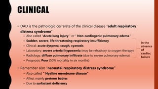
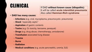
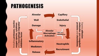
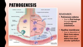
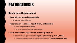
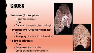
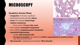
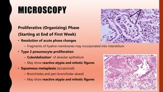
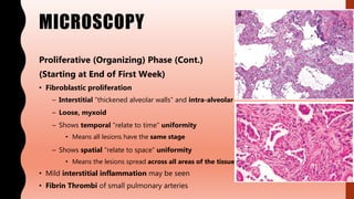
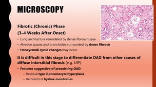
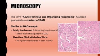
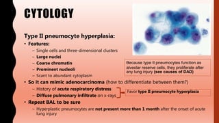
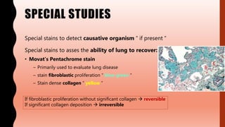
FLASHPATH
H A Z E M A L I
DIFFUSE ALVEOLAR
DAMAGE
/
ACUTE INTERSTITIAL
PNEUMONIAH A Z E M A L I
CLINICAL
• DAD is the pathologic correlate of the clinical disease “adult respiratory
distress syndrome”
– Also called “Acute lung injury ” or “ Non-cardiogenic pulmonary edema ”
– Sudden, severe, life threatening respiratory insufficiency
– Clinical: acute dyspnea, cough, cyanosis
– Laboratory: severe arterial hypoxemia (may be refractory to oxygen therapy)
– Radiology: diffuse pulmonary infiltrate (due to severe pulmonary edema)
– Prognosis: Poor (50% mortality in six months)
• Remember also “neonatal respiratory distress syndrome”
– Also called “ Hyaline membrane disease”
– Affect mainly preterm babies
– Due to surfactant deficiency
in the
absence
of
cardiac
failure
CLINICAL
• DAD has many causes:
– Infections (e.g. viral, mycoplasma, pneumocystis pneumonia)
– Shock “especially septic”
– Aspiration of gastric contents
– Toxins (e.g. O2 toxicity, kerosene, paraquat)
– Drugs (e.g. drug abuse, chemotherapy, amiodarone)
– Transfusion-associated lung disease
– Trauma
– Burns
– Radiation
– Medical conditions (e.g. acute pancreatitis, uremia, SLE)
If DAD without known cause (idiopathic)
It will be called acute interstitial pneumonia
Also called Hamman-Rich syndrome
CLINICAL
• Nitric oxide (vasodilator)
– Decreases pulmonary vascular resistance
– Reduces ventilation-perfusion mismatch
• Supportive care
– O2 therapy, mechanical ventilation
• Treat underlying cause
– E.g. infection, pancreatitis, etc.
PATHOGENESIS
Capillary
Endothelial
Injury
Neutrophils
Recruitment
Inflammatory
Mediators
Release
Alveolar
Wall
Damage
Expressadhesion
molecules,pro-coagulant
proteinsandchemokines.
Includingproteases,reactive
oxygenspecies,cytokines
andmacrophagemigration
inhibitionfactor
Alveolar
Macrophage
Activation
TNF, IL-1,
chemokines
PATHOGENESIS
REMEMBER:
• Pulmonary edema
• Due to fluid leakage
through injured
pulmonary
endothelium
• Hyaline membrane
• Due to deposition of
fibrin from edema
fluid mixed with
debris from dead
alveolar epithelium
PATHOGENESIS
Resolution (Organization)
• Resorption of intra-alveolar debris
– By alveolar macrophages
• Regeneration of damaged epithelium / endothelium
– May show regenerative atypia
• Be carful not to misinterpret it as malignancy
• Fibro-proliferative organization of damaged tissues
– Alveolar macrophages release fibrogenic cytokines (e.g. TGF-β, PDGF)
• Stimulate fibroblast growth and collagen deposition  thickened alveolar walls
GROSS
Exudative (Acute) phase
– Heavy (edematous)
– Firm
– Dark red (congested, hemorrhagic)
• Proliferative (Organizing) phase
– Firm
– Pale-gray (fibroblastic proliferation)
• Fibrotic (chronic)
– Firm
– Grayish-white (fibrosis)
– Cystic changes (honeycombing)
MICROSCOPY
Exudative (Acute) Phase
• Congestion of alveolar capillaries
• Interstitial and intra-alveolar edema
• Focal intra-alveolar hemorrhage
• Hyaline membranes
– More evident 72 hours after onset
• Intra-capillary neutrophil margination
• Intra-capillary megakaryocytes entrapment
• Interstitial inflammation
– Usually mild (scattered cells)
– Increased neutrophils may indicate that DAD is superimposed on another process
(e.g. bacterial pneumonia)
• Microvascular “capillaries and arterioles” fibrin thrombi
MICROSCOPY
Proliferative (Organizing) Phase
(Starting at End of First Week)
• Resolution of acute phase changes
– Fragments of hyaline membranes may incorporated into interstitium
• Type 2 pneumocyte proliferation
– Cuboidalization” of alveolar epithelium
– May show reactive atypia and mitotic figures
• Squamous metaplasia (occasional)
– Bronchioles and peri-bronchiolar alveoli
– May show reactive atypia and mitotic figures
MICROSCOPY
Proliferative (Organizing) Phase (Cont.)
(Starting at End of First Week)
• Fibroblastic proliferation
– Interstitial “thickened alveolar walls” and intra-alveolar
– Loose, myxoid
– Shows temporal “relate to time” uniformity
• Means all lesions have the same stage
– Shows spatial “relate to space” uniformity
• Means the lesions spread across all areas of the tissue
• Mild interstitial inflammation may be seen
• Fibrin Thrombi of small pulmonary arteries
MICROSCOPY
Fibrotic (Chronic) Phase
(3–4 Weeks After Onset)
• Lung architecture remodeled by dense fibrous tissue
• Alveolar spaces and bronchioles surrounded by dense fibrosis
• Honeycomb cystic changes may occur
It is difficult in this stage to differentiate DAD from other causes of
diffuse interstitial fibrosis (e.g. UIP)
• Features suggestive of preexisting DAD:
– Residual type II pneumocyte hyperplasia
– Remnants of hyaline membranes
MICROSCOPY
The term “Acute Fibrinous and Organizing Pneumonia” has been
proposed as a variant of DAD
Similar to DAD except:
• Patchy involvement (Intervening lung is normal)
– rather than diffuse pattern of DAD
• Alveoli are filled with balls of fibrin
– No hyaline membranes as seen in DAD
CYTOLOGY
Type II pneumocyte hyperplasia:
• Features:
– Single cells and three-dimensional clusters
– Large nuclei
– Coarse chromatin
– Prominent nucleoli
– Scant to abundant cytoplasm
• So it can mimic adenocarcinoma (how to differentiate between them?)
– History of acute respiratory distress
– Diffuse pulmonary infiltrate on x-rays
• Repeat BAL to be sure
– Hyperplastic pneumocytes are not present more than 1 month after the onset of acute
lung injury
Because type II pneumocytes function as
alveolar reserve cells, they proliferate after
any lung injury (see causes of DAD)
Favor type II pneumocyte hyperplasia
CYTOLOGY
Reparative epithelium:
• Features:
– Flat, cohesive sheets
– Abundant cytoplasm
– Enlarged, often hypochromatic nuclei
– Enlarged nucleoli
– Mitoses
• So it can mimic squamous cell carcinoma (how to differentiate between them?)
– Clinical and radiological data
– Malignant cells are usually less cohesive and more numerous than reparative
epithelium
SPECIAL STUDIES
Special stains to detect causative organism “ if present “
Special stains to asses the ability of lung to recover:
• Movat’s Pentachrome stain
– Primarily used to evaluate lung disease
– stain fibroblastic proliferation “ blue-green “
– Stain dense collagen “ yellow “
If fibroblastic proliferation without significant collagen  reversible
If significant collagen deposition  irreversible
DIFFERENTIAL DIAGNOSIS
Other common interstitial lung diseases:
• Usual interstitial pneumonia
– Insidious onset, chronic disease
– Temporal heterogeneity
– Prominent honeycomb change, present earlier
– No hyaline membranes
• Nonspecific interstitial pneumonia
– Insidious onset, chronic disease
– Predominantly interstitial
• Rare / focal intra-alveolar changes
– No / rare honeycomb change
– No hyaline membranes
DIFFERENTIAL DIAGNOSIS
DAD pattern may occur in the setting of acute exacerbation of
a chronic interstitial lung disease
1. Clinical and radiographic history is important
2. Defining feature of DAD
– Hyaline membranes, intact or organizing
3. Features suggest superimposed DAD
– Squamous metaplasia of respiratory bronchioles
– Fibrin thrombi in small arteries
– Type II pneumocyte hyperplasia
DIFFERENTIAL DIAGNOSIS
Other acute lung injury patterns:
• Cryptogenic organizing pneumonia
– Mainly bronchiolar (sometimes extends to peri-bronchiolar alveoli)
– Patchy, Nodular on low power (Masson bodies)
– Organization is intraluminal
– No hyaline membranes
– No collagen deposition (No honeycombing)
Sometimes the differentiation between DAD and COP is so difficult when examining
transbronchial biopsies, at which they may share similar findings (e.g. organizing airspace fibrosis
and type II pnuemocyte proliferation)
In these case, it’s better to use the term “ organizing acute lung injury “ which refers to an acute
injury with attending lung response, as opposite to a chronic interstitial lung disease
DIFFERENTIAL DIAGNOSIS
• Also DAD pattern can be seen in Acute eosinophilic pneumonia
– Prominent eosinophils
– Steroid responsive
DIFFERENTIAL DIAGNOSIS
Do not misinterpret the regenerative atypia seen in organizing
phase as a sign of malignancy
Be carful and conservative on calling malignancy in acutely ill
patients with diffuse pulmonary infiltrate on x-ray/ CT
WWW.
DO NOT FORGET TO SEARCH FOR MORE PICS
AND VIRTUAL SLIDES
THANK YOU
H A Z E M A L I

FlashPath- Lung - Diffuse Alveolar Damage / Acute Interstitial Pneumonia

  • 1.
    FLASHPATH H A ZE M A L I
  • 2.
  • 3.
    CLINICAL • DAD isthe pathologic correlate of the clinical disease “adult respiratory distress syndrome” – Also called “Acute lung injury ” or “ Non-cardiogenic pulmonary edema ” – Sudden, severe, life threatening respiratory insufficiency – Clinical: acute dyspnea, cough, cyanosis – Laboratory: severe arterial hypoxemia (may be refractory to oxygen therapy) – Radiology: diffuse pulmonary infiltrate (due to severe pulmonary edema) – Prognosis: Poor (50% mortality in six months) • Remember also “neonatal respiratory distress syndrome” – Also called “ Hyaline membrane disease” – Affect mainly preterm babies – Due to surfactant deficiency in the absence of cardiac failure
  • 4.
    CLINICAL • DAD hasmany causes: – Infections (e.g. viral, mycoplasma, pneumocystis pneumonia) – Shock “especially septic” – Aspiration of gastric contents – Toxins (e.g. O2 toxicity, kerosene, paraquat) – Drugs (e.g. drug abuse, chemotherapy, amiodarone) – Transfusion-associated lung disease – Trauma – Burns – Radiation – Medical conditions (e.g. acute pancreatitis, uremia, SLE) If DAD without known cause (idiopathic) It will be called acute interstitial pneumonia Also called Hamman-Rich syndrome
  • 5.
    CLINICAL • Nitric oxide(vasodilator) – Decreases pulmonary vascular resistance – Reduces ventilation-perfusion mismatch • Supportive care – O2 therapy, mechanical ventilation • Treat underlying cause – E.g. infection, pancreatitis, etc.
  • 6.
  • 7.
    PATHOGENESIS REMEMBER: • Pulmonary edema •Due to fluid leakage through injured pulmonary endothelium • Hyaline membrane • Due to deposition of fibrin from edema fluid mixed with debris from dead alveolar epithelium
  • 8.
    PATHOGENESIS Resolution (Organization) • Resorptionof intra-alveolar debris – By alveolar macrophages • Regeneration of damaged epithelium / endothelium – May show regenerative atypia • Be carful not to misinterpret it as malignancy • Fibro-proliferative organization of damaged tissues – Alveolar macrophages release fibrogenic cytokines (e.g. TGF-β, PDGF) • Stimulate fibroblast growth and collagen deposition  thickened alveolar walls
  • 9.
    GROSS Exudative (Acute) phase –Heavy (edematous) – Firm – Dark red (congested, hemorrhagic) • Proliferative (Organizing) phase – Firm – Pale-gray (fibroblastic proliferation) • Fibrotic (chronic) – Firm – Grayish-white (fibrosis) – Cystic changes (honeycombing)
  • 10.
    MICROSCOPY Exudative (Acute) Phase •Congestion of alveolar capillaries • Interstitial and intra-alveolar edema • Focal intra-alveolar hemorrhage • Hyaline membranes – More evident 72 hours after onset • Intra-capillary neutrophil margination • Intra-capillary megakaryocytes entrapment • Interstitial inflammation – Usually mild (scattered cells) – Increased neutrophils may indicate that DAD is superimposed on another process (e.g. bacterial pneumonia) • Microvascular “capillaries and arterioles” fibrin thrombi
  • 11.
    MICROSCOPY Proliferative (Organizing) Phase (Startingat End of First Week) • Resolution of acute phase changes – Fragments of hyaline membranes may incorporated into interstitium • Type 2 pneumocyte proliferation – Cuboidalization” of alveolar epithelium – May show reactive atypia and mitotic figures • Squamous metaplasia (occasional) – Bronchioles and peri-bronchiolar alveoli – May show reactive atypia and mitotic figures
  • 12.
    MICROSCOPY Proliferative (Organizing) Phase(Cont.) (Starting at End of First Week) • Fibroblastic proliferation – Interstitial “thickened alveolar walls” and intra-alveolar – Loose, myxoid – Shows temporal “relate to time” uniformity • Means all lesions have the same stage – Shows spatial “relate to space” uniformity • Means the lesions spread across all areas of the tissue • Mild interstitial inflammation may be seen • Fibrin Thrombi of small pulmonary arteries
  • 13.
    MICROSCOPY Fibrotic (Chronic) Phase (3–4Weeks After Onset) • Lung architecture remodeled by dense fibrous tissue • Alveolar spaces and bronchioles surrounded by dense fibrosis • Honeycomb cystic changes may occur It is difficult in this stage to differentiate DAD from other causes of diffuse interstitial fibrosis (e.g. UIP) • Features suggestive of preexisting DAD: – Residual type II pneumocyte hyperplasia – Remnants of hyaline membranes
  • 14.
    MICROSCOPY The term “AcuteFibrinous and Organizing Pneumonia” has been proposed as a variant of DAD Similar to DAD except: • Patchy involvement (Intervening lung is normal) – rather than diffuse pattern of DAD • Alveoli are filled with balls of fibrin – No hyaline membranes as seen in DAD
  • 15.
    CYTOLOGY Type II pneumocytehyperplasia: • Features: – Single cells and three-dimensional clusters – Large nuclei – Coarse chromatin – Prominent nucleoli – Scant to abundant cytoplasm • So it can mimic adenocarcinoma (how to differentiate between them?) – History of acute respiratory distress – Diffuse pulmonary infiltrate on x-rays • Repeat BAL to be sure – Hyperplastic pneumocytes are not present more than 1 month after the onset of acute lung injury Because type II pneumocytes function as alveolar reserve cells, they proliferate after any lung injury (see causes of DAD) Favor type II pneumocyte hyperplasia
  • 16.
    CYTOLOGY Reparative epithelium: • Features: –Flat, cohesive sheets – Abundant cytoplasm – Enlarged, often hypochromatic nuclei – Enlarged nucleoli – Mitoses • So it can mimic squamous cell carcinoma (how to differentiate between them?) – Clinical and radiological data – Malignant cells are usually less cohesive and more numerous than reparative epithelium
  • 17.
    SPECIAL STUDIES Special stainsto detect causative organism “ if present “ Special stains to asses the ability of lung to recover: • Movat’s Pentachrome stain – Primarily used to evaluate lung disease – stain fibroblastic proliferation “ blue-green “ – Stain dense collagen “ yellow “ If fibroblastic proliferation without significant collagen  reversible If significant collagen deposition  irreversible
  • 18.
    DIFFERENTIAL DIAGNOSIS Other commoninterstitial lung diseases: • Usual interstitial pneumonia – Insidious onset, chronic disease – Temporal heterogeneity – Prominent honeycomb change, present earlier – No hyaline membranes • Nonspecific interstitial pneumonia – Insidious onset, chronic disease – Predominantly interstitial • Rare / focal intra-alveolar changes – No / rare honeycomb change – No hyaline membranes
  • 19.
    DIFFERENTIAL DIAGNOSIS DAD patternmay occur in the setting of acute exacerbation of a chronic interstitial lung disease 1. Clinical and radiographic history is important 2. Defining feature of DAD – Hyaline membranes, intact or organizing 3. Features suggest superimposed DAD – Squamous metaplasia of respiratory bronchioles – Fibrin thrombi in small arteries – Type II pneumocyte hyperplasia
  • 20.
    DIFFERENTIAL DIAGNOSIS Other acutelung injury patterns: • Cryptogenic organizing pneumonia – Mainly bronchiolar (sometimes extends to peri-bronchiolar alveoli) – Patchy, Nodular on low power (Masson bodies) – Organization is intraluminal – No hyaline membranes – No collagen deposition (No honeycombing) Sometimes the differentiation between DAD and COP is so difficult when examining transbronchial biopsies, at which they may share similar findings (e.g. organizing airspace fibrosis and type II pnuemocyte proliferation) In these case, it’s better to use the term “ organizing acute lung injury “ which refers to an acute injury with attending lung response, as opposite to a chronic interstitial lung disease
  • 21.
    DIFFERENTIAL DIAGNOSIS • AlsoDAD pattern can be seen in Acute eosinophilic pneumonia – Prominent eosinophils – Steroid responsive
  • 22.
    DIFFERENTIAL DIAGNOSIS Do notmisinterpret the regenerative atypia seen in organizing phase as a sign of malignancy Be carful and conservative on calling malignancy in acutely ill patients with diffuse pulmonary infiltrate on x-ray/ CT
  • 23.
    WWW. DO NOT FORGETTO SEARCH FOR MORE PICS AND VIRTUAL SLIDES
  • 24.
    THANK YOU H AZ E M A L I