Case report - discussion about presentation and managements of laryngoceles.
Published in Journal of Evolution of Medical and Dental Sciences 2015; Vol. 4, Issue 32, April 20; Page: 5586-5591
Abstract: Laryngoceles are rare, cystic dilatation of saccule of ventricle of larynx. Three types are recognized –internal, external and mixed types. Many of the laryngoceles are asymptomatic; few require surgical excision via internal/endoscopic or external approach. Contrast CT is the investigation of choice. A 40year old male presented to our OPD with a neck Scar, later diagnosed as laryngocele. Here is the case report about presentation, diagnosis and management of a large mixed layngocele.
KEYWORDS: Layngocele, Neck swelling, Saccule, Ventricle of larynx.
ENT Nasal septal perforation..... for best rhinoplasty and nose reshape surgery contact
Dr Junaid Ahmad (MBBS FCPS) is the best plastic surgeon in Lahore. He is a well known, trained and expert in his field. He is MBBS and FCPS in Plastic and Recosntructive Surgery. He is a post graduate of the College of Physicians and Surgeons Pakistan which is oldest and best institute for post graduation in this area of the world. He is doing his practice in Lahore, Pakistan. He is always kind to the patients and listens them carefully as it is part of modern clinical skill and training. He is expert in both cosmetic as well as reconstructive surgery. He is also skin cancer and burn expert. A few of Dr Junaid Ahmad expertise are listed here..... call 03104037071
This is a seminar presentation conducted by 4th year medical students under supervision of a lecturer. Reference were not attached here, but all information are from google, few textbooks and also from previous ENT posting's seminar.
Abstract: We report a case of sinonasal paraganglioma presenting with episodes of epistaxis. A 55 year old male presented with a
nasal mass. It is an uncommon site of presentation and in an uncommon age group. A high grade of suspicion is required to diagnose
sino nasal paraganglioma. However, CT Scan and histopathology helps in early diagnosis and treatment. Surgical excision done with
cranialization of frontal sinus with fascia lata graft, followed up for 1 year without any evidence of disease recurrence.
Keywords: Sinonasal; Paraganglioma; Fascia Lata.
Unusual Presentation of Tuberculosis in Head and Neck RegionSachender Tanwar
Abstract: 3 case reports of tuberculosis at uncommon sites within head and neck region. Diagnosed on the basis of various clinical,
histopathological and imaging studies. Managed either with DOTS regimen only or both surgery & antitubercular treatment. None of
the cases showed non-compliance to treatment or recurrence of disease.
Keywords: tuberculosis, intra parotid lymphadenitis, branchial cyst, jugular chain lymphadenitis level II-III.
Papillary carcinoma of the thyroid gland is the most common, accounting for 75% of all thyroid malignancies, and the most indolent with a survival rate of 98%. Usually it presents as hypoechoic nodules in the thyroid gland. It is very rare for papillary carcinoma to present with large neck mass compromising airway and invading surrounding tissues. These features are more characteristic of anaplastic thyroid carcinoma.
There are many challenges in treating such patients.
1) Airway access to overcome obstruction.
2) Anesthesia concerns.
3) Surgical clearance (as there is soft tissue invasion).
4) Preservation of the recurrent laryngeal nerve.
5) Preserving parathyroids to prevent post-operative hypocalcaemia.
6) Hypopharyngeal and cervical oesophageal integrity and
continuity.
Dr. Ahmed M. Adawy, Professor Emeritus, Dep. Oral & Maxillofacial Surgery. Former Dean, Faculty of Dental Medicine
Al-Azhar University. There are four pairs of air sinuses making the boundaries of the nasal cavity. Maxillary sinus is the largest air cell. Anatomy and physiology of the maxillary sinus are given. Maxillary sinusitis is an inflammation of the sinus. Odontogenic causes represent nearly 30% of the etiology. Clinical and radiographic examinations are discussed together with treatment plan.
Oro-antral fistula is a rare complication of surgery at the posterior maxillary region. Several techniques for closure are presented. Additionally, information about sinus lift procedure is given.
Role of CT Mastoids in the Diagnosis and Management of Chronic Inflammatory E...Dr.Juveria Majeed
This study emphasises the role of CT Temporal bone in the diagnosis and management of chronic inflammatory ear disease...published in the Indian Journal of Otolaryngology Head and Neck surgery.
PAPILLARY CARCINOMA IN THYROGLOSSAL CYST-By Dr.Juveria Majeed MS ENT.Dr.Juveria Majeed
Carcinoma of thyroglossal duct cyst is a very rare entity. Here we present a case report of papillary carcinoma in thyroglossal cyst in a 21 year old female patient and discussion regarding dilemmas in the management, whether total thyroidectomy should be done or not.
Knee anatomy and clinical tests 2024.pdfvimalpl1234
This includes all relevant anatomy and clinical tests compiled from standard textbooks, Campbell,netter etc..It is comprehensive and best suited for orthopaedicians and orthopaedic residents.
Ozempic: Preoperative Management of Patients on GLP-1 Receptor Agonists Saeid Safari
Preoperative Management of Patients on GLP-1 Receptor Agonists like Ozempic and Semiglutide
ASA GUIDELINE
NYSORA Guideline
2 Case Reports of Gastric Ultrasound
263778731218 Abortion Clinic /Pills In Harare ,sisternakatoto
263778731218 Abortion Clinic /Pills In Harare ,ABORTION WOMEN’S CLINIC +27730423979 IN women clinic we believe that every woman should be able to make choices in her pregnancy. Our job is to provide compassionate care, safety,affordable and confidential services. That’s why we have won the trust from all generations of women all over the world. we use non surgical method(Abortion pills) to terminate…Dr.LISA +27730423979women Clinic is committed to providing the highest quality of obstetrical and gynecological care to women of all ages. Our dedicated staff aim to treat each patient and her health concerns with compassion and respect.Our dedicated group ABORTION WOMEN’S CLINIC +27730423979 IN women clinic we believe that every woman should be able to make choices in her pregnancy. Our job is to provide compassionate care, safety,affordable and confidential services. That’s why we have won the trust from all generations of women all over the world. we use non surgical method(Abortion pills) to terminate…Dr.LISA +27730423979women Clinic is committed to providing the highest quality of obstetrical and gynecological care to women of all ages. Our dedicated staff aim to treat each patient and her health concerns with compassion and respect.Our dedicated group of receptionists, nurses, and physicians have worked together as a teamof receptionists, nurses, and physicians have worked together as a team wwww.lisywomensclinic.co.za/
Basavarajeeyam is a Sreshta Sangraha grantha (Compiled book ), written by Neelkanta kotturu Basavaraja Virachita. It contains 25 Prakaranas, First 24 Chapters related to Rogas& 25th to Rasadravyas.
Title: Sense of Taste
Presenter: Dr. Faiza, Assistant Professor of Physiology
Qualifications:
MBBS (Best Graduate, AIMC Lahore)
FCPS Physiology
ICMT, CHPE, DHPE (STMU)
MPH (GC University, Faisalabad)
MBA (Virtual University of Pakistan)
Learning Objectives:
Describe the structure and function of taste buds.
Describe the relationship between the taste threshold and taste index of common substances.
Explain the chemical basis and signal transduction of taste perception for each type of primary taste sensation.
Recognize different abnormalities of taste perception and their causes.
Key Topics:
Significance of Taste Sensation:
Differentiation between pleasant and harmful food
Influence on behavior
Selection of food based on metabolic needs
Receptors of Taste:
Taste buds on the tongue
Influence of sense of smell, texture of food, and pain stimulation (e.g., by pepper)
Primary and Secondary Taste Sensations:
Primary taste sensations: Sweet, Sour, Salty, Bitter, Umami
Chemical basis and signal transduction mechanisms for each taste
Taste Threshold and Index:
Taste threshold values for Sweet (sucrose), Salty (NaCl), Sour (HCl), and Bitter (Quinine)
Taste index relationship: Inversely proportional to taste threshold
Taste Blindness:
Inability to taste certain substances, particularly thiourea compounds
Example: Phenylthiocarbamide
Structure and Function of Taste Buds:
Composition: Epithelial cells, Sustentacular/Supporting cells, Taste cells, Basal cells
Features: Taste pores, Taste hairs/microvilli, and Taste nerve fibers
Location of Taste Buds:
Found in papillae of the tongue (Fungiform, Circumvallate, Foliate)
Also present on the palate, tonsillar pillars, epiglottis, and proximal esophagus
Mechanism of Taste Stimulation:
Interaction of taste substances with receptors on microvilli
Signal transduction pathways for Umami, Sweet, Bitter, Sour, and Salty tastes
Taste Sensitivity and Adaptation:
Decrease in sensitivity with age
Rapid adaptation of taste sensation
Role of Saliva in Taste:
Dissolution of tastants to reach receptors
Washing away the stimulus
Taste Preferences and Aversions:
Mechanisms behind taste preference and aversion
Influence of receptors and neural pathways
Impact of Sensory Nerve Damage:
Degeneration of taste buds if the sensory nerve fiber is cut
Abnormalities of Taste Detection:
Conditions: Ageusia, Hypogeusia, Dysgeusia (parageusia)
Causes: Nerve damage, neurological disorders, infections, poor oral hygiene, adverse drug effects, deficiencies, aging, tobacco use, altered neurotransmitter levels
Neurotransmitters and Taste Threshold:
Effects of serotonin (5-HT) and norepinephrine (NE) on taste sensitivity
Supertasters:
25% of the population with heightened sensitivity to taste, especially bitterness
Increased number of fungiform papillae
These simplified slides by Dr. Sidra Arshad present an overview of the non-respiratory functions of the respiratory tract.
Learning objectives:
1. Enlist the non-respiratory functions of the respiratory tract
2. Briefly explain how these functions are carried out
3. Discuss the significance of dead space
4. Differentiate between minute ventilation and alveolar ventilation
5. Describe the cough and sneeze reflexes
Study Resources:
1. Chapter 39, Guyton and Hall Textbook of Medical Physiology, 14th edition
2. Chapter 34, Ganong’s Review of Medical Physiology, 26th edition
3. Chapter 17, Human Physiology by Lauralee Sherwood, 9th edition
4. Non-respiratory functions of the lungs https://academic.oup.com/bjaed/article/13/3/98/278874
Integrating Ayurveda into Parkinson’s Management: A Holistic ApproachAyurveda ForAll
Explore the benefits of combining Ayurveda with conventional Parkinson's treatments. Learn how a holistic approach can manage symptoms, enhance well-being, and balance body energies. Discover the steps to safely integrate Ayurvedic practices into your Parkinson’s care plan, including expert guidance on diet, herbal remedies, and lifestyle modifications.
Triangles of Neck and Clinical Correlation by Dr. RIG.pptx
Laryngocele
1. DOI: 10.14260/jemds/2015/817
CASE REPORT
J of Evolution of Med and Dent Sci/ eISSN- 2278-4802, pISSN- 2278-4748/ Vol. 4/ Issue 32/ Apr 20, 2015 Page 5586
LARYNGOCELE: A CASE REPORT
B. Umakanth Goud1, A. V. S. Hanumantha Rao2, D. Satyanarayana3, Manish Kumar Gupta4,
Juveria Majeed5
HOW TO CITE THIS ARTICLE:
B. Umakanth Goud, A. V. S. Hanumantha Rao, D. Satyanarayana, Manish Kumar Gupta, Juveria Majeed. “Laryn-
gocele: A Case Report”. Journal of Evolution of Medical and Dental Sciences 2015; Vol. 4, Issue 32, April 20;
Page: 5586-5591, DOI: 10.14260/jemds/2015/817
ABSTRACT: Laryngoceles are rare, cystic dilatation of saccule of ventricle of larynx. Three types are
recognized –internal, external and mixed types. Many of the laryngoceles are asymptomatic; few
require surgical excision via internal/endoscopic or external approach. Contrast CT is the
investigation of choice. A 40year old male presented to our OPD with a neck Scar, later diagnosed as
laryngocele. Here is the case report about presentation, diagnosis and management of a large mixed
layngocele.
KEYWORDS: Layngocele, Neck swelling, Saccule, Ventricle of larynx.
CASE REPORT: 40yrs old Male Agricultural laborer presented with history of swelling on right side
of Neck increasing in size associated with cough from last 3yrs. Patient was operated 3 times earlier.
On examination a horizontal scar mark of about 4cm size with sinus opening was found. CT Scan Neck
was suggestive of air filled sac bulging into the neck through the Thyrohyoid membrane of about 6cm
X 3cm X 2cm size. Video Laryngoscopy shows bulge above the right vocal card. Cervical exploration
was done by the vertical incision of about 8cm, 1cm lateral to midline in right side extending from
hyoid bone to adam’s apple. Dissection was carried out air filled saccule was identified the laryngeal
opening was closed with 00 proline. Superior Laryngeal nerve, Recurrent Laryngeal nerve, Ansa
cervicalis and hypoglossal nerve where identified and preserved. Postoperative period was
uneventful. Histopathology report of excised sac is not suggestive of any malignancy. Patient is under
follow up after surgery from last one month.
DISCUSSION: A LARYNGOCELE is an abnormal saccular dilatation of the appendix of the laryngeal
ventricle of Morgagni forming an air sac lined with pseudo-stratified, ciliated, columnar epithelium,
which maintains its communication with the ventricle by means of a narrow stalk.1 The laryngeal
ventricle of Morgagni is normally a small elliptical recess located between the false cords above and
the true cords below. The anterosuperior aspect of this recess ends in a blind pouch, which is called
the appendix or appendage of the ventricle of Morgagni.1,2 According to Burke and Goldlen3 if the
appendage extends above the upper border of the thyroid cartilage, it is abnormally long. Such a long
appendage is considered to be congenital in origin and is found in I5 to 30 per cent of adults.3
It is uncommon lesion which usually occurs in the middle age but may be rarely seen in
infancy, when it can produce respiratory distress which typically becomes worse on crying due to
increased distension of laryngocele with air.4
A laryngocele is classified as internal it is contained entirely with the laryngeal framework,
external if it pierces the thyrohyoid membrane and combined, if there are both external and internal
components.4
2. DOI: 10.14260/jemds/2015/817
CASE REPORT
J of Evolution of Med and Dent Sci/ eISSN- 2278-4802, pISSN- 2278-4748/ Vol. 4/ Issue 32/ Apr 20, 2015 Page 5587
Many laryngoceles are asymptomatic; sometimes they may cause a cough, hoarseness,
stridor, sore throat, globus sensation and may present as a swelling on one or both sides of the neck.5
More commonly unilateral6,7 and rarely bilateral internal8 and bilateral external laryngoceles9 also
exist. Sometimes a laryngocele may obstruct and becomes filled with mucus or become infected
(laryngopyocele), thus becomes indistinguishable from saccular cyst.4
Pre-Operative Photo showing
scar mark and Sinus opening
Pre-Operative VLS
photo showing bulge
3. DOI: 10.14260/jemds/2015/817
CASE REPORT
J of Evolution of Med and Dent Sci/ eISSN- 2278-4802, pISSN- 2278-4748/ Vol. 4/ Issue 32/ Apr 20, 2015 Page 5588
Intra operative photo showing external
Laryngeal nerve recurrent Laryngeal
nerve, Hypoglossal nerve & Ansa cervicalis
Intra operative photo
showing the sac
sac
CT Images showing
mixed Laryngocele
CT Images showing
mixed Laryngocele
Photo Showing the
incision mark
Intra operative
Photo showing the
sac
4. DOI: 10.14260/jemds/2015/817
CASE REPORT
J of Evolution of Med and Dent Sci/ eISSN- 2278-4802, pISSN- 2278-4748/ Vol. 4/ Issue 32/ Apr 20, 2015 Page 5589
ETIOPATHOGENESIS: For development of a laryngocele, its maintenance and its further
enlargement, certain predisposing and concomitant factors are necessary. One prerequisite is a
longer than normal ventricular appendage. Since laryngoceles occur in middle age or later, perhaps
tissue changes associated with age also predispose to this condition. Functional factors which
increase the intralaryngeal pressure, such as coughing, straining, singing and blowing wind
instruments also are of importance in the etiopathogenesis of laryngoceles (Acquired type of
Laryngocele).
Another mechanism is the appearance of a valve-like closure at the neck of the ventricular
appendage, which allows the entrance of air, but prevents its exit. This valve-like action results from
narrowing of the neck by inflammatory or neoplastic processes and may even be aggravated by
pressure of the distended sac itself on the neck.
It is of interest to note that the reported incidence of laryngocele in cases of laryngeal cancers
is between 1 per cent and 10 per cent. Therefore, if a laryngocele is detected clinically or
radiologically, a carcinoma must be taken into consideration and appropriate tests be performed.10,11
While the diagnosis of laryngocele is usually made on clinical grounds, it has to be
differentiated from all other neck swellings which includes any cystic swelling in the upper part of
the neck like branchial cyst, submandibular salivary gland duct cyst, cystic hygroma, saccular cyst,
Photo showing
excised Laryngocele
sac
Photo after excision of sac
showing different nerves
sac
Post-Operative Photo
showing the healing scar
sac
5. DOI: 10.14260/jemds/2015/817
CASE REPORT
J of Evolution of Med and Dent Sci/ eISSN- 2278-4802, pISSN- 2278-4748/ Vol. 4/ Issue 32/ Apr 20, 2015 Page 5590
mucous retention cyst, thyroglossal duct cyst and neck swellings which increase in size on valsalva
manoeuvre like Jugular vein phlebectasia. A saccular cyst should also be kept in mind when an
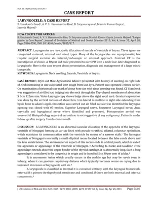
internal layngocele is diagnosed. A pharyngocele also presents as a cystic swelling in the upper part
of the neck.12
Computerised Tomography scan shows a well-defined, smooth, air filled sac in the lateral
aspect of the superior paralaryngeal space. The connection between the air sac and the airway helps
to establish the diagnosis. Magnetic resonance imaging, because of its multiplanner capability
provides high definition of soft tissues, offers detailed information on the boundaries of the air-filled
sac and, is useful when laryngomucocoele or laryngopyocele are suspected. MRI is also helpful to
distinguish obstructed mucus and inflammation from neoplastic disease.13
Management of laryngoceles depands on the size of the laryngocele and patients complaints.
Incidentally discovered or very small asymptomatic laryngoceles are observed, no active intervention
done. Very small to small laryngocele can be dealt endoscopically or Endoscopic marsupialization
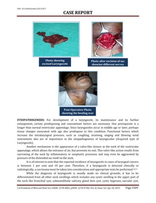
with co2 laser.14 Large laryngoceles are excised via external approach or to say precisely only the
external or combined laryngoceles has to accessed via external approach. Two main external
approach: Transthyrohyoid membrane and V shaped thyrotomies. A lateral cervical incision
approach extending through the thyrohyoid membrane at the superior margin of the ala of the
thyroid cartilage, with subperichondrial resection of a portion of upper part of the ala.4,14
CONCLUSION: For a upper neck swelling, in middle age group a diagnosis of laryngocele should be
kept in mind. Diagnosis is mostly on clinical grounds, which is confirmed by CT neck. The traditional
treatment of a laryngocele was excision using an external approach. Advances in endoscopic
techniques and laser surgery have modified the treatment strategy. Microlaryngoscopy with use of a
CO2 laser has become the main therapeutic procedure for the treatment of internal laryngoceles.
However, an external approach still remains the main therapeutic approach for the treatment of
combined laryngoceles.
REFERENCES:
1. Joseph Giovanniello, R. Vincent Grieco and Noel F. Bartone. LARYNGOCELE. APRIL, 1970.
www. ajronline. org/doi/pdf/10. 2214/ajr. 108. 4. 825.
2. Bateman, G. H. Case of bilateral external laryngocele. Journal of Larvngology & Otology, 1957,
71, 769-772.
3. Burke, E. N., and Golden, J. L. External ventricular laryngocele. AM. J. ROENTGENOL., RAD.
THERAPY & NUCLEAR MED., 1958, 8o, 4953.
4. Brown S. Benign conditions of larynx, 7th edition, pg no. 1136-1137.
5. Lancella A, Abbate G, and Dosdegani R, Mixed Layngolocele- case report Acta
Otorhinolaryngol Ital. 2007 Oct; 27(5): 255–257.
6. Erdogmus B, Yazici B, Ozturk O, Ataoglu S, Yazici S Wien Klin Wochenschr.Laryngocele in
association with ankylosing spondylitis. 2005 Oct; 117(19-20): 718-20.
7. The association of laryngoceles with ventricular phonation. Dray TG, Waugh PF, Hillel AD J
Voice. 2000 Jun; 14(2): 278-81.
8. Luzzago F, Nicolai P, Tomenzoli D, Maroldi R, Antonelli AR. Laryngocele: analysis of 18 cases
and review of the literature]. Acta Otorhinolaryngol Ital. 1990 Jul-Aug; 10(4): 399-412.
6. DOI: 10.14260/jemds/2015/817
CASE REPORT
J of Evolution of Med and Dent Sci/ eISSN- 2278-4802, pISSN- 2278-4748/ Vol. 4/ Issue 32/ Apr 20, 2015 Page 5591
9. Meda, P. Symptomatic laryngoceie in cancer of larynx. A. M. A. Arch. Otolaryng., 1952, 56, 512-
520.
10. Harney M, Patil N, Walsh R, Brennan P, Walsh M. Laryngocele and squamous cell carcinoma of
the larynx. J Laryngol Otol 2001; 115: 590-2.
11. Taylor, H. M. Ventricular laryngoceie. ‘Jr. Am. Laryng. A., 1944, 66, 114-126.
12. Binu R. George, Suhel Hasan, Naveen Kumar G, Salem MS. Tobook, Manjula Dhinakar. A Rare
Case of Laryngocele in a Young Omani Male. George BR, et al. OMJ. 25, (2010);
doi: 10. 5001/omj. 2010. 45.
13. J Gulia, S Yadav, A Khaowas, S Basur, A Agrawal. Laryngocele: A Case Report and Review of
Literature. Indian Journal of otorhinolaryngology, 14(1).
https: //ispub. com/IJORL/14/1/13884.
14. Prasad K C, Vijayalaxmi S, Prasad S C, Laryngoceles- Presentations and management, Indian J. of
Otolaryngology Head Neck Surgery, Oct-Dec 2008; 60; 303-306.
AUTHORS:
1. B. Umakanth Goud
2. A.V.S. Hanumantha Rao
3. D. Satyanarayana
4. Manish Kumar Gupta
5. Juveria Majeed
PARTICULARS OF CONTRIBUTORS:
1. Associate Professor, Department of
Surgical Oncology, MNJ Cancer Hospital,
Hyderabad.
2. Associate Professor, Department of ENT,
Kakatiya, Medical College, Warangal.
3. Associate Professor, Department of ENT,
Kakatiya, Medical College, Warangal.
FINANCIAL OR OTHER
COMPETING INTERESTS: None
4. Assistant Professor, Department of ENT,
Osmania Medical College, Hyderabad.
5. Post Graduate, Department of ENT,
Osmania Medical College, Hyderabad.
NAME ADDRESS EMAIL ID OF THE
CORRESPONDING AUTHOR:
Dr. Manish Kumar Gupta,
H. No. 564, Road No. 12,
Banjara Hills,
Hyderabad-500034.
E-mail: drmanishgupta003@gmail.com
Date of Submission: 25/03/2015.
Date of Peer Review: 26/03/2015.
Date of Acceptance: 08/04/2015.
Date of Publishing: 20/04/2015.