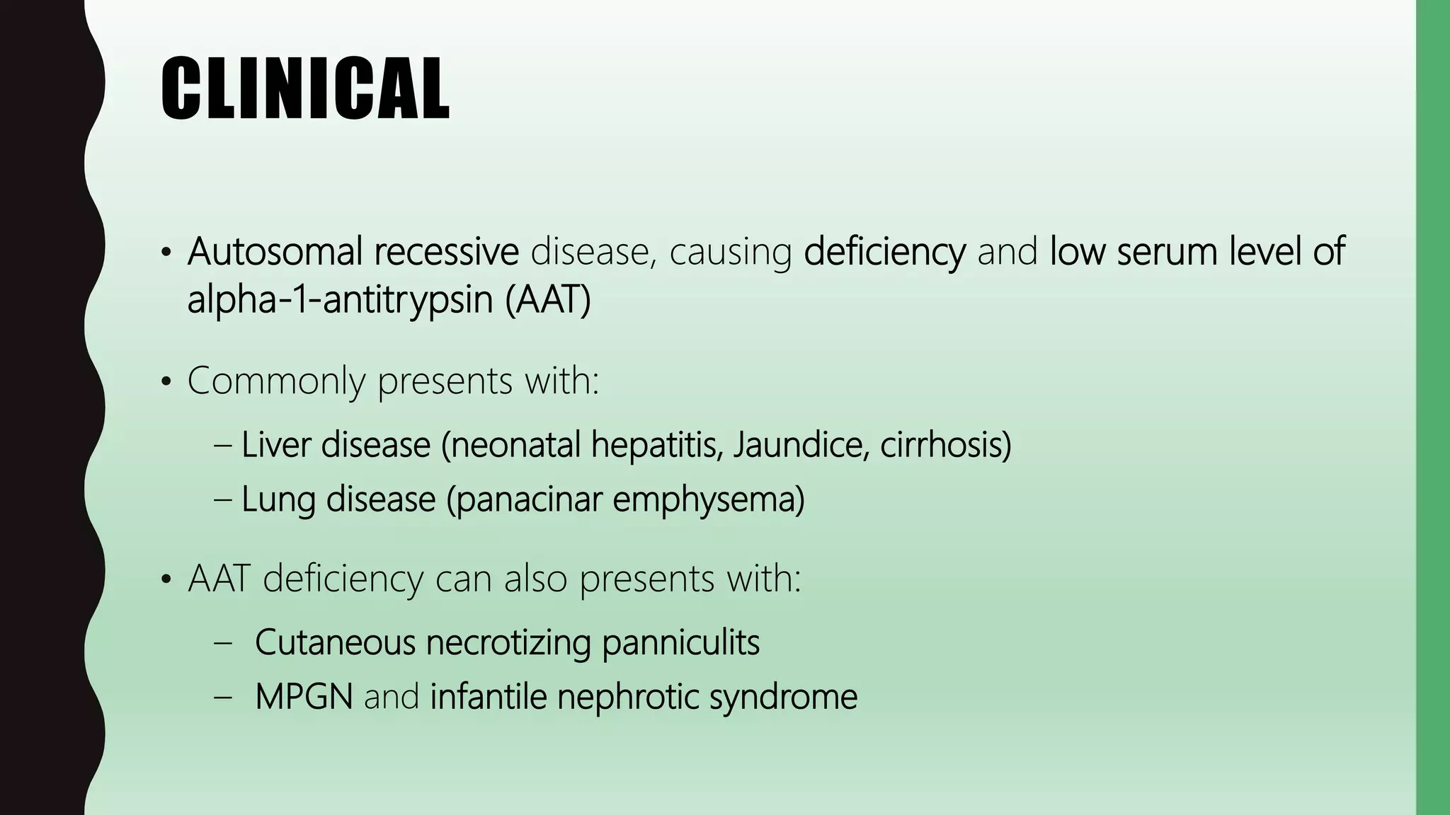
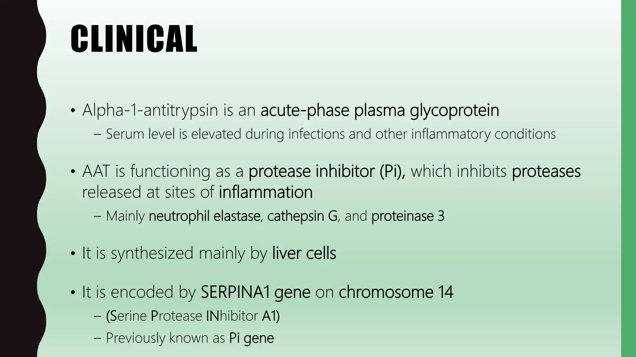
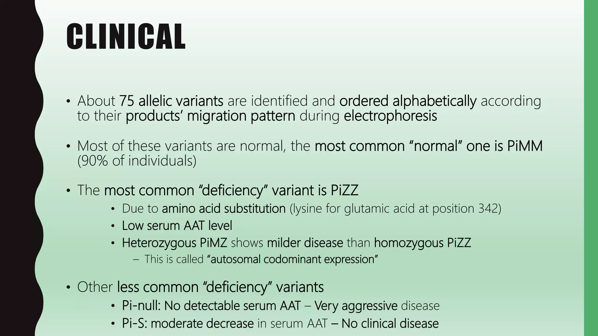
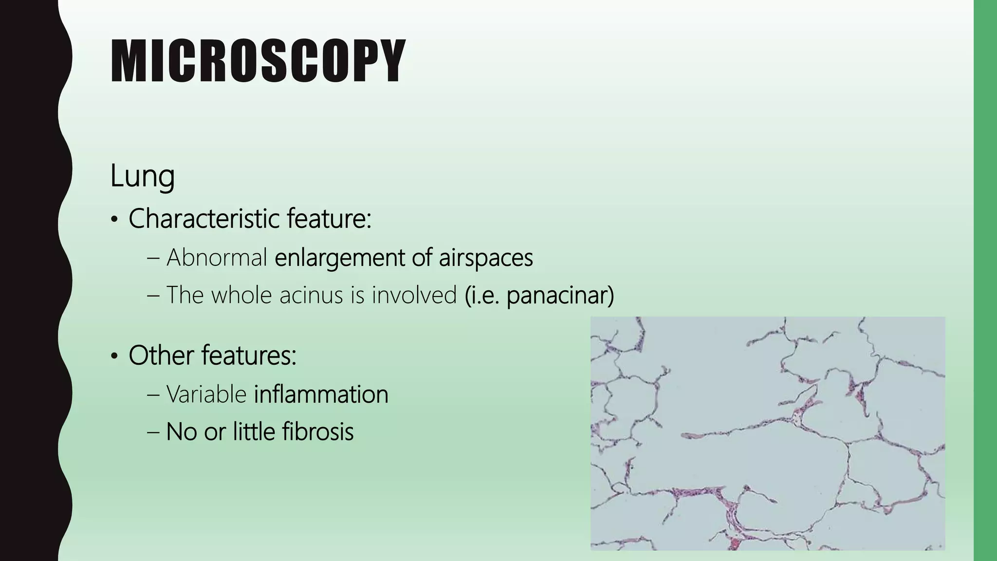
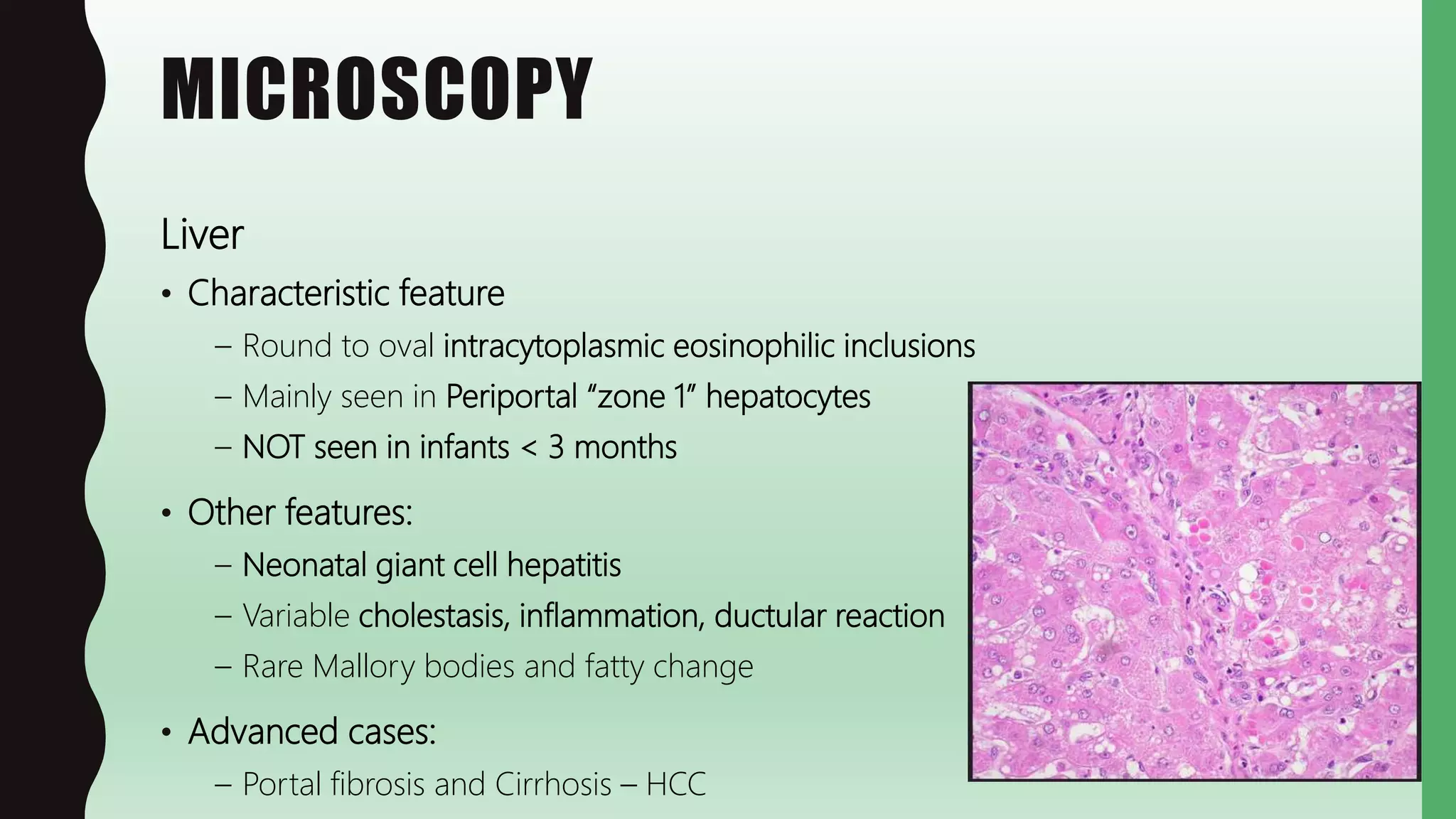
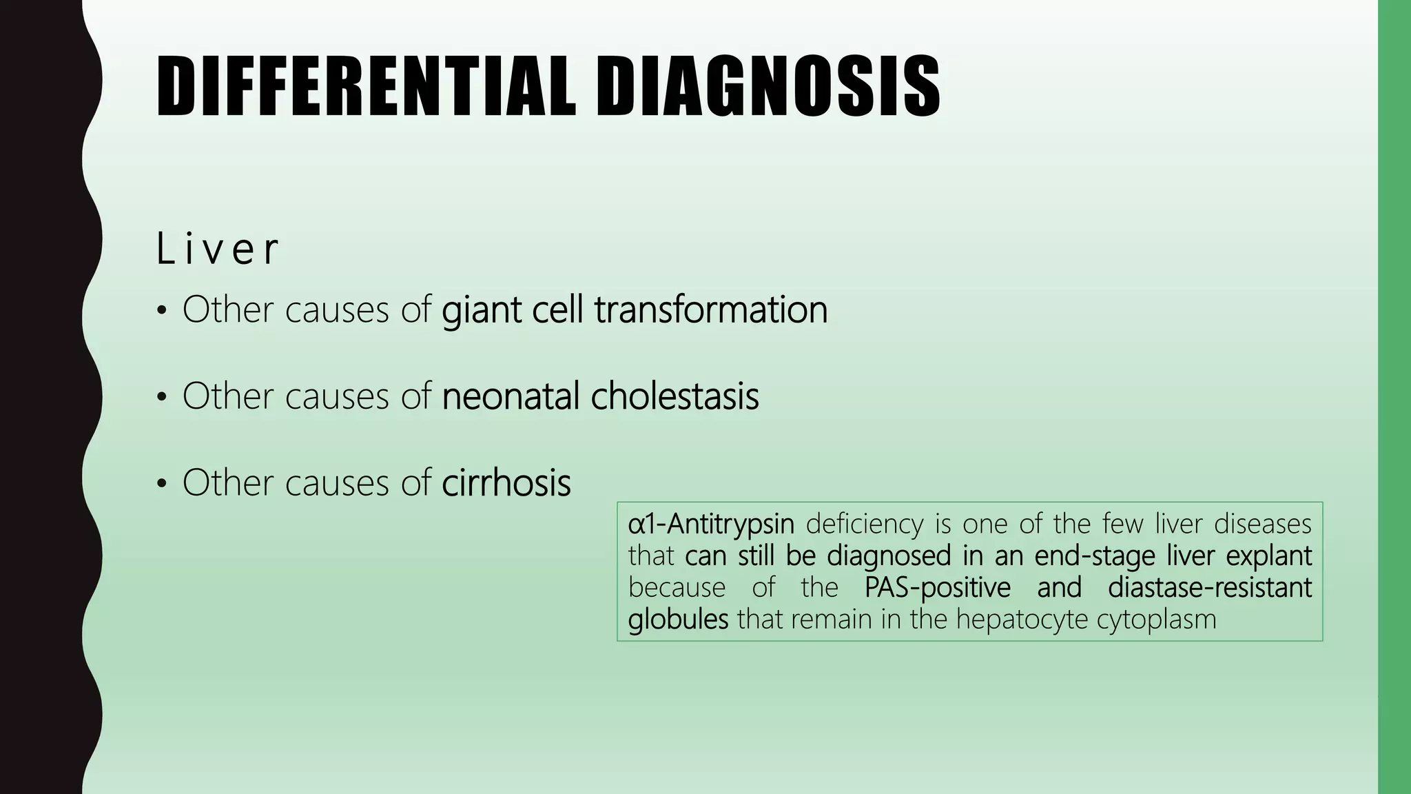
FLASHPATH
H a z e m A l i
ALPHA-1
ANTITRYPSIN
DEFICIENCY
H a z e m A l i
CLINICAL
• Autosomal recessive disease, causing deficiency and low serum level of
alpha-1-antitrypsin (AAT)
• Commonly presents with:
– Liver disease (neonatal hepatitis, Jaundice, cirrhosis)
– Lung disease (panacinar emphysema)
• AAT deficiency can also presents with:
– Cutaneous necrotizing panniculits
– MPGN and infantile nephrotic syndrome
CLINICAL
• Alpha-1-antitrypsin is an acute-phase plasma glycoprotein
– Serum level is elevated during infections and other inflammatory conditions
• AAT is functioning as a protease inhibitor (Pi), which inhibits proteases
released at sites of inflammation
– Mainly neutrophil elastase, cathepsin G, and proteinase 3
• It is synthesized mainly by liver cells
• It is encoded by SERPINA1 gene on chromosome 14
– (Serine Protease INhibitor A1)
– Previously known as Pi gene
CLINICAL
• About 75 allelic variants are identified and ordered alphabetically according
to their products’ migration pattern during electrophoresis
• Most of these variants are normal, the most common “normal” one is PiMM
(90% of individuals)
• The most common “deficiency” variant is PiZZ
• Due to amino acid substitution (lysine for glutamic acid at position 342)
• Low serum AAT level
• Heterozygous PiMZ shows milder disease than homozygous PiZZ
– This is called “autosomal codominant expression”
• Other less common “deficiency” variants
• Pi-null: No detectable serum AAT – Very aggressive disease
• Pi-S: moderate decrease in serum AAT – No clinical disease
PATHOGENESIS
• Due to missense mutation (amino acid substitution), causing abnormal
protein folding and resulting in:
– Blockage of protein transfer from the endoplasmic reticulum to Golgi apparatus
– Prevention of protein secretion into the circulation
• Accumulation of misfolded AAT in hepatocytes results in formation of
cytoplasmic inclusions and apoptosis
• Absent/low circulatory AAT results in unopposed leukocyte proteases activity
during any inflammatory process
– In lungs, destruction of connective tissues causes emphysema
• This can be aggravated by smoking (by increases the activation and influx of
neutrophils)
CLINICAL
Treatment
• Lung:
– "Augmentation therapy“
• infusion of purified AAT from pooled human plasma
– Avoid cigarette smoking
• Liver:
– Mainly symptomatic and supportive
–Liver transplantation in end-stage disease
GROSS
Lung:
• Panacinar emphysema (enlarged cystic lung)
Liver:
• Non-specific
• Advanced disease  cirrhosis
MICROSCOPY
Lung
• Characteristic feature:
– Abnormal enlargement of airspaces
– The whole acinus is involved (i.e. panacinar)
• Other features:
– Variable inflammation
– No or little fibrosis
MICROSCOPY
Liver
• Characteristic feature
– Round to oval intracytoplasmic eosinophilic inclusions
– Mainly seen in Periportal “zone 1” hepatocytes
– NOT seen in infants < 3 months
• Other features:
– Neonatal giant cell hepatitis
– Variable cholestasis, inflammation, ductular reaction
– Rare Mallory bodies and fatty change
• Advanced cases:
– Portal fibrosis and Cirrhosis – HCC
MICROSCOPY
Neonatal giant cell hepatitis
• An injury pattern seen in neonates
• Associated with wide variety of liver diseases
– The most frequently associated disorder is alpha-1 antitrypsin deficiency
• The hallmark is syncytial giant cell transformation of hepatocytes
– Thought to reflect hepatocyte cell fusion and/or mitotic inhibition
Ductular reaction is usually mild in AAT deficiency, but:
– in some case it may predominate (simulate biliary atresia)
– In other cases, there may be paucity of intrahepatic bile ducts
SPECIAL STUDIES
• Laboratory:
Detection of abnormal protein by electrophoresis
• Special stains:
Liver cytoplasmic inclusions are PAS positive and Diastase resistant
• IHC:
Liver cytoplasmic inclusions are AAT positive
• Electron microscopy:
Hepatocytes shows granular material “misfolded protein”
in dilated endoplasmic reticulum
SPECIAL STUDIES
Periodic acid – Schiff (PAS) can also stain:
• Glycogen – red
– E.g. Acinar carcinoma and pancreatic serous cystadenoma
– Also Alveolar soft parts sarcoma (PAS+ intracytoplasmic crystals)
– Also Ewing sarcoma/PNET and Rhabdomyosarcoma (DD from lymphomas)
• Basement membranes – red
– E.g. in Glomerular diseases
• Mucin – red
– E.g. metaplasia and adenocarcinoma
• Colloid – red
• Fungi – red
SPECIAL STUDIES
PAS conjugated with other stains/substances:
• PAS/Diastase:
– Glycogen is digested by diastase “diastase sensitive” (absence of red stain)
– Mucin is not digested by diastase “diastase resistant” (persistence of red stain)
– Also used to detect fungi (in glycogen-rich background e.g. skin)
• PAS/Alcian Blue:
– “pan-mucin” stain, used routinely in all GIT biopsies
– Stains both neutral mucin (PAS+ = red) and acid mucin (AB+ = blue)
• “neutral”  gastric mucin cell metaplasia in small intestine
• “acid”  intestinal metaplasia with goblet cells in stomach or Barrett’s esophagus
• PAS/Light Green:
– Stains fungi (red) and background (green)
SPECIAL STUDIES
Alpha 1-antitrypsin (AAT) is also positive in:
• Normal cells:
– Histiocytes and Liver cells
• Tumors:
– Histiocytic neoplasms
– Many GIT, liver and pancreas neoplasms
– Salivary gland neoplasms
– Yolk sac tumor
– Giant cell tumor of bone
Verylowspecificity
DIFFERENTIAL DIAGNOSIS
L u n g
• Other types of emphysema
• Other causes of obstructive lung diseases
• Other causes of congenital / cystic lung diseases
DIFFERENTIAL DIAGNOSIS
Chronic
bronchitis
Bronchiectasis Asthma
Small-airway
disease
“bronchiolitis”
Emphysema
Site B r o n c h u s Bronchioles Alveoli
Major
pathology
• Mucous gland
hyperplasia
• Excess mucus
• Inflammation
• Airway
dilation &
scarring
• Smooth
muscle
hyperplasia
• Excess mucus
• Inflammation
(eosinophils)
• Inflammatory
scarring &
obliteration
• Airspace
enlargement
• Wall
destruction
• No fibrosis
Other obstructive lung diseases:
DIFFERENTIAL DIAGNOSIS
O t h e r c o n g e n i t a l / c y s t i c l u n g d i s e a s e s :
• Congenital:
– Bronchogenic cysts
– Congenital pulmonary cysts
– Congenital pulmonary airway malformation
– Congenital lobar emphysema
– Pulmonary sequestration
• Acquired:
– Healed abscess
– Honeycombing
• Mixed:
– Cystic fibrosis
No destruction of alveoli
Fibrosis
DIFFERENTIAL DIAGNOSIS
L i v e r
• Other causes of giant cell transformation
• Other causes of neonatal cholestasis
• Other causes of cirrhosis
α1-Antitrypsin deficiency is one of the few liver diseases
that can still be diagnosed in an end-stage liver explant
because of the PAS-positive and diastase-resistant
globules that remain in the hepatocyte cytoplasm
WWW.
DO NOT FORGET TO SEARCH FOR MORE PICS
AND VIRTUAL SLIDES
THANK YOU
H a z e m A l i

FlashPath - Alpha-1-Antitrypsin Deficiency

  • 1.
    FLASHPATH H a ze m A l i
  • 2.
  • 3.
    CLINICAL • Autosomal recessivedisease, causing deficiency and low serum level of alpha-1-antitrypsin (AAT) • Commonly presents with: – Liver disease (neonatal hepatitis, Jaundice, cirrhosis) – Lung disease (panacinar emphysema) • AAT deficiency can also presents with: – Cutaneous necrotizing panniculits – MPGN and infantile nephrotic syndrome
  • 4.
    CLINICAL • Alpha-1-antitrypsin isan acute-phase plasma glycoprotein – Serum level is elevated during infections and other inflammatory conditions • AAT is functioning as a protease inhibitor (Pi), which inhibits proteases released at sites of inflammation – Mainly neutrophil elastase, cathepsin G, and proteinase 3 • It is synthesized mainly by liver cells • It is encoded by SERPINA1 gene on chromosome 14 – (Serine Protease INhibitor A1) – Previously known as Pi gene
  • 5.
    CLINICAL • About 75allelic variants are identified and ordered alphabetically according to their products’ migration pattern during electrophoresis • Most of these variants are normal, the most common “normal” one is PiMM (90% of individuals) • The most common “deficiency” variant is PiZZ • Due to amino acid substitution (lysine for glutamic acid at position 342) • Low serum AAT level • Heterozygous PiMZ shows milder disease than homozygous PiZZ – This is called “autosomal codominant expression” • Other less common “deficiency” variants • Pi-null: No detectable serum AAT – Very aggressive disease • Pi-S: moderate decrease in serum AAT – No clinical disease
  • 6.
    PATHOGENESIS • Due tomissense mutation (amino acid substitution), causing abnormal protein folding and resulting in: – Blockage of protein transfer from the endoplasmic reticulum to Golgi apparatus – Prevention of protein secretion into the circulation • Accumulation of misfolded AAT in hepatocytes results in formation of cytoplasmic inclusions and apoptosis • Absent/low circulatory AAT results in unopposed leukocyte proteases activity during any inflammatory process – In lungs, destruction of connective tissues causes emphysema • This can be aggravated by smoking (by increases the activation and influx of neutrophils)
  • 7.
    CLINICAL Treatment • Lung: – "Augmentationtherapy“ • infusion of purified AAT from pooled human plasma – Avoid cigarette smoking • Liver: – Mainly symptomatic and supportive –Liver transplantation in end-stage disease
  • 8.
    GROSS Lung: • Panacinar emphysema(enlarged cystic lung) Liver: • Non-specific • Advanced disease  cirrhosis
  • 9.
    MICROSCOPY Lung • Characteristic feature: –Abnormal enlargement of airspaces – The whole acinus is involved (i.e. panacinar) • Other features: – Variable inflammation – No or little fibrosis
  • 10.
    MICROSCOPY Liver • Characteristic feature –Round to oval intracytoplasmic eosinophilic inclusions – Mainly seen in Periportal “zone 1” hepatocytes – NOT seen in infants < 3 months • Other features: – Neonatal giant cell hepatitis – Variable cholestasis, inflammation, ductular reaction – Rare Mallory bodies and fatty change • Advanced cases: – Portal fibrosis and Cirrhosis – HCC
  • 11.
    MICROSCOPY Neonatal giant cellhepatitis • An injury pattern seen in neonates • Associated with wide variety of liver diseases – The most frequently associated disorder is alpha-1 antitrypsin deficiency • The hallmark is syncytial giant cell transformation of hepatocytes – Thought to reflect hepatocyte cell fusion and/or mitotic inhibition Ductular reaction is usually mild in AAT deficiency, but: – in some case it may predominate (simulate biliary atresia) – In other cases, there may be paucity of intrahepatic bile ducts
  • 12.
    SPECIAL STUDIES • Laboratory: Detectionof abnormal protein by electrophoresis • Special stains: Liver cytoplasmic inclusions are PAS positive and Diastase resistant • IHC: Liver cytoplasmic inclusions are AAT positive • Electron microscopy: Hepatocytes shows granular material “misfolded protein” in dilated endoplasmic reticulum
  • 13.
    SPECIAL STUDIES Periodic acid– Schiff (PAS) can also stain: • Glycogen – red – E.g. Acinar carcinoma and pancreatic serous cystadenoma – Also Alveolar soft parts sarcoma (PAS+ intracytoplasmic crystals) – Also Ewing sarcoma/PNET and Rhabdomyosarcoma (DD from lymphomas) • Basement membranes – red – E.g. in Glomerular diseases • Mucin – red – E.g. metaplasia and adenocarcinoma • Colloid – red • Fungi – red
  • 14.
    SPECIAL STUDIES PAS conjugatedwith other stains/substances: • PAS/Diastase: – Glycogen is digested by diastase “diastase sensitive” (absence of red stain) – Mucin is not digested by diastase “diastase resistant” (persistence of red stain) – Also used to detect fungi (in glycogen-rich background e.g. skin) • PAS/Alcian Blue: – “pan-mucin” stain, used routinely in all GIT biopsies – Stains both neutral mucin (PAS+ = red) and acid mucin (AB+ = blue) • “neutral”  gastric mucin cell metaplasia in small intestine • “acid”  intestinal metaplasia with goblet cells in stomach or Barrett’s esophagus • PAS/Light Green: – Stains fungi (red) and background (green)
  • 15.
    SPECIAL STUDIES Alpha 1-antitrypsin(AAT) is also positive in: • Normal cells: – Histiocytes and Liver cells • Tumors: – Histiocytic neoplasms – Many GIT, liver and pancreas neoplasms – Salivary gland neoplasms – Yolk sac tumor – Giant cell tumor of bone Verylowspecificity
  • 16.
    DIFFERENTIAL DIAGNOSIS L un g • Other types of emphysema • Other causes of obstructive lung diseases • Other causes of congenital / cystic lung diseases
  • 17.
    DIFFERENTIAL DIAGNOSIS Chronic bronchitis Bronchiectasis Asthma Small-airway disease “bronchiolitis” Emphysema SiteB r o n c h u s Bronchioles Alveoli Major pathology • Mucous gland hyperplasia • Excess mucus • Inflammation • Airway dilation & scarring • Smooth muscle hyperplasia • Excess mucus • Inflammation (eosinophils) • Inflammatory scarring & obliteration • Airspace enlargement • Wall destruction • No fibrosis Other obstructive lung diseases:
  • 18.
    DIFFERENTIAL DIAGNOSIS O th e r c o n g e n i t a l / c y s t i c l u n g d i s e a s e s : • Congenital: – Bronchogenic cysts – Congenital pulmonary cysts – Congenital pulmonary airway malformation – Congenital lobar emphysema – Pulmonary sequestration • Acquired: – Healed abscess – Honeycombing • Mixed: – Cystic fibrosis No destruction of alveoli Fibrosis
  • 19.
    DIFFERENTIAL DIAGNOSIS L iv e r • Other causes of giant cell transformation • Other causes of neonatal cholestasis • Other causes of cirrhosis α1-Antitrypsin deficiency is one of the few liver diseases that can still be diagnosed in an end-stage liver explant because of the PAS-positive and diastase-resistant globules that remain in the hepatocyte cytoplasm
  • 20.
    WWW. DO NOT FORGETTO SEARCH FOR MORE PICS AND VIRTUAL SLIDES
  • 21.
    THANK YOU H az e m A l i