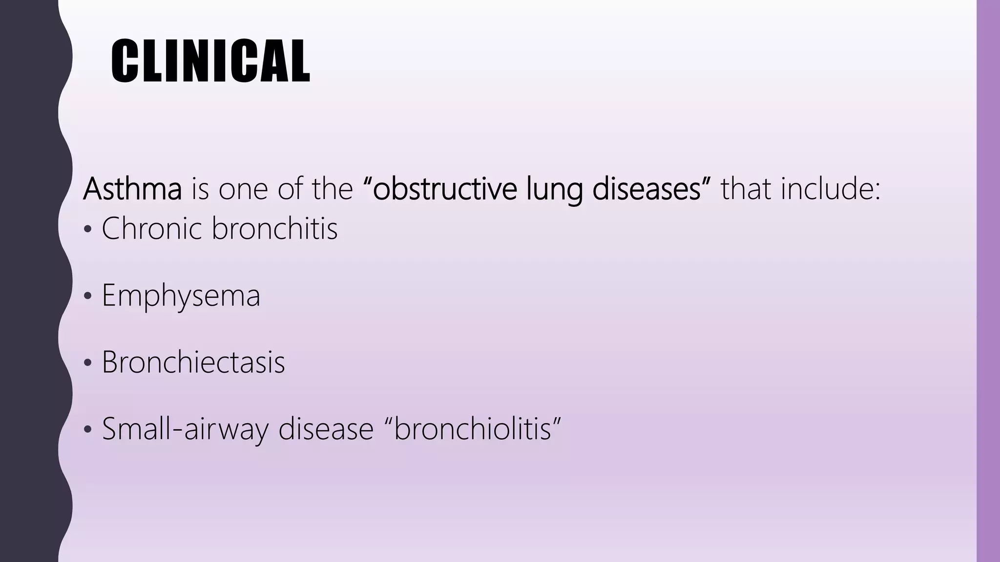
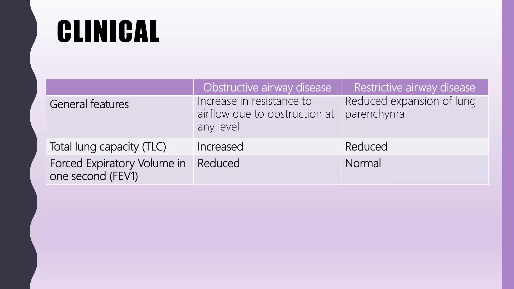
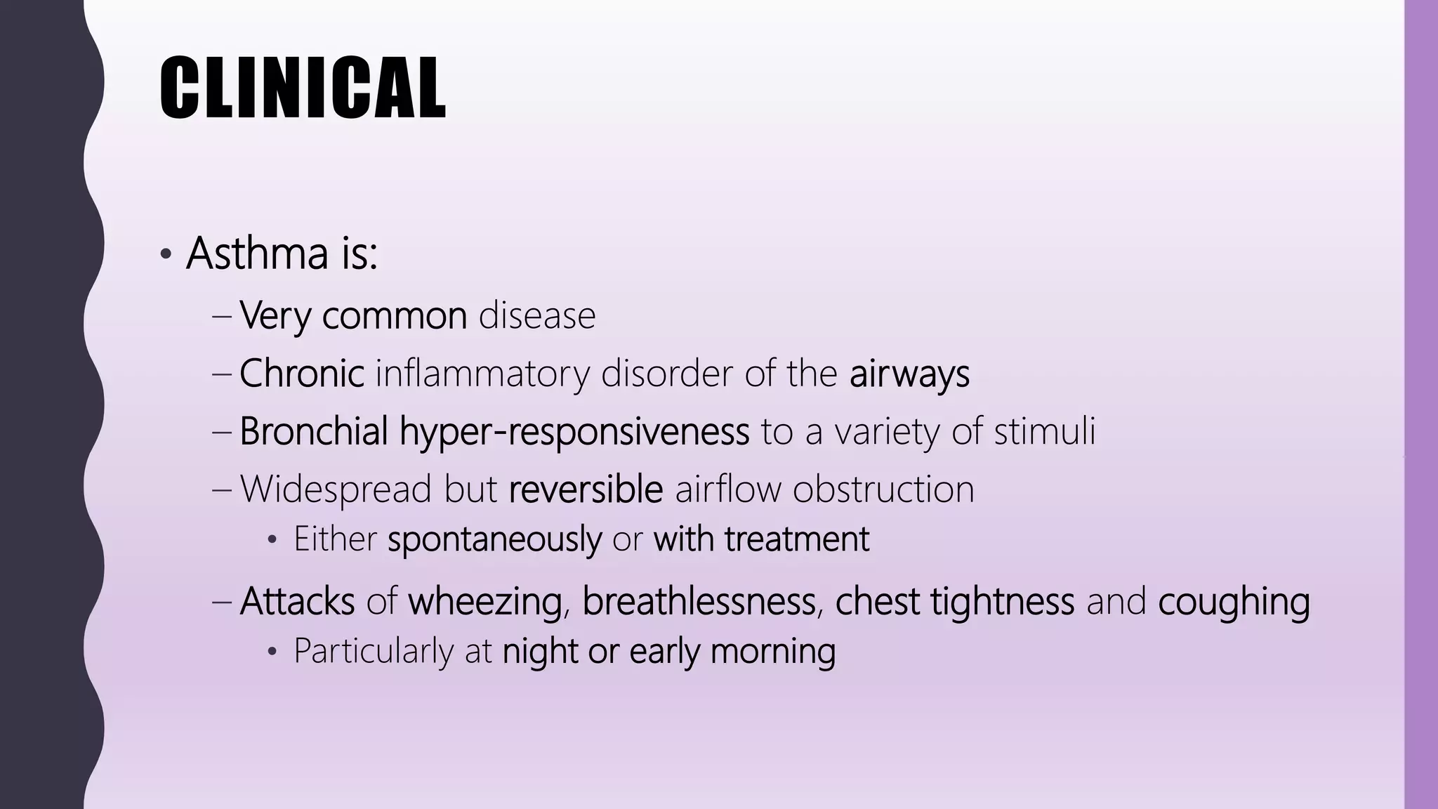
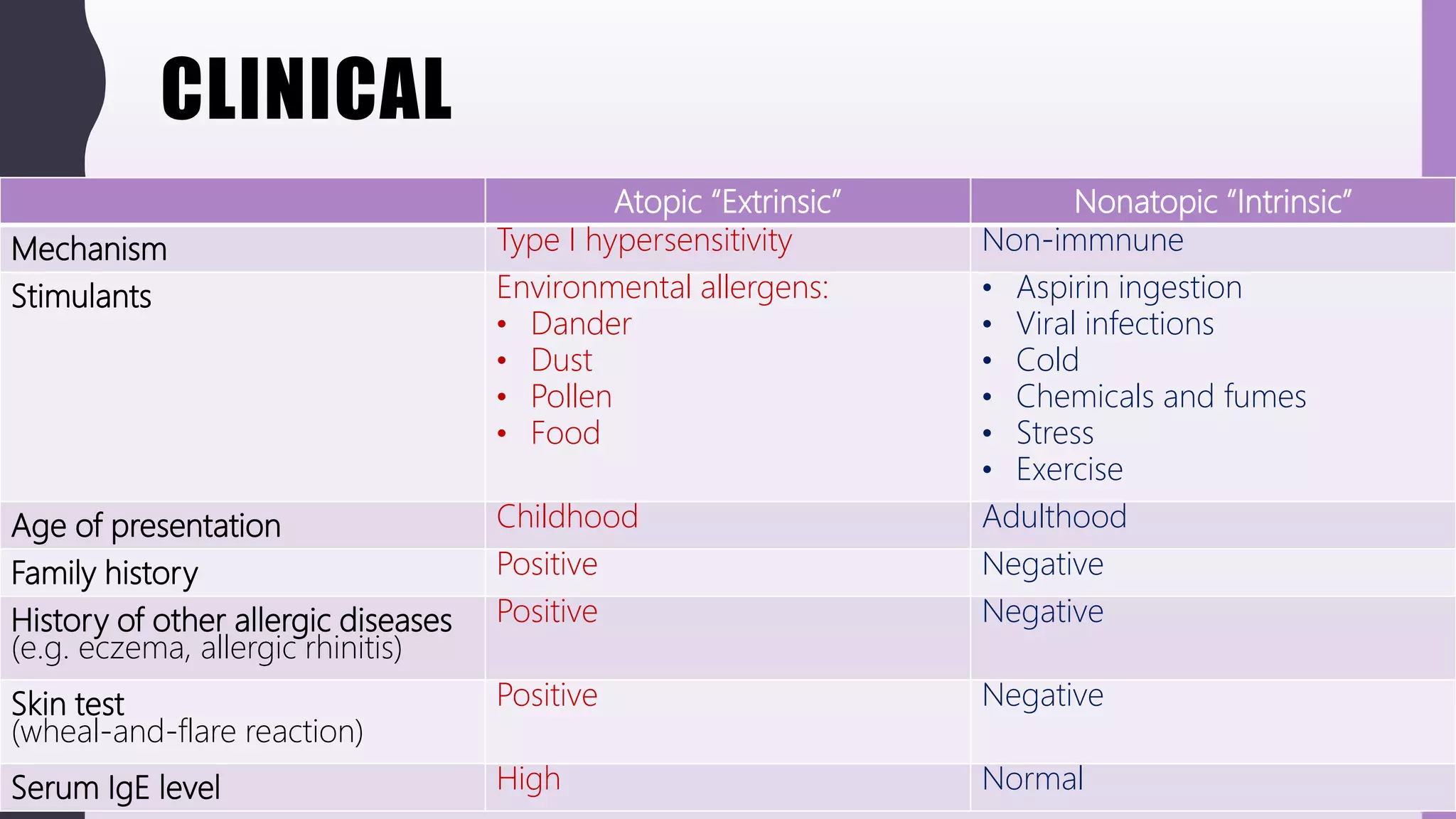
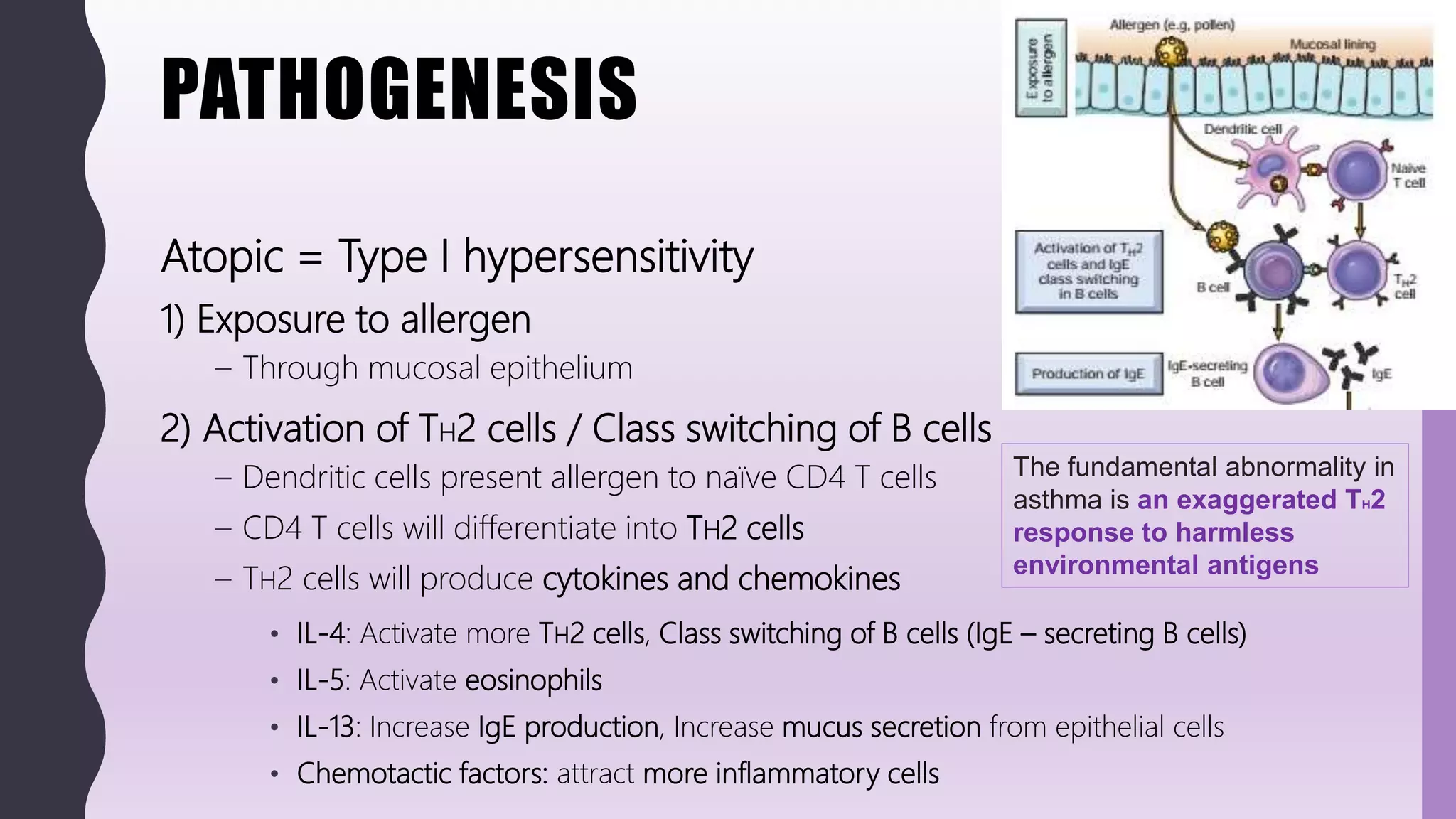
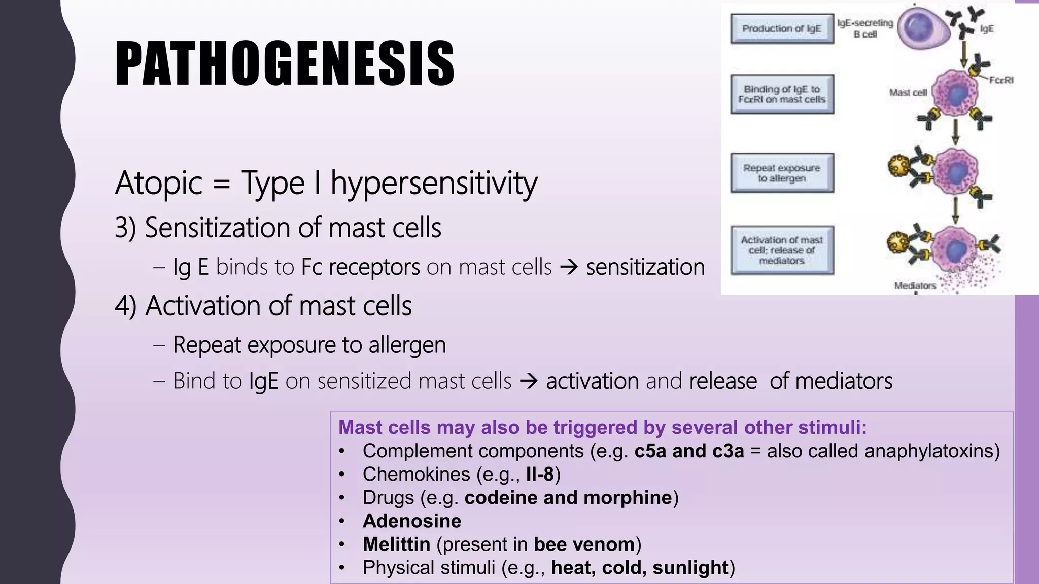
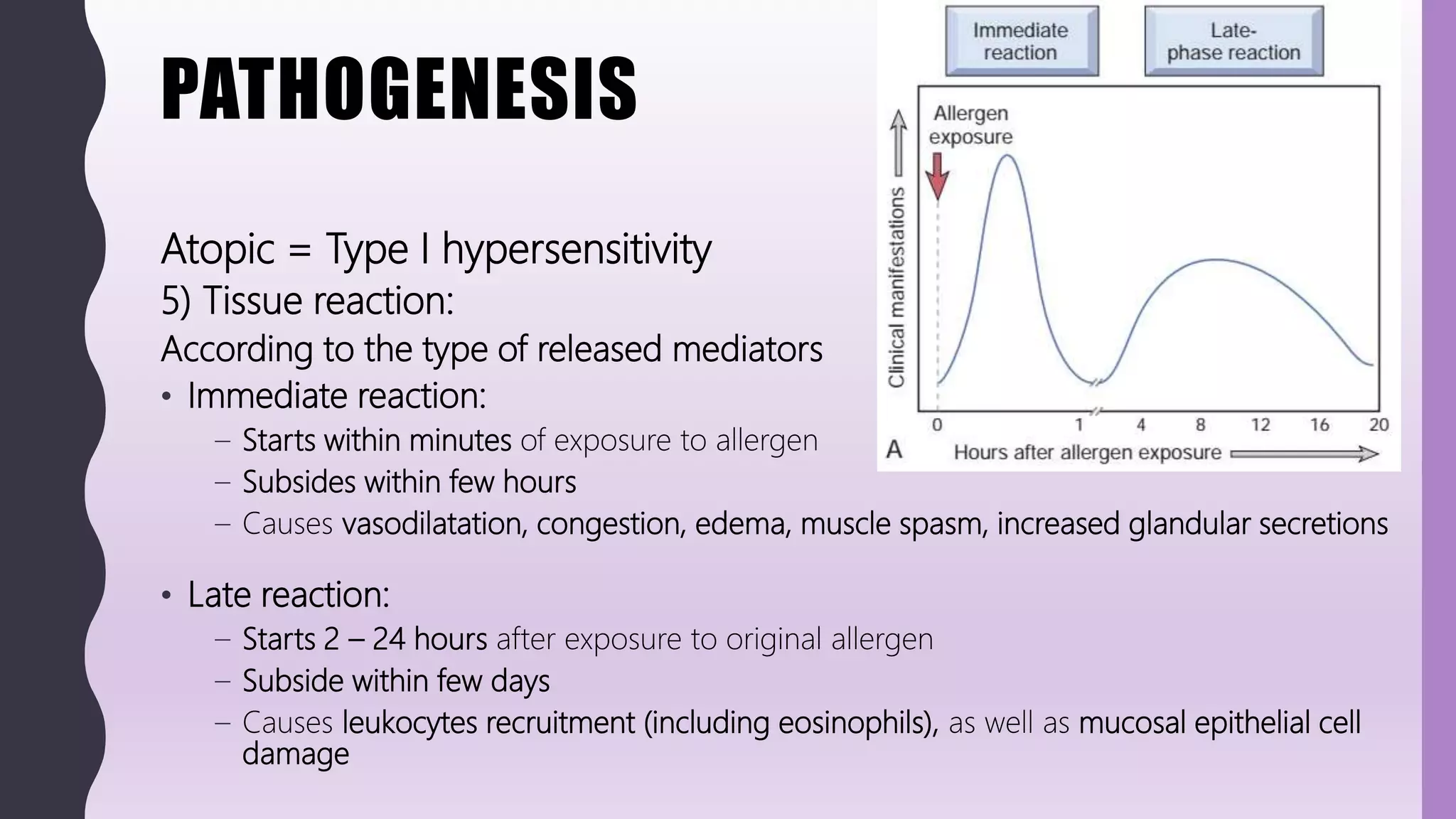
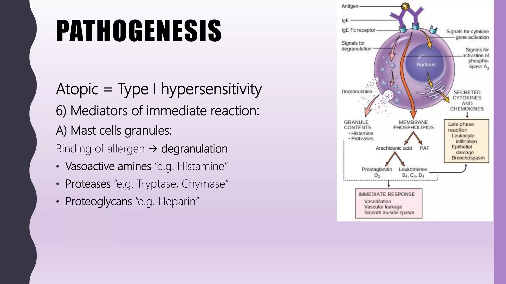
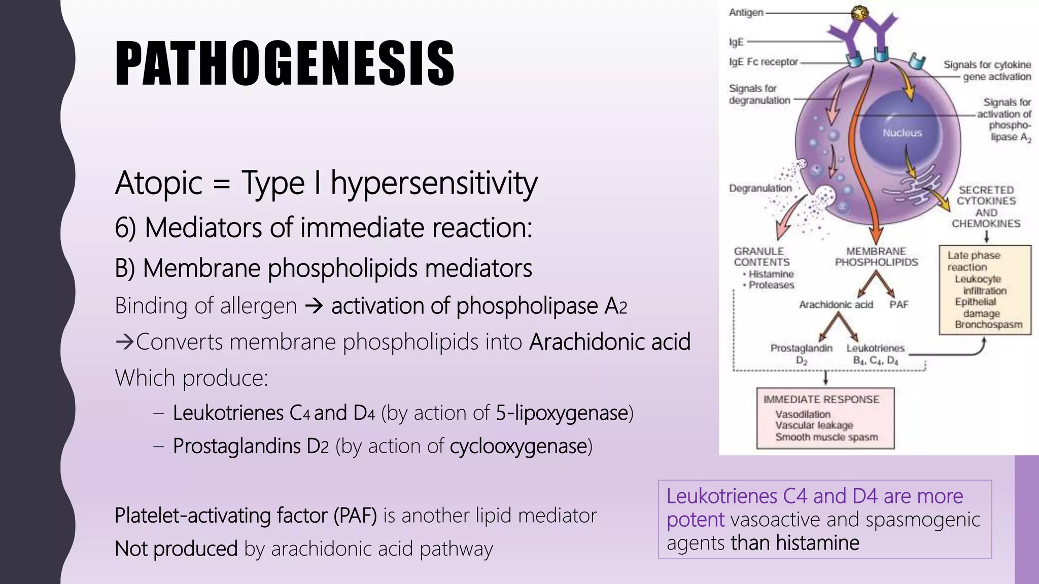
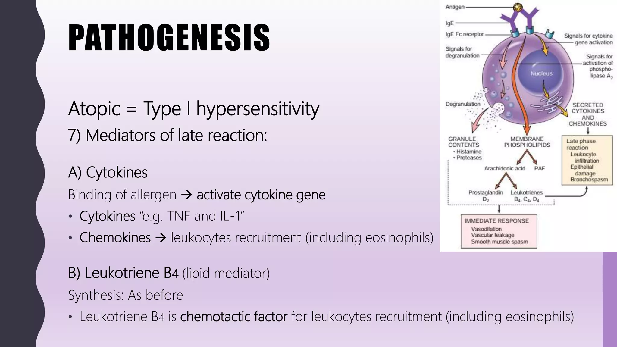
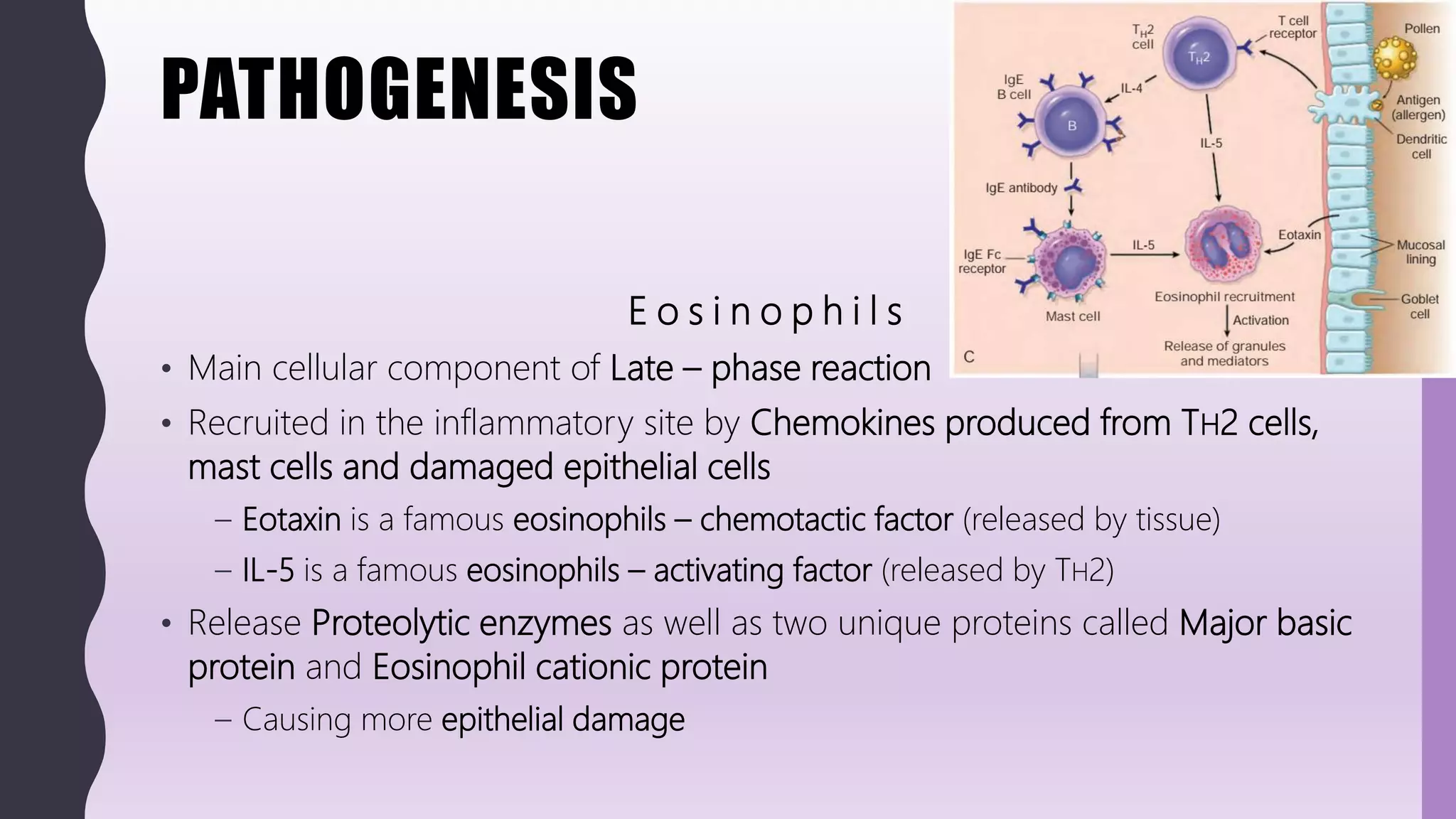
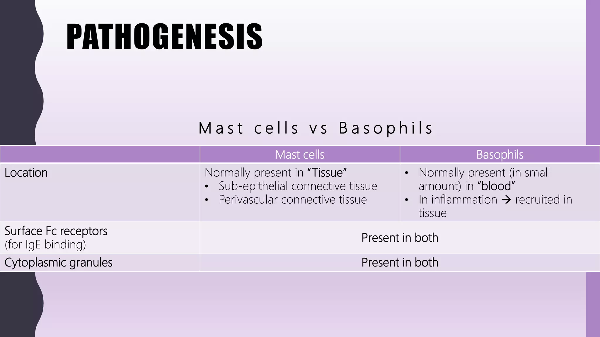
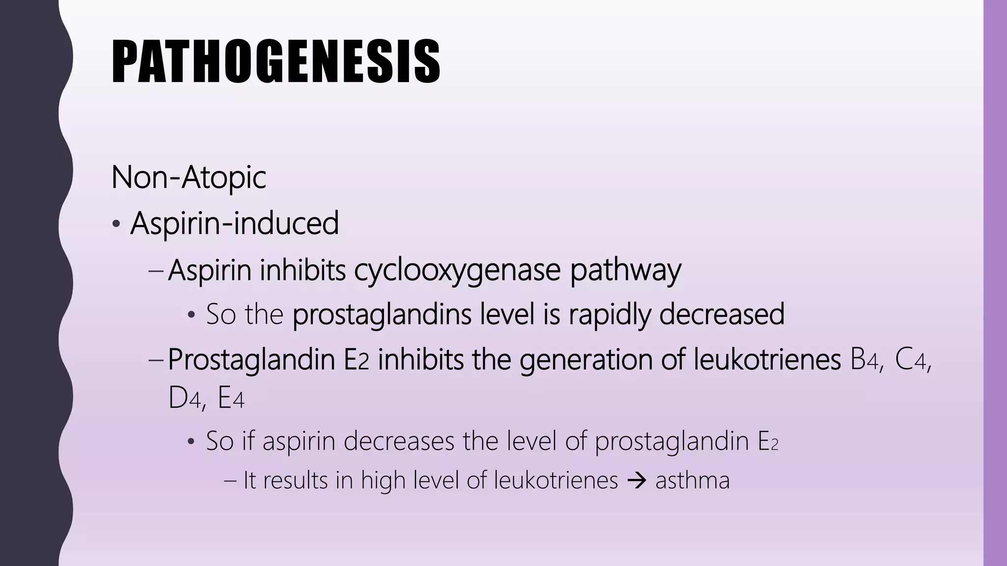
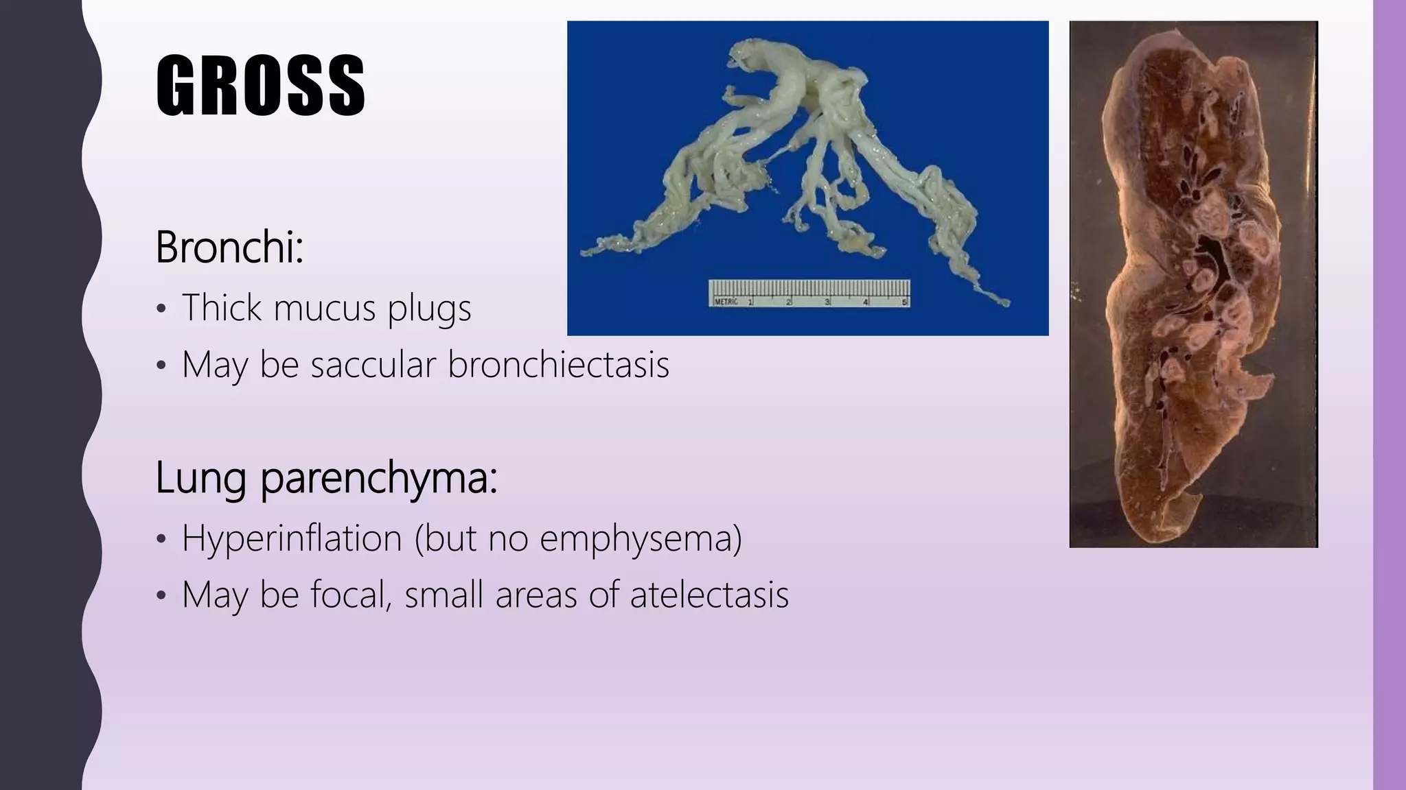
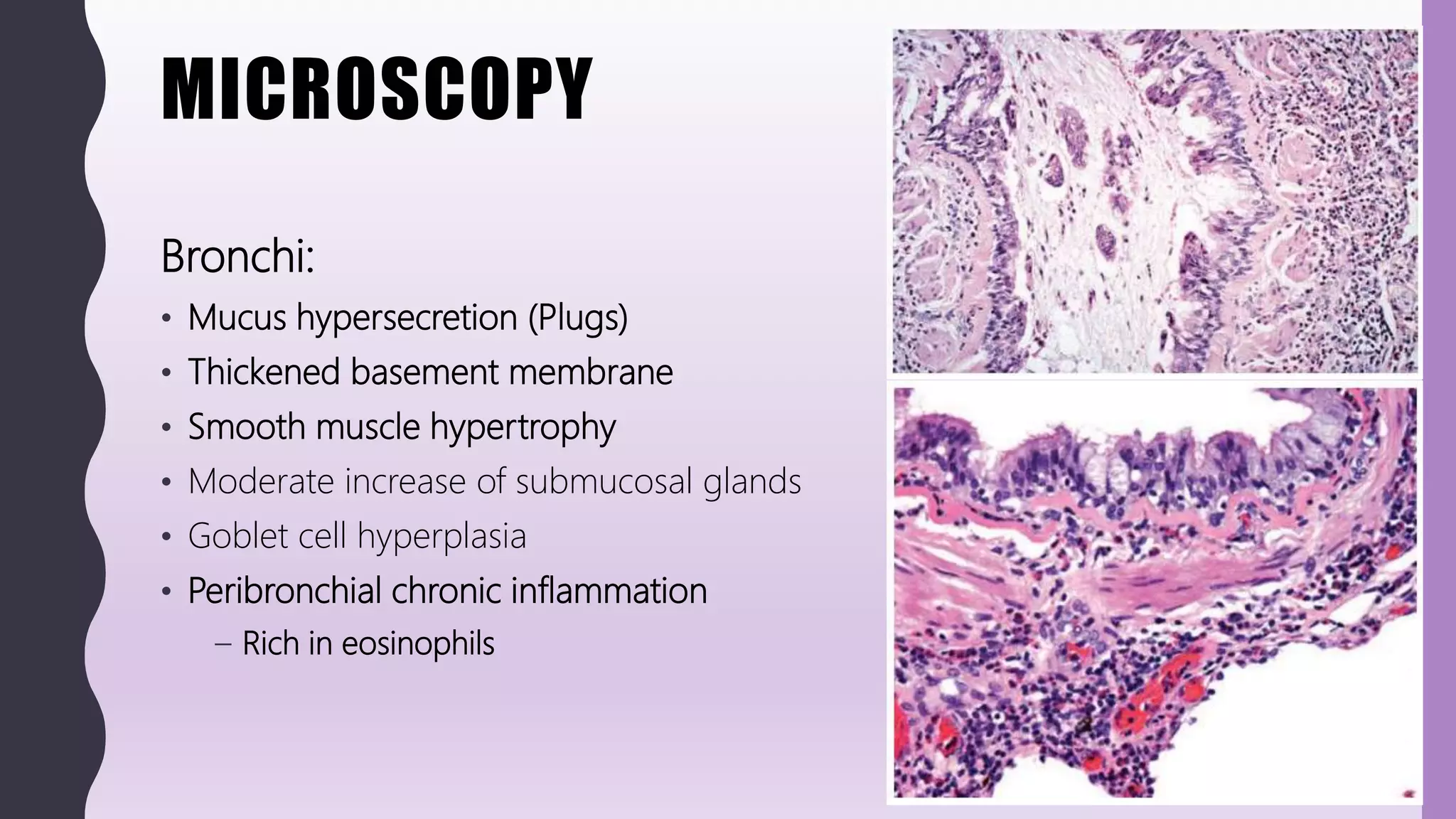
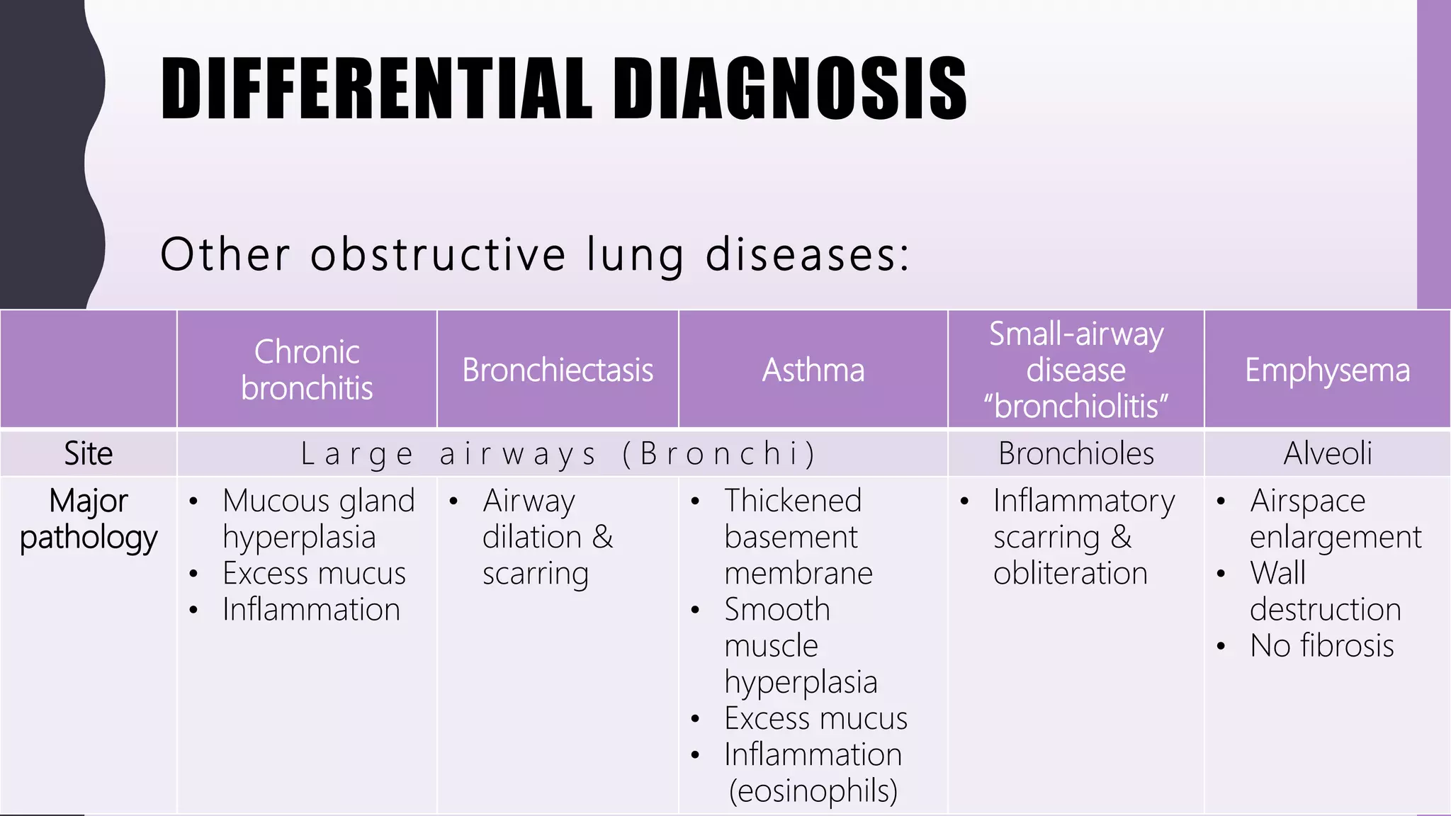
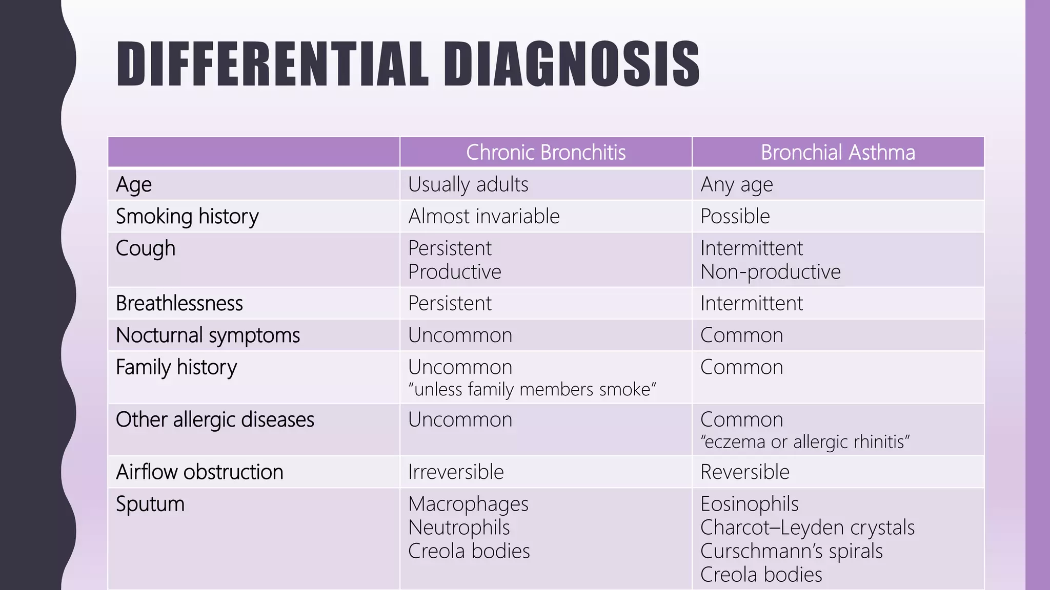
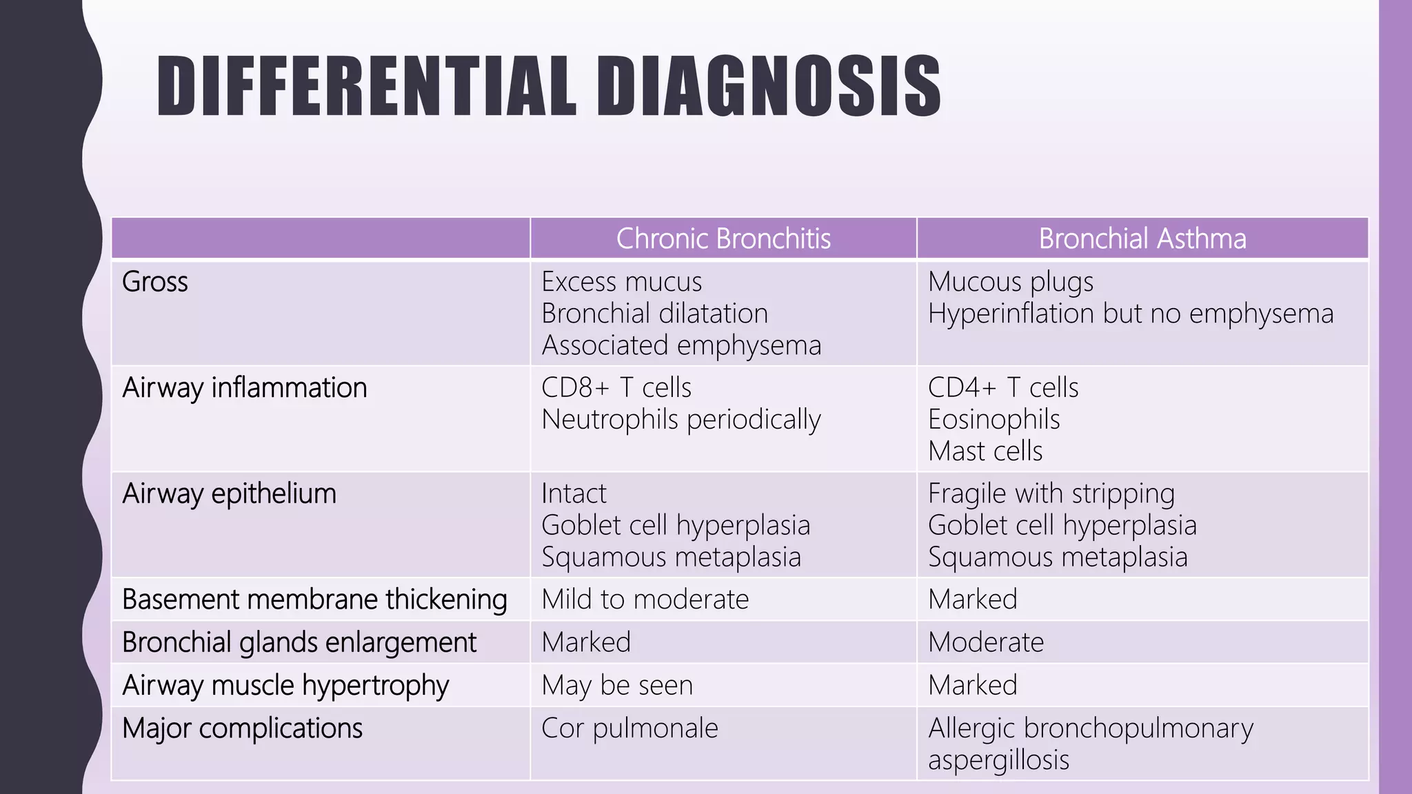
FLASHPATH
H a z e m A l i
ASTHMA
H a z e m A l i
CLINICAL
Asthma is one of the “obstructive lung diseases” that include:
• Chronic bronchitis
• Emphysema
• Bronchiectasis
• Small-airway disease “bronchiolitis”
CLINICAL
Obstructive airway disease Restrictive airway disease
General features Increase in resistance to
airflow due to obstruction at
any level
Reduced expansion of lung
parenchyma
Total lung capacity (TLC) Increased Reduced
Forced Expiratory Volume in
one second (FEV1)
Reduced Normal
CLINICAL
• Asthma is:
– Very common disease
– Chronic inflammatory disorder of the airways
– Bronchial hyper-responsiveness to a variety of stimuli
– Widespread but reversible airflow obstruction
• Either spontaneously or with treatment
– Attacks of wheezing, breathlessness, chest tightness and coughing
• Particularly at night or early morning
CLINICAL
Atopic “Extrinsic” Nonatopic “Intrinsic”
Mechanism Type I hypersensitivity Non-immnune
Stimulants Environmental allergens:
• Dander
• Dust
• Pollen
• Food
• Aspirin ingestion
• Viral infections
• Cold
• Chemicals and fumes
• Stress
• Exercise
Age of presentation Childhood Adulthood
Family history Positive Negative
History of other allergic diseases
(e.g. eczema, allergic rhinitis)
Positive Negative
Skin test
(wheal-and-flare reaction)
Positive Negative
Serum IgE level High Normal
CLINICAL
Treatment:
• Avoidance of trigger agents
• Desensitization to specific allergens
• Bronchodilator drugs and corticosteroids
• Leukotriene and leukotriene receptor antagonists
• Antibodies directed against IgE
Status asthmaticus:
• Unremitting acute severe asthmatic attacks, may be fatal
• Usually in patients with a long history of asthma
PATHOGENESIS
Atopic = Type I hypersensitivity
1) Exposure to allergen
– Through mucosal epithelium
2) Activation of TH2 cells / Class switching of B cells
– Dendritic cells present allergen to naïve CD4 T cells
– CD4 T cells will differentiate into TH2 cells
– TH2 cells will produce cytokines and chemokines
• IL-4: Activate more TH2 cells, Class switching of B cells (IgE – secreting B cells)
• IL-5: Activate eosinophils
• IL-13: Increase IgE production, Increase mucus secretion from epithelial cells
• Chemotactic factors: attract more inflammatory cells
The fundamental abnormality in
asthma is an exaggerated TH2
response to harmless
environmental antigens
PATHOGENESIS
Atopic = Type I hypersensitivity
3) Sensitization of mast cells
– Ig E binds to Fc receptors on mast cells  sensitization
4) Activation of mast cells
– Repeat exposure to allergen
– Bind to IgE on sensitized mast cells  activation and release of mediators
Mast cells may also be triggered by several other stimuli:
• Complement components (e.g. c5a and c3a = also called anaphylatoxins)
• Chemokines (e.g., Il-8)
• Drugs (e.g. codeine and morphine)
• Adenosine
• Melittin (present in bee venom)
• Physical stimuli (e.g., heat, cold, sunlight)
PATHOGENESIS
Atopic = Type I hypersensitivity
5) Tissue reaction:
According to the type of released mediators
• Immediate reaction:
– Starts within minutes of exposure to allergen
– Subsides within few hours
– Causes vasodilatation, congestion, edema, muscle spasm, increased glandular secretions
• Late reaction:
– Starts 2 – 24 hours after exposure to original allergen
– Subside within few days
– Causes leukocytes recruitment (including eosinophils), as well as mucosal epithelial cell
damage
PATHOGENESIS
Atopic = Type I hypersensitivity
6) Mediators of immediate reaction:
A) Mast cells granules:
Binding of allergen  degranulation
• Vasoactive amines “e.g. Histamine”
• Proteases “e.g. Tryptase, Chymase”
• Proteoglycans “e.g. Heparin”
PATHOGENESIS
Atopic = Type I hypersensitivity
6) Mediators of immediate reaction:
B) Membrane phospholipids mediators
Binding of allergen  activation of phospholipase A2
Converts membrane phospholipids into Arachidonic acid
Which produce:
– Leukotrienes C4 and D4 (by action of 5-lipoxygenase)
– Prostaglandins D2 (by action of cyclooxygenase)
Platelet-activating factor (PAF) is another lipid mediator
Not produced by arachidonic acid pathway
Leukotrienes C4 and D4 are more
potent vasoactive and spasmogenic
agents than histamine
PATHOGENESIS
Atopic = Type I hypersensitivity
7) Mediators of late reaction:
A) Cytokines
Binding of allergen  activate cytokine gene
• Cytokines “e.g. TNF and IL-1”
• Chemokines  leukocytes recruitment (including eosinophils)
B) Leukotriene B4 (lipid mediator)
Synthesis: As before
• Leukotriene B4 is chemotactic factor for leukocytes recruitment (including eosinophils)
PATHOGENESIS
E o s i n o p h i l s
• Main cellular component of Late – phase reaction
• Recruited in the inflammatory site by Chemokines produced from TH2 cells,
mast cells and damaged epithelial cells
– Eotaxin is a famous eosinophils – chemotactic factor (released by tissue)
– IL-5 is a famous eosinophils – activating factor (released by TH2)
• Release Proteolytic enzymes as well as two unique proteins called Major basic
protein and Eosinophil cationic protein
– Causing more epithelial damage
PATHOGENESIS
M a s t c e l l s v s B a s o p h i l s
Mast cells Basophils
Location Normally present in “Tissue”
• Sub-epithelial connective tissue
• Perivascular connective tissue
• Normally present (in small
amount) in “blood”
• In inflammation  recruited in
tissue
Surface Fc receptors
(for IgE binding)
Present in both
Cytoplasmic granules Present in both
PATHOGENESIS
Non-Atopic
• Aspirin-induced
– Aspirin inhibits cyclooxygenase pathway
• So the prostaglandins level is rapidly decreased
– Prostaglandin E2 inhibits the generation of leukotrienes B4, C4,
D4, E4
• So if aspirin decreases the level of prostaglandin E2
– It results in high level of leukotrienes  asthma
GROSS
Bronchi:
• Thick mucus plugs
• May be saccular bronchiectasis
Lung parenchyma:
• Hyperinflation (but no emphysema)
• May be focal, small areas of atelectasis
MICROSCOPY
Bronchi:
• Mucus hypersecretion (Plugs)
• Thickened basement membrane
• Smooth muscle hypertrophy
• Moderate increase of submucosal glands
• Goblet cell hyperplasia
• Peribronchial chronic inflammation
– Rich in eosinophils
MICROSCOPY
Complications / associations:
• Churg-Strauss Syndrome
– Small vessels vasculitis
– Mainly affect skin, GIT, kidneys, heart and lungs
– Vasculitis / Necrotizing granulomas near blood vessels (angio-centric)
– Also eosinophilic infiltration and peripheral eosinophilia
– P-ANCAs are seen in 50% of cases
MICROSCOPY
Complications / associations:
• Allergic Bronchopulmonary Aspergillosis
– Hypersensitivity reaction to Aspergillus fumigatus
– Necrotizing granulomas around airways (broncho-centric)
• Necrotic material contain aspergillus hyphae
– Also mucus impaction and eosinophilic pneumonia
– Later on, bronchial dilatation and bronchiectasis
– Elevated IgE directed against aspergillus antigens
CYTOLOGY
Eosinophils
Charcot-Leyden crystals
• Rhomboid-shaped crystalloids
• Composed of galectin-10
– Lysophospholipase
– Derived from degenerating eosinophils
Curschmann spirals
• Excess Mucus
• Also seen in Chronic bronchitis
– No Charcot-leyden crystals
– No Eosinophils
CYTOLOGY
Benign bronchial cell changes
1. Reactive changes
– Columnar, ciliated bronchial cells
– Nuclear enlargement
– Coarse chromatin
– Prominent nucleoli
2. Creola bodies
– Spherical 3D Clusters
– Columnar, ciliated bronchial cells
– Also seen in Chronic bronchitis
• No Charcot-leyden crystals
• No Eosinophils
Retainedciliaisanevidenceofbenign
natureofthebronchialcells
CYTOLOGY
3. Goblet cell hyperplasia
– Large sheets or round clusters
– Composed almost exclusively of goblet cells
• Abundant mucin-filled cytoplasm
– Benign columnar, ciliated bronchial cells are also seen
• The bronchi are lined with ciliated or columnar epithelium with scattered goblet
cells. Goblet cell hyperplasia is an indication of irritation, such as in bronchitis or asthma
Benign bronchial cells changes can mimic Adenocarcinoma:
– LOSS OF CILIA
– LESS COHESIVE
– MARKED MITOSIS / NECROSIS
– CLINICAL HISTORY
• Do not rush for calling adenocarcinoma on just few
small group of atypical cells
- especially with inflammatory background OR history of
COPD, Asthma
• Reactive changes resolve within 1 month
 treat, wait and repeat cytology
CYTOLOGY
Other injury-associated findings
1. Bronchial reserve cell hyperplasia
– Tightly packed small cells
– Scant cytoplasm
– Smudged dark chromatin
– Nuclear molding may be seen
– No mitoses or necrosis
Bronchial reserve cell hyperplasia can mimic Small cell carcinoma:
– LESS COHESIVE
– MARKED MITOSIS / NECROSIS
– CLINICAL HISTORY
CYTOLOGY
Other injury-associated findings
2. Reparative “re-epithelialization” of respiratory tract
– Flat, cohesive sheets
– Abundant cytoplasm
– Enlarged nuclei
– prominent nucleoli
– Mitoses
Reparative epithelium can mimic Non-Small cell carcinoma:
– LESS COHESIVE
– MARKED MITOSIS / NECROSIS
– CLINICAL HISTORY
DIFFERENTIAL DIAGNOSIS
Chronic
bronchitis
Bronchiectasis Asthma
Small-airway
disease
“bronchiolitis”
Emphysema
Site L a r g e a i r w a y s ( B r o n c h i ) Bronchioles Alveoli
Major
pathology
• Mucous gland
hyperplasia
• Excess mucus
• Inflammation
• Airway
dilation &
scarring
• Thickened
basement
membrane
• Smooth
muscle
hyperplasia
• Excess mucus
• Inflammation
(eosinophils)
• Inflammatory
scarring &
obliteration
• Airspace
enlargement
• Wall
destruction
• No fibrosis
Other obstructive lung diseases:
DIFFERENTIAL DIAGNOSIS
Chronic Bronchitis Bronchial Asthma
Age Usually adults Any age
Smoking history Almost invariable Possible
Cough Persistent
Productive
Intermittent
Non-productive
Breathlessness Persistent Intermittent
Nocturnal symptoms Uncommon Common
Family history Uncommon
“unless family members smoke”
Common
Other allergic diseases Uncommon Common
“eczema or allergic rhinitis”
Airflow obstruction Irreversible Reversible
Sputum Macrophages
Neutrophils
Creola bodies
Eosinophils
Charcot–Leyden crystals
Curschmann’s spirals
Creola bodies
DIFFERENTIAL DIAGNOSIS
Chronic Bronchitis Bronchial Asthma
Gross Excess mucus
Bronchial dilatation
Associated emphysema
Mucous plugs
Hyperinflation but no emphysema
Airway inflammation CD8+ T cells
Neutrophils periodically
CD4+ T cells
Eosinophils
Mast cells
Airway epithelium Intact
Goblet cell hyperplasia
Squamous metaplasia
Fragile with stripping
Goblet cell hyperplasia
Squamous metaplasia
Basement membrane thickening Mild to moderate Marked
Bronchial glands enlargement Marked Moderate
Airway muscle hypertrophy May be seen Marked
Major complications Cor pulmonale Allergic bronchopulmonary
aspergillosis
WWW.
DO NOT FORGET TO SEARCH FOR MORE PICS
AND VIRTUAL SLIDES
THANK YOU
H a z e m A l i

FlashPath - Lung - Asthma

  • 1.
    FLASHPATH H a ze m A l i
  • 2.
    ASTHMA H a ze m A l i
  • 3.
    CLINICAL Asthma is oneof the “obstructive lung diseases” that include: • Chronic bronchitis • Emphysema • Bronchiectasis • Small-airway disease “bronchiolitis”
  • 4.
    CLINICAL Obstructive airway diseaseRestrictive airway disease General features Increase in resistance to airflow due to obstruction at any level Reduced expansion of lung parenchyma Total lung capacity (TLC) Increased Reduced Forced Expiratory Volume in one second (FEV1) Reduced Normal
  • 5.
    CLINICAL • Asthma is: –Very common disease – Chronic inflammatory disorder of the airways – Bronchial hyper-responsiveness to a variety of stimuli – Widespread but reversible airflow obstruction • Either spontaneously or with treatment – Attacks of wheezing, breathlessness, chest tightness and coughing • Particularly at night or early morning
  • 6.
    CLINICAL Atopic “Extrinsic” Nonatopic“Intrinsic” Mechanism Type I hypersensitivity Non-immnune Stimulants Environmental allergens: • Dander • Dust • Pollen • Food • Aspirin ingestion • Viral infections • Cold • Chemicals and fumes • Stress • Exercise Age of presentation Childhood Adulthood Family history Positive Negative History of other allergic diseases (e.g. eczema, allergic rhinitis) Positive Negative Skin test (wheal-and-flare reaction) Positive Negative Serum IgE level High Normal
  • 7.
    CLINICAL Treatment: • Avoidance oftrigger agents • Desensitization to specific allergens • Bronchodilator drugs and corticosteroids • Leukotriene and leukotriene receptor antagonists • Antibodies directed against IgE Status asthmaticus: • Unremitting acute severe asthmatic attacks, may be fatal • Usually in patients with a long history of asthma
  • 8.
    PATHOGENESIS Atopic = TypeI hypersensitivity 1) Exposure to allergen – Through mucosal epithelium 2) Activation of TH2 cells / Class switching of B cells – Dendritic cells present allergen to naïve CD4 T cells – CD4 T cells will differentiate into TH2 cells – TH2 cells will produce cytokines and chemokines • IL-4: Activate more TH2 cells, Class switching of B cells (IgE – secreting B cells) • IL-5: Activate eosinophils • IL-13: Increase IgE production, Increase mucus secretion from epithelial cells • Chemotactic factors: attract more inflammatory cells The fundamental abnormality in asthma is an exaggerated TH2 response to harmless environmental antigens
  • 9.
    PATHOGENESIS Atopic = TypeI hypersensitivity 3) Sensitization of mast cells – Ig E binds to Fc receptors on mast cells  sensitization 4) Activation of mast cells – Repeat exposure to allergen – Bind to IgE on sensitized mast cells  activation and release of mediators Mast cells may also be triggered by several other stimuli: • Complement components (e.g. c5a and c3a = also called anaphylatoxins) • Chemokines (e.g., Il-8) • Drugs (e.g. codeine and morphine) • Adenosine • Melittin (present in bee venom) • Physical stimuli (e.g., heat, cold, sunlight)
  • 10.
    PATHOGENESIS Atopic = TypeI hypersensitivity 5) Tissue reaction: According to the type of released mediators • Immediate reaction: – Starts within minutes of exposure to allergen – Subsides within few hours – Causes vasodilatation, congestion, edema, muscle spasm, increased glandular secretions • Late reaction: – Starts 2 – 24 hours after exposure to original allergen – Subside within few days – Causes leukocytes recruitment (including eosinophils), as well as mucosal epithelial cell damage
  • 11.
    PATHOGENESIS Atopic = TypeI hypersensitivity 6) Mediators of immediate reaction: A) Mast cells granules: Binding of allergen  degranulation • Vasoactive amines “e.g. Histamine” • Proteases “e.g. Tryptase, Chymase” • Proteoglycans “e.g. Heparin”
  • 12.
    PATHOGENESIS Atopic = TypeI hypersensitivity 6) Mediators of immediate reaction: B) Membrane phospholipids mediators Binding of allergen  activation of phospholipase A2 Converts membrane phospholipids into Arachidonic acid Which produce: – Leukotrienes C4 and D4 (by action of 5-lipoxygenase) – Prostaglandins D2 (by action of cyclooxygenase) Platelet-activating factor (PAF) is another lipid mediator Not produced by arachidonic acid pathway Leukotrienes C4 and D4 are more potent vasoactive and spasmogenic agents than histamine
  • 13.
    PATHOGENESIS Atopic = TypeI hypersensitivity 7) Mediators of late reaction: A) Cytokines Binding of allergen  activate cytokine gene • Cytokines “e.g. TNF and IL-1” • Chemokines  leukocytes recruitment (including eosinophils) B) Leukotriene B4 (lipid mediator) Synthesis: As before • Leukotriene B4 is chemotactic factor for leukocytes recruitment (including eosinophils)
  • 14.
    PATHOGENESIS E o si n o p h i l s • Main cellular component of Late – phase reaction • Recruited in the inflammatory site by Chemokines produced from TH2 cells, mast cells and damaged epithelial cells – Eotaxin is a famous eosinophils – chemotactic factor (released by tissue) – IL-5 is a famous eosinophils – activating factor (released by TH2) • Release Proteolytic enzymes as well as two unique proteins called Major basic protein and Eosinophil cationic protein – Causing more epithelial damage
  • 15.
    PATHOGENESIS M a st c e l l s v s B a s o p h i l s Mast cells Basophils Location Normally present in “Tissue” • Sub-epithelial connective tissue • Perivascular connective tissue • Normally present (in small amount) in “blood” • In inflammation  recruited in tissue Surface Fc receptors (for IgE binding) Present in both Cytoplasmic granules Present in both
  • 16.
    PATHOGENESIS Non-Atopic • Aspirin-induced – Aspirininhibits cyclooxygenase pathway • So the prostaglandins level is rapidly decreased – Prostaglandin E2 inhibits the generation of leukotrienes B4, C4, D4, E4 • So if aspirin decreases the level of prostaglandin E2 – It results in high level of leukotrienes  asthma
  • 17.
    GROSS Bronchi: • Thick mucusplugs • May be saccular bronchiectasis Lung parenchyma: • Hyperinflation (but no emphysema) • May be focal, small areas of atelectasis
  • 18.
    MICROSCOPY Bronchi: • Mucus hypersecretion(Plugs) • Thickened basement membrane • Smooth muscle hypertrophy • Moderate increase of submucosal glands • Goblet cell hyperplasia • Peribronchial chronic inflammation – Rich in eosinophils
  • 19.
    MICROSCOPY Complications / associations: •Churg-Strauss Syndrome – Small vessels vasculitis – Mainly affect skin, GIT, kidneys, heart and lungs – Vasculitis / Necrotizing granulomas near blood vessels (angio-centric) – Also eosinophilic infiltration and peripheral eosinophilia – P-ANCAs are seen in 50% of cases
  • 20.
    MICROSCOPY Complications / associations: •Allergic Bronchopulmonary Aspergillosis – Hypersensitivity reaction to Aspergillus fumigatus – Necrotizing granulomas around airways (broncho-centric) • Necrotic material contain aspergillus hyphae – Also mucus impaction and eosinophilic pneumonia – Later on, bronchial dilatation and bronchiectasis – Elevated IgE directed against aspergillus antigens
  • 21.
    CYTOLOGY Eosinophils Charcot-Leyden crystals • Rhomboid-shapedcrystalloids • Composed of galectin-10 – Lysophospholipase – Derived from degenerating eosinophils Curschmann spirals • Excess Mucus • Also seen in Chronic bronchitis – No Charcot-leyden crystals – No Eosinophils
  • 22.
    CYTOLOGY Benign bronchial cellchanges 1. Reactive changes – Columnar, ciliated bronchial cells – Nuclear enlargement – Coarse chromatin – Prominent nucleoli 2. Creola bodies – Spherical 3D Clusters – Columnar, ciliated bronchial cells – Also seen in Chronic bronchitis • No Charcot-leyden crystals • No Eosinophils Retainedciliaisanevidenceofbenign natureofthebronchialcells
  • 23.
    CYTOLOGY 3. Goblet cellhyperplasia – Large sheets or round clusters – Composed almost exclusively of goblet cells • Abundant mucin-filled cytoplasm – Benign columnar, ciliated bronchial cells are also seen • The bronchi are lined with ciliated or columnar epithelium with scattered goblet cells. Goblet cell hyperplasia is an indication of irritation, such as in bronchitis or asthma Benign bronchial cells changes can mimic Adenocarcinoma: – LOSS OF CILIA – LESS COHESIVE – MARKED MITOSIS / NECROSIS – CLINICAL HISTORY • Do not rush for calling adenocarcinoma on just few small group of atypical cells - especially with inflammatory background OR history of COPD, Asthma • Reactive changes resolve within 1 month  treat, wait and repeat cytology
  • 24.
    CYTOLOGY Other injury-associated findings 1.Bronchial reserve cell hyperplasia – Tightly packed small cells – Scant cytoplasm – Smudged dark chromatin – Nuclear molding may be seen – No mitoses or necrosis Bronchial reserve cell hyperplasia can mimic Small cell carcinoma: – LESS COHESIVE – MARKED MITOSIS / NECROSIS – CLINICAL HISTORY
  • 25.
    CYTOLOGY Other injury-associated findings 2.Reparative “re-epithelialization” of respiratory tract – Flat, cohesive sheets – Abundant cytoplasm – Enlarged nuclei – prominent nucleoli – Mitoses Reparative epithelium can mimic Non-Small cell carcinoma: – LESS COHESIVE – MARKED MITOSIS / NECROSIS – CLINICAL HISTORY
  • 26.
    DIFFERENTIAL DIAGNOSIS Chronic bronchitis Bronchiectasis Asthma Small-airway disease “bronchiolitis” Emphysema SiteL a r g e a i r w a y s ( B r o n c h i ) Bronchioles Alveoli Major pathology • Mucous gland hyperplasia • Excess mucus • Inflammation • Airway dilation & scarring • Thickened basement membrane • Smooth muscle hyperplasia • Excess mucus • Inflammation (eosinophils) • Inflammatory scarring & obliteration • Airspace enlargement • Wall destruction • No fibrosis Other obstructive lung diseases:
  • 27.
    DIFFERENTIAL DIAGNOSIS Chronic BronchitisBronchial Asthma Age Usually adults Any age Smoking history Almost invariable Possible Cough Persistent Productive Intermittent Non-productive Breathlessness Persistent Intermittent Nocturnal symptoms Uncommon Common Family history Uncommon “unless family members smoke” Common Other allergic diseases Uncommon Common “eczema or allergic rhinitis” Airflow obstruction Irreversible Reversible Sputum Macrophages Neutrophils Creola bodies Eosinophils Charcot–Leyden crystals Curschmann’s spirals Creola bodies
  • 28.
    DIFFERENTIAL DIAGNOSIS Chronic BronchitisBronchial Asthma Gross Excess mucus Bronchial dilatation Associated emphysema Mucous plugs Hyperinflation but no emphysema Airway inflammation CD8+ T cells Neutrophils periodically CD4+ T cells Eosinophils Mast cells Airway epithelium Intact Goblet cell hyperplasia Squamous metaplasia Fragile with stripping Goblet cell hyperplasia Squamous metaplasia Basement membrane thickening Mild to moderate Marked Bronchial glands enlargement Marked Moderate Airway muscle hypertrophy May be seen Marked Major complications Cor pulmonale Allergic bronchopulmonary aspergillosis
  • 29.
    WWW. DO NOT FORGETTO SEARCH FOR MORE PICS AND VIRTUAL SLIDES
  • 30.
    THANK YOU H az e m A l i