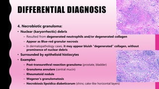
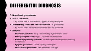
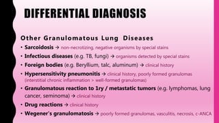
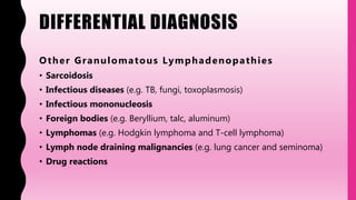
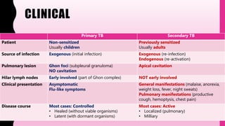
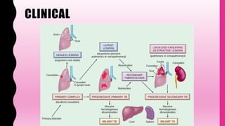
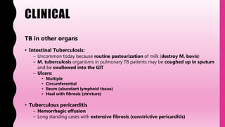
3. CLINICAL
• Serious chronic pulmonary disease (but can affect any organ)
– More than 1 billion infected individuals worldwide
– About 9 million new cases a year
– Estimated 1.5 million deaths each year
• Caused by Mycobacteria tuberculosis
• Transmitted by airborne droplets (sputum)
– Needs contact with active case
• Risk factors:
– General: poverty, overcrowding, malnutrition
– Chronic diseases: RA, SLE, DM, chronic renal failure
– Low immunity: AIDS, immunosuppression
– Chronic pulmonary diseases: especially Silicosis
Other common mycobacterium species:
M. Bovis Intestinal TB (unpasteurized milk)
M. Lepra Leprosy
M. Avium – Intracellulare (MAI) Atypical TB
– common in AIDS
4. CLINICAL
Primary TB Secondary TB
Patient Non-sensitized
Usually children
Previously sensitized
Usually adults
Source of infection Exogenous (initial infection) Exogenous (re-infection)
Endogenous (re-activation)
Pulmonary lesion Ghon foci (subpleural granuloma)
NO cavitation
Apical cavitation
Hilar lymph nodes Early involved (part of Ghon complex) NOT early involved
Clinical presentation Asymptomatic
Flu-like symptoms
General manifestations (malaise, anorexia,
weight loss, fever, night sweats)
Pulmonary manifestations (productive
cough, hemoptysis, chest pain)
Disease course Most cases: Controlled
• Healed (without viable organisms)
• Latent (with dormant organisms)
Most cases: Active
• Localized (pulmonary)
• Milliary
6. CLINICAL
TB in other organs
• Intestinal Tuberculosis:
– Uncommon today because routine pasteurization of milk (destroy M. bovis)
– M. tuberculosis organisms in pulmonary TB patients may be coughed up in sputum
and be swallowed into the GIT
– Ulcers:
• Multiple
• Circumferential
• Ileum (abundant lymphoid tissue)
• Heal with fibrosis (stricture)
• Tuberculous pericarditis
– Hemorrhagic effusion
– Long standing cases with extensive fibrosis (constrictive pericarditis)
7. CLINICAL
TB in other organs
• CNS Tuberculosis
– Tuberculous meningitis (CSF shows a high protein, low glucose, and
lymphocytosis)
– The base of the brain is often involved (various cranial nerve affection)
– Rarely, a solitary granuloma, or "tuberculoma“ (manifest with seizures)
• Tuberculous spondylitis (Pott's disease)
– Involves mainly the thoracic and lumbar vertebrae
– Extensive bony destruction with compressed fractures (scoliosis – kyphosis –
neurological deficits)
– Extension to soft tissues (including psoas cold abscess)
• Scrofula
– Tuberculous lymphadenitis of the cervical nodes
– Firm mass of matted nodes just under the mandible
– There can be chronic draining fistulous tracts to overlying skin
8. CLINICAL
TB in other organs
• Renal Tuberculosis:
– “Sterile pyuria" with WBC's present in urine but a negative routine bacterial culture
– Progressive destruction of renal parenchyma occurs if not treated
– Drainage to the ureters (ureteral stricture, tuberculous cystitis)
• Adrenal Tuberculosis:
– Usually bilateral
– Adrenals are markedly enlarged
– Destruction of cortex (Addison's disease)
• Genital Tract Tuberculosis
– Tuberculous salpingitis and endometritis (irregular menstrual bleeding and
infertility)
– Tuberculous prostatitis and epididymitis (non-tender induration and infertility)
9. CLINICAL
TB and Amyloidosis
• Secondary amyloidosis or AA amyloidosis (amyloid A protein)
• Associate long-standing chronic inflammatory diseases
– e.g., chronic osteomyelitis, rheumatoid arthritis, tuberculosis
11. CLINICAL
BCG vaccine
• Contains mycobacteria called “bacille Calmette-Guérin”
– Attenuated strain of Mycobacterium bovis
• Often given to people in countries where TB is common
– South America, Africa, Asia, Eastern Europe, and Russia
• Most effective in babies and children
– Less effective in adults older than 20 years
• Most effective in preventing TB from spreading outside of the lungs (extrapulmonary
TB)
• Contraindicated in AIDS and immunosuppressed patients
• Interferes with tuberculin skin test results (false positivity)
• Can be also used as a treatment for early-stage bladder cancer
12. PATHOGENESIS
Primary TB infection
A) Before T-cell sensitization:
( Macrop h age - mediated )
• Bacillary recognition and engulfment
– (Macrophage) Mannose receptor (Bacillary) Mannose
– (Macrophage) CR3 (Bacillary) C3b
• Phagosomal manipulation
– TB Inhibition phagosomes maturation
– TB Inhibition phagosomes fusion with lysosomes
• Unchecked bacillary replication
– Bacteremia and seeding of multiple sites can occur
TB Virulence
• Cell envelope components
• (mycolic acids, peptidoglycan,
arabin galactan)
• Lipopolysaccharide lipoarabinomannan
(LAM)
• Mycobacterial cell entry protein (Mcep)
encoded by mce1A
13. PATHOGENESIS
Primary TB infection
B) T-cell sensitization:
( Antigen - p resenting cells ( AP Cs) - mediated )
• Recognition of bacillary antigens
– (Bacillary) lipoproteins and glycolipids (APCs) Toll-like receptors (TLR)
• Introduction of bacillary antigens to T cells
– (APCs) MHC class II A n t i g e n (T cells) receptor
• T cells activation and differentiation
– (APCs) IL-12 (T cells) differentiation into TH1 cells
14. PATHOGENESIS
Primary TB infection
C) After T-cell sensitization:
( T h elp er 1 ( T H 1) - mediated )
• TH1-mediated macrophage activation
– (TH1) IFN-γ (Macrophages) activation
• Bacillary destruction by activated macrophages
– Phagolysosome maturation and activation
– Production of nitric oxide
– Production of reactive oxygen species
– Autophagy
• Positive skin test (Tuberculin test)
– T cell-mediated reaction
Immunity to M. tuberculosis is primarily
mediated by TH1 cells, which stimulate
macrophages to kill the bacteria
15. PATHOGENESIS
Primary TB infection
D) Granulomas formation:
( Activ ated macrop h ages - mediated )
• Monocytes recruitment
– Due to TNF and chemokines (produced by macrophages)
• Differentiation into epithelioid macrophages
– Due to IFN-γ (produced by TH1)
• Giant cells formation
– Due to fusion of epithelioid cells
• Necrosis (caseation)
– Due to TNF, IL-1, others (produced by macrophages)
16. PATHOGENESIS
Secondary TB infection
• Occurs in previously sensitized host
• Re-activation of latent infection / Re-exposure to the bacilli
• Rapid immune response than primary TB
– TH1 cells is already present (rapid macrophages activation and bacilli destruction)
• But causes more tissue necrosis than primary TB
17. GROSS
Primary pulmonary TB
– Ghon foci:
• Shape: gray-white, caseous nodule
• Size: small (1 – 1.5 cm in diameter)
• Location: subpleural
– Typically seen at lower part of the upper lobe or the upper part of the lower lobe
• Outcome:
– Most cases Good health / receiving therapy: heals with fibrosis/calcification
– Some cases Bad health conditions: progressive pulmonary TB – miliary TB
– Ghon complex:
• Ghon foci (red arrow) + enlarged, caseous hilar lymph nodes (blue arrow)
18. GROSS
Secondary pulmonary TB
– Initial lesion:
• Shape: gray-white, caseous nodule
• Size: small (1 - 1.5 cm in diameter)
• Location: apical lobes
• Outcome:
– Some cases Good health / receiving therapy: heals with fibrosis/calcification
– Most cases Bad health conditions: progressive pulmonary TB – miliary TB
– Enlarged hilar LN: not evident
20. GROSS
Miliary TB
• Seen in bad health conditions
• Due to hematogenous spread
• Shape: yellowish - grayish white nodules
• Number: innumerable
• Size: very small ( 1 – 3 mm)
– But may coalescence to form larger lesions
• Location: Any organ (including lungs)
LUNG
SPLEEN
21. MICROSCOPY
Granulomas
• Necrotizing
• Aggregates of:
– Epithelioid macrophages
– Langhans giant cells
• Nuclei arranged in horseshoe shape
– Lymphocytes, plasma cells, maybe few PMN's
• Fibroblasts with collagen at periphery
22. MICROSCOPY
• Sometimes (even in immunocompetent patients) the tubercular granulomas may
NOT show central necrosis
• Hence using special stains for organisms detection is mandatory in any
granulomatous lesion, regardless the presence or absence of necrosis
23. MICROSCOPY
In Immunocompromised people (AIDS):
• Characteristic granulomas is not formed
• No CD4 T cells = no sensitization and macrophage activation
• Macrophages contain many bacilli
• Due to phagosomal manipulation and unchecked replication
24. CYTOLOGY
• Granulomas = aggregates of epithelioid histiocytes
– Nuclei are round, oval, curved (boomerang-shaped), or spindled
– Cytoplasm is abundant
– Cell border is indistinct (Forming pseudo-syncytial aggregates)
• Also giant cells and lymphocytes
– Isolated multinucleated giant cells should prompt
a thorough search for granulomas
• +/- Acellular necrosis (caseation) and neutrophils
• Need special studies to detect organism
– Acid fast stains – culture and sensitivity
25. CYTOLOGY
The “negative image” phenomenon
• Seen especially when using Romanowsky stain
• Occurs because the lipid coat of the bacillus resists staining
• Bacilli are seen as optically clear rods or striations
• Surrounded by stained proteinaceous or necrotic material
• Confirmed by acid fast stain
26. SPECIAL STUDIES
Pathological:
• Acid fast stains
• Auramine – Rhodamine stain (using fluorescence microscopy)
• Culture and sensitivity of sputum
– The most sensitive test to detect bacilli
– Takes 3 or more weeks to develop colonies
Molecular:
• PCR amplification of M. tuberculosis RNA
– More rapid than culture
– But less sensitive
Clinical:
• Tuberculin skin testing
• Interferon-Gamma release assay
27. SPECIAL STUDIES
Acid fast stains
• Types / Methods:
– Ziehl-Neelsen
– Kinyoun
– Fite-Faraco
• Appearance:
– M. tuberculosis & MAI: red, rod-like bacilli in blue background
– M. leprae: weak acid fast and NOT stained by standard method
• Need Fite-Faraco method
28. SPECIAL STUDIES
Acid fast stains
• Principle:
– Glycolipids and mycolic acid components of mycobacteria cell wall making them retain
red dye (carbol fuchsin = lipid soluble) and resist decolorization with acid and alcohol
– Weakly acid fast organisms (e.g. Nocardia, M. leprae) get decolorized by strong acids in
standard AFB stains, But are able to retain the dye when treated with weaker acids in
modified AFB stain (Fite)
• How to screen:
– Use thick section (e.g. 10 micron)
– Use high power (oil lens or 40x)
– Screen for 20 minutes (after which the concentration is reduced)
– Focus up and down (mycobacteria may stick up perpendicular to the plane of the section)
29. SPECIAL STUDIES
Auramine – Rhodamine stain
• More sensitive than an acid fast stain
• Requires fluorescence microscopy
• Yellow-orange, rod-like organisms
30. SPECIAL STUDIES
Tuberculin skin test
• Other names: PPD skin test or Mantoux skin test
• Principle: T-cell–mediated immunity (delayed hypersensitivity) to mycobacterial
antigens
• Method: intradermal injection of purified protein derivative (PPD) of M. tuberculosis
bacteria
– A small wheal appears on the skin immediately after injection
– The patient should be instructed NOT to scratch the wheal (but it tends to itch)
• Results: Reddish induration > 10 mm in diameter that peaks in 48 to 72 hours
– It is the induration (firm area) that is gently palpated that determines the size, not the area of
redness
• Disadvantages:
– Can not differentiate controlled infection from active progressive disease
– False-negative and false-positive results
31. SPECIAL STUDIES
Tuberculin skin test
False-negative False-Positive
Some viral infections BCG vaccination
Sarcoidosis Atypical mycobacteria
Hodgkin lymphoma
Immunosuppression
Malnutrition
Overwhelming active TB
32. SPECIAL STUDIES
• HIV usually show false-negative sputum smears and tuberculin tests
• This is because the lack of CD4 T cells resulting in:
– Markedly decreased cell-mediated immunity
– Markedly decreased tissue destruction
• Without tissue destruction (cavitation and communication with bronchi),
there is a little chance for tuberculous bacilli to be coughed up in
sputum
33. SPECIAL STUDIES
Infectious Agents and Their Diagnostic Tests
Method Organism
Gram stains
(Brown & Hopps) (Brown & Brenn)
(Gram-Weigert)
Brown & Hopps / Brown & Brenn for most bacteria
• Gram-negative (red)
• Gram-positive (blue)
Gram-Weigert for Gram positive ONLY (blue) & PCP (blue)
Acid-fast stain
(Ziehl-Neelsen) (Kinyoun)
(Fite-Faraco)
Ziehl-Neelsen / Kinyoun for most mycobacteria (red)
Fite-Faraco for M. leprae and Nocardia (red)
Auramine-Rhodamine stain Need fluorescent microscope
Mycobacteria (yellow – orange)
Giemsa stain
(Diff Quick = modified Giemsa)
• Bacteria (blue)
• Campylobacter
• Helicobacter pylori
• Blood parasites (blue)
• Leishmania
• Malaria (plasmodium)
• Toxoplasma gondii
• Rickettsia
35. DIFFERENTIAL DIAGNOSIS
G e n e r a l C a t e g o r i e s o f G r a n u l o m a s
Classic
(Non-necrotizing)
Necrotizing
(Caseating)
Necrobiotic
Suppurative Non-classic
36. DIFFERENTIAL DIAGNOSIS
1. Classic (non-necrotizing):
• Round aggregate of histiocytes
– Abundant cytoplasm (epithelioid)
– Indistinct cell borders (syncytial growth)
• Giant cells are usually (but not always) present
• Examples:
– Foreign body or particles (suture, beryllium)
– Sarcoidosis
– Infection (some cases are non-necrotizing)
– Autoimmune (Crohn’s, hypersensitivity pneumonitis, primary biliary cirrhosis)
– Drug reactions
– Chronic granulomatous disease (rare)
Better to called it granulomatous
inflammation (rather than granuloma) if
syncytial aggregates of
histiocytes/giant cells are in sheets
(rather than in a discreet nodule)
37. DIFFERENTIAL DIAGNOSIS
2. Necrotizing (caseating) granuloma:
• Caseating:
– Cheese-like appearance
– Macroscopic term only
• Necrotizing:
– Microscopic counterpart to “caseating”
• Examples:
– Infection (TB, fungus)
– Sarcoidosis (minimal necrosis is allowed)
– Necrobiotic granulomas (see later)
38. DIFFERENTIAL DIAGNOSIS
3. Suppurative granuloma:
• Granulomas with central collections of neutrophils
• Examples:
– Cat scratch disease (Bartonella henselae)
– Lymphogranuloma venereum (Chlamydia trachomatis)
– Tularemia
– Yersinia
• You should rule out Mycobacteria, Fungi at first
Coalescent abscesses in granulomas have been
termed “stellate microabscesses”
39. DIFFERENTIAL DIAGNOSIS
4. Necrobiotic granuloma:
• Nuclear (karyorrhectic) debris
– Resulted from degenerated neutrophils and/or degenerated collagen
– Appear as Blue-red granular necrosis
– In dermatopathology cases, it may appear bluish “degenerated” collagen, without
prominence of nuclear debris
• Surrounded by epithelioid histiocytes
• Examples:
– Post-transurethral resection granuloma (prostate, bladder)
– Granuloma annulare (central mucin)
– Rheumatoid nodule
– Wegener’s granulomatosis
– Necrobiosis lipoidica diabeticorum (shins; cake-like horizontal layers)
40. DIFFERENTIAL DIAGNOSIS
5. Non-classic granulomas:
• Either a “misnomer”
– E.g. clinical term of “nodule/mass”, applied by non-pathologists
• Or Not strictly follow the “classic definition” of granulomas
– E.g. lesions where histiocytes are present but are not prominent
• Examples:
– Plasma cell granuloma (lung) = Inflammatory myofibroblastic tumor
– Eosinophilic granuloma (lung) = Langerhans cell histiocytosis
– Pulmonary hyalinizing granuloma = sclerosing lesion analogous to sclerosing
mediastinitis
– Pyogenic granuloma = Lobular capillary hemangioma
– Lethal midline granuloma = NK/T lymphoma nasal type
41. DIFFERENTIAL DIAGNOSIS
Other Granulomatous Lung Diseases
• Sarcoidosis non-necrotizing, negative organisms by special stains
• Infectious diseases (e.g. TB, fungi) organisms detected by special stains
• Foreign bodies (e.g. Beryllium, talc, aluminum) clinical history
• Hypersensitivity pneumonitis clinical history, poorly formed granulomas
(interstitial chronic inflammation > well-formed granulomas)
• Granulomatous reaction to 1ry / metastatic tumors (e.g. lymphomas, lung
cancer, seminoma) clinical history
• Drug reactions clinical history
• Wegener's granulomatosis poorly formed granulomas, vasculitis, necrosis, c-ANCA
42. DIFFERENTIAL DIAGNOSIS
Other Granulomatous Lymphadenopathies
• Sarcoidosis
• Infectious diseases (e.g. TB, fungi, toxoplasmosis)
• Infectious mononucleosis
• Foreign bodies (e.g. Beryllium, talc, aluminum)
• Lymphomas (e.g. Hodgkin lymphoma and T-cell lymphoma)
• Lymph node draining malignancies (e.g. lung cancer and seminoma)
• Drug reactions
43. DIFFERENTIAL DIAGNOSIS
Other Causes of Granulomas in Other Organs
Other Causes of GI ulceration ( esp ecially small intestine)
Other Causes of Head and Neck Lymphadenopathy
Other Causes of Pericarditis
Other Causes of Adrenal hypofunction
Other Causes of Meningitis
45. HIGH RISK AUTOPSY / DISSECTION
General Guidelines for handling highly infectious specimens
• Protect yourself
– Protective cloths (double-gloving, aprons, face masks, and eye protection)
– Wash your hands before leaving
– Do not bring food / drinks to the gross room
• Protect the others
– Immediately remove your gloves after finishing the specimen
• Do not touch anything with contaminated gloves
– Clean the workstation by removing all blood, tissue, and sharps (scalpel blades, syringe needles)
• must be discarded in designated, tightly sealed, leak-proof containers
– Clean and disinfect all used tools (forceps, rulers, scalpel handles)
46. HIGH RISK AUTOPSY / DISSECTION
Special Guidelines for handling TB specimens
• Frozen sections should be avoided if possible
– Freezing does NOT inactivate infectious agents
– If performed, the cryostat must be clearly marked as contaminated and should be
decontaminated before further use
– Spraying tissue blocks with compressed gas coolant can transmit TB and should NOT be used
• Mycobacteria can survive in tissue fixed in formalin
– Thus, even fixed tissue must be regarded as potentially infectious
• Special respiratory protective devices are recommended when dealing with TB
• Treatment should be giving to
– Immunocompromised healthcare workers
– Personnel who have positive tuberculin test (convertors)