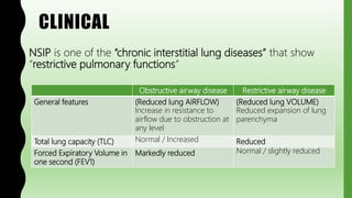
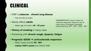
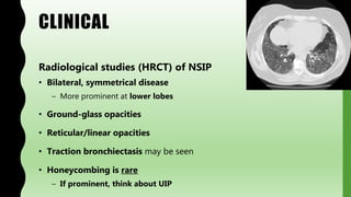
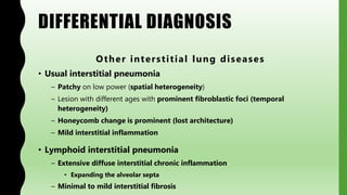
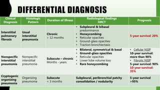
FLASHPATH
H A Z E M A L I
NONSPECIFIC
INTERSTITIAL
PNEUMONIA
H A Z E M A L I
CLINICAL
NSIP is one of the “chronic interstitial lung diseases” that show
“restrictive pulmonary functions”
Obstructive airway disease Restrictive airway disease
General features (Reduced lung AIRFLOW)
Increase in resistance to
airflow due to obstruction at
any level
(Reduced lung VOLUME)
Reduced expansion of lung
parenchyma
Total lung capacity (TLC) Normal / Increased Reduced
Forced Expiratory Volume in
one second (FEV1)
Markedly reduced Normal / slightly reduced
CLINICAL
• NSIP is subacute - chronic lung disease
– Few months to years
• Mainly affects adults
– Mean age of onset is 40 – 50 years
• History of smoking in many cases
• Presenting with chronic cough, dyspnea, fatigue
• Prognosis: GOOD  corticosteroids responsive
– Mean 5 years survival: 80 – 90%
– Cellular NSIP is better than fibrotic NSIP
NONSSPECIFIC means it does not
meet the diagnostic criteria for any of
other forms of idiopathic interstitial
pneumonia (e.g. UIP – DIP – DAD – HP)
CLINICAL
Radiological studies (HRCT) of NSIP
• Bilateral, symmetrical disease
– More prominent at lower lobes
• Ground-glass opacities
• Reticular/linear opacities
• Traction bronchiectasis may be seen
• Honeycombing is rare
– If prominent, think about UIP
GROSS
• Distribution:
– Bilateral, symmetrical disease
• More prominent at lower lobes
• Lung parenchyma:
– More homogenous fibrosis than UIP
• Firm, greyish white
– Traction bronchiectasis may be seen
– Honeycomb changes are very rare
MICROSCOPY
NSIP has two main subtypes:
• Cellular type
– Changes are predominantly cellular, with scant fibrosis
• Interstitial chronic inflammation
– Better prognosis
• Fibrotic type
– Changes are predominantly fibrotic, with less cellularity
• Uniform interstitial fibrosis
– Worse prognosis
• Mixed patterns can be seen in some cases
MICROSCOPY
Interstitial fibrosis in NSIP
• Distribution:
– Bilateral, symmetrical
• More prominent in lower lobes
• The whole lobule (periphery and center) is involved uniformly
• Lung architecture:
– Preserved architecture
• No/very rare honeycomb changes can be seen
MICROSCOPY
Interstitial fibrosis in NSIP
• Showing spatial uniformity
– Diffuse involvement on low power
• Diffuse = affects the whole lobule homogenously
– Mainly alveolar septa and bronchovascular bundles
– Sub-pleural involvement is less severe than UIP
• Showing temporal uniformity
– Loose to dense interstitial fibrosis
• No/very rare fibroblastic foci can be seen
• No lesions with different ages
MICROSCOPY
Interstitial inflammation in NSIP
• Diffuse or patchy distribution
• Mild – moderate infiltrate
• Chronic inflammation
• More in cellular phase > fibrotic phase
• Mainly lymphocytes > plasma cells”
MICROSCOPY
Other associated lesions / complications:
• Traction bronchiectasis may be seen
– Due to peri-bronchial fibrosis
• Smoking-related lesions
(( can be seen focally, but if extensive  reconsider your diagnosis ))
– Intra-luminal macrophages: Desquamative interstitial pneumonia,
respiratory bronchiolitis
– Emphysema / chronic bronchitis
MICROSCOPY
Features NOT SEEN in NSIP
(( should be excluded before diagnosing NSIP ))
• Bronchiolo-centric distribution
• Acute lung injury / hyaline membranes
• Prominent fibroblastic foci / honeycomb changes
• Significant eosinophilic infiltrate
• Granulomas or giant cells
• Evidence of infection (confirmed by special studies)
MICROSCOPY
Discordant histologic features may be seen i.e. UIP on one biopsy,
NSIP on another one
• In this case, the final diagnosis is based on the worst lesion seen (UIP)
• Multiple biopsies are recommended to solve these cases
SPECIAL STUDIES
Special stains:
• Verhoeff’s Van Gieson “Elastic stain”
– Asses the lung architecture (preservation of elastic tissue)
– Stain elastic fibers “black”
– Stain collagen “red”
– Stain other tissue elements “yellow”
Verhoerff’s Van Gieson is also used in:
• Confirm invasion of elastic fibers in the visceral pleura
• For lung cancer staging
• Differentiate arteries from veins
• Evaluate vascular elastic tissue in vascular diseases
SPECIAL STUDIES
Special stains:
• Movat’s Pentachrome stain
– Asses the stage of the disease
• (degree of fibrosis/fibroblastic foci)
– Stain fibroblastic proliferation “blue-green“
– Stain dense collagen “yellow“
• Also Stains to exclude infections:
– Acid fast stains
– Fungal stains
Movat’s Pentachrome also stains:
• Elastic laminae “ black “
• Smooth muscle fibers “ dull red “
• Fibrinoid necrosis “ bright red “
SPECIAL STUDIES
Laboratory studies:
• Increased serum KL-6
– High-molecular weight glycoprotein
– Secreted by type 2 pneumonocytes
– Increased in interstitial lung diseases
• Also Negative serum markers for:
– Antibodies of connective tissue diseases
– Antibodies of hypersensitive pneumonia
DIFFERENTIAL DIAGNOSIS
Other interstitial lung diseases
• Usual interstitial pneumonia
– Patchy on low power (spatial heterogeneity)
– Lesion with different ages with prominent fibroblastic foci (temporal
heterogeneity)
– Honeycomb change is prominent (lost architecture)
– Mild interstitial inflammation
• Lymphoid interstitial pneumonia
– Extensive diffuse interstitial chronic inflammation
• Expanding the alveolar septa
– Minimal to mild interstitial fibrosis
DIFFERENTIAL DIAGNOSIS
Other interstitial lung diseases
(Organizing acute lung injury)
• Diffuse alveolar damage
– Temporal uniformity
– Diffuse
– Prominent hyaline membrane in early stages
– Edematous, fibroblasts proliferation more than dense fibrosis in organizing
stage
– Honeycomb changes only in late stages
• Cryptogenic organizing pneumonia
– Temporally uniform
– Bronchiolo-centeric
– Intraluminal fibroblastic proliferation
– No collagenous fibrosis
DIFFERENTIAL DIAGNOSIS
Other interstitial lung diseases
(Smoking-related)
• Desquamative interstitial pneumonia
– Diffuse intra-alveolar macrophages
– Mild – moderate interstitial fibrosis (with temporal uniformity)
– No/rare honeycomb changes
– No fibroblastic foci
• Respiratory bronchiolitis-related interstitial lung disease
– Patchy, bronchiolo-centric intraluminal macrophages
– Mild – moderate interstitial fibrosis (with temporal uniformity)
– No/rare honeycomb changes
– No fibroblastic foci
DIFFERENTIAL DIAGNOSIS
Clinical
Diagnosis
Histologic
Pattern
Duration of Illness
Radiological findings
HRCT
Prognosis
Interstitial
pulmonary
fibrosis
Usual
interstitial
pneumonia
Chronic
> 12 months
• Subpleural & bibasal
predominance
• Honeycombing
• Reticular opacities
• Ground-glass opacities
• Traction bronchiectasis
5-year survival: 20%
Nonspecific
interstitial
pneumonia
Nonspecific
interstitial
pneumonia
Subacute – chronic
Months - years
• Bilateral, symmetrical & basal
• Ground-glass opacities
• Reticular opacities
• Lower lobe volume loss
• Rare honeycombing
• Cellular NSIP
10-year survival:
more than 90%
• Fibrotic NSIP
5-year survival: 90%
10-year survival:
35%
Cryptogenic
organizing
pneumonia
Organizing
pneumonia
Subacute
< 3 months
Subpleural, peribronchial patchy
consolidation / nodularity
5-year survival
>95%
DIFFERENTIAL DIAGNOSIS
Clinical
Diagnosis
Histologic
Pattern
Duration of
Illness
Radiological findings
HRCT
Prognosis
Acute
respiratory
distress
syndrome
Diffuse
alveolar
damage
Acute
1 – 2 weeks
• Diffuse pulmonary infiltrate
• Ground-glass opacities
40%-60%
mortality rate in
< 6 months
Desquamative
interstitial
pneumonia
Desquamative
interstitial
pneumonia
Subacute
Weeks - months
• Subpleural predominance
• Ground-glass opacities
• Reticular opacities
• Thin-walled cysts
• Rare honeycombing
5-year survival:
more than 95%
Respiratory
bronchiolitis–
associated
interstitial lung
disease
Respiratory
bronchiolitis
Subacute
Weeks - months
• Diffuse bronchial wall thickening
• Centrilobular nodules
• Patchy ground-glass opacity
No deaths
reported
DIFFERENTIAL DIAGNOSIS
Other interstitial lung diseases (Granulomatous)
• Sarcoidosis
– Well formed granulomas (Epithelioid histiocytes, multinucleated giant cells)
– Fibrosis of granulomas is late change
– Less honeycomb changes
– Other organs involvement
• Hypersensitivity pneumonitis
– Bronchiolo-centric distribution
– Poorly formed granulomas or scattered giant cells
– Less interstitial fibrosis
– Less honeycomb changes
DIFFERENTIAL DIAGNOSIS
Other interstitial lung diseases
• Langerhans cell histiocytosis
– Stellate, bronchiolo-centric nodules of Langerhans cells
• Positive for CD1a
– Fibrosis of nodules is a late change
– No/rare honeycomb changes
– No fibroblastic foci
DIFFERENTIAL DIAGNOSIS
Other interstitial lung diseases
(that may show NSIP-Like pattern)
• Connective tissue diseases - associated interstitial lung diseases
• Hypersensitivity pneumonitis
– NSIP may be the sole histologic lesion of hypersensitivity pneumonia
• Positive exposure history
• Positive serum antibodies
• Drug induced injury
• Immunodeficiency (HIV)
DIFFERENTIAL DIAGNOSIS
Connective tissue disease-associated
• Common associated patterns: NSIP and LIP
• Less common: UIP (especially with Rheumatoid arthritis)
• Think about connective tissue diseases if:
– More Interstitial chronic inflammation
• Prominent plasma cells differentiation
• Lymphoid aggregates with germinal center
– Less fibrosis / honeycomb changes
– Extensive pleuritic
– Dense perivascular collagen
• Also positive serum antibodies
DIFFERENTIAL DIAGNOSIS
Other restrictive lung diseases
 Chest Wall / Pleura
• Neuromuscular diseases (e.g. poliomyelitis)
• Severe obesity
• Pleural diseases
• Kyphoscoliosis
WWW.
DO NOT FORGET TO SEARCH FOR MORE PICS
AND VIRTUAL SLIDES
THANK YOU
H A Z E M A L I

FlashPath - Lung - Nonspecific Interstital Pneumonia

  • 1.
    FLASHPATH H A ZE M A L I
  • 2.
  • 3.
    CLINICAL NSIP is oneof the “chronic interstitial lung diseases” that show “restrictive pulmonary functions” Obstructive airway disease Restrictive airway disease General features (Reduced lung AIRFLOW) Increase in resistance to airflow due to obstruction at any level (Reduced lung VOLUME) Reduced expansion of lung parenchyma Total lung capacity (TLC) Normal / Increased Reduced Forced Expiratory Volume in one second (FEV1) Markedly reduced Normal / slightly reduced
  • 4.
    CLINICAL • NSIP issubacute - chronic lung disease – Few months to years • Mainly affects adults – Mean age of onset is 40 – 50 years • History of smoking in many cases • Presenting with chronic cough, dyspnea, fatigue • Prognosis: GOOD  corticosteroids responsive – Mean 5 years survival: 80 – 90% – Cellular NSIP is better than fibrotic NSIP NONSSPECIFIC means it does not meet the diagnostic criteria for any of other forms of idiopathic interstitial pneumonia (e.g. UIP – DIP – DAD – HP)
  • 5.
    CLINICAL Radiological studies (HRCT)of NSIP • Bilateral, symmetrical disease – More prominent at lower lobes • Ground-glass opacities • Reticular/linear opacities • Traction bronchiectasis may be seen • Honeycombing is rare – If prominent, think about UIP
  • 6.
    GROSS • Distribution: – Bilateral,symmetrical disease • More prominent at lower lobes • Lung parenchyma: – More homogenous fibrosis than UIP • Firm, greyish white – Traction bronchiectasis may be seen – Honeycomb changes are very rare
  • 7.
    MICROSCOPY NSIP has twomain subtypes: • Cellular type – Changes are predominantly cellular, with scant fibrosis • Interstitial chronic inflammation – Better prognosis • Fibrotic type – Changes are predominantly fibrotic, with less cellularity • Uniform interstitial fibrosis – Worse prognosis • Mixed patterns can be seen in some cases
  • 8.
    MICROSCOPY Interstitial fibrosis inNSIP • Distribution: – Bilateral, symmetrical • More prominent in lower lobes • The whole lobule (periphery and center) is involved uniformly • Lung architecture: – Preserved architecture • No/very rare honeycomb changes can be seen
  • 9.
    MICROSCOPY Interstitial fibrosis inNSIP • Showing spatial uniformity – Diffuse involvement on low power • Diffuse = affects the whole lobule homogenously – Mainly alveolar septa and bronchovascular bundles – Sub-pleural involvement is less severe than UIP • Showing temporal uniformity – Loose to dense interstitial fibrosis • No/very rare fibroblastic foci can be seen • No lesions with different ages
  • 10.
    MICROSCOPY Interstitial inflammation inNSIP • Diffuse or patchy distribution • Mild – moderate infiltrate • Chronic inflammation • More in cellular phase > fibrotic phase • Mainly lymphocytes > plasma cells”
  • 11.
    MICROSCOPY Other associated lesions/ complications: • Traction bronchiectasis may be seen – Due to peri-bronchial fibrosis • Smoking-related lesions (( can be seen focally, but if extensive  reconsider your diagnosis )) – Intra-luminal macrophages: Desquamative interstitial pneumonia, respiratory bronchiolitis – Emphysema / chronic bronchitis
  • 12.
    MICROSCOPY Features NOT SEENin NSIP (( should be excluded before diagnosing NSIP )) • Bronchiolo-centric distribution • Acute lung injury / hyaline membranes • Prominent fibroblastic foci / honeycomb changes • Significant eosinophilic infiltrate • Granulomas or giant cells • Evidence of infection (confirmed by special studies)
  • 13.
    MICROSCOPY Discordant histologic featuresmay be seen i.e. UIP on one biopsy, NSIP on another one • In this case, the final diagnosis is based on the worst lesion seen (UIP) • Multiple biopsies are recommended to solve these cases
  • 14.
    SPECIAL STUDIES Special stains: •Verhoeff’s Van Gieson “Elastic stain” – Asses the lung architecture (preservation of elastic tissue) – Stain elastic fibers “black” – Stain collagen “red” – Stain other tissue elements “yellow” Verhoerff’s Van Gieson is also used in: • Confirm invasion of elastic fibers in the visceral pleura • For lung cancer staging • Differentiate arteries from veins • Evaluate vascular elastic tissue in vascular diseases
  • 15.
    SPECIAL STUDIES Special stains: •Movat’s Pentachrome stain – Asses the stage of the disease • (degree of fibrosis/fibroblastic foci) – Stain fibroblastic proliferation “blue-green“ – Stain dense collagen “yellow“ • Also Stains to exclude infections: – Acid fast stains – Fungal stains Movat’s Pentachrome also stains: • Elastic laminae “ black “ • Smooth muscle fibers “ dull red “ • Fibrinoid necrosis “ bright red “
  • 16.
    SPECIAL STUDIES Laboratory studies: •Increased serum KL-6 – High-molecular weight glycoprotein – Secreted by type 2 pneumonocytes – Increased in interstitial lung diseases • Also Negative serum markers for: – Antibodies of connective tissue diseases – Antibodies of hypersensitive pneumonia
  • 17.
    DIFFERENTIAL DIAGNOSIS Other interstitiallung diseases • Usual interstitial pneumonia – Patchy on low power (spatial heterogeneity) – Lesion with different ages with prominent fibroblastic foci (temporal heterogeneity) – Honeycomb change is prominent (lost architecture) – Mild interstitial inflammation • Lymphoid interstitial pneumonia – Extensive diffuse interstitial chronic inflammation • Expanding the alveolar septa – Minimal to mild interstitial fibrosis
  • 18.
    DIFFERENTIAL DIAGNOSIS Other interstitiallung diseases (Organizing acute lung injury) • Diffuse alveolar damage – Temporal uniformity – Diffuse – Prominent hyaline membrane in early stages – Edematous, fibroblasts proliferation more than dense fibrosis in organizing stage – Honeycomb changes only in late stages • Cryptogenic organizing pneumonia – Temporally uniform – Bronchiolo-centeric – Intraluminal fibroblastic proliferation – No collagenous fibrosis
  • 19.
    DIFFERENTIAL DIAGNOSIS Other interstitiallung diseases (Smoking-related) • Desquamative interstitial pneumonia – Diffuse intra-alveolar macrophages – Mild – moderate interstitial fibrosis (with temporal uniformity) – No/rare honeycomb changes – No fibroblastic foci • Respiratory bronchiolitis-related interstitial lung disease – Patchy, bronchiolo-centric intraluminal macrophages – Mild – moderate interstitial fibrosis (with temporal uniformity) – No/rare honeycomb changes – No fibroblastic foci
  • 20.
    DIFFERENTIAL DIAGNOSIS Clinical Diagnosis Histologic Pattern Duration ofIllness Radiological findings HRCT Prognosis Interstitial pulmonary fibrosis Usual interstitial pneumonia Chronic > 12 months • Subpleural & bibasal predominance • Honeycombing • Reticular opacities • Ground-glass opacities • Traction bronchiectasis 5-year survival: 20% Nonspecific interstitial pneumonia Nonspecific interstitial pneumonia Subacute – chronic Months - years • Bilateral, symmetrical & basal • Ground-glass opacities • Reticular opacities • Lower lobe volume loss • Rare honeycombing • Cellular NSIP 10-year survival: more than 90% • Fibrotic NSIP 5-year survival: 90% 10-year survival: 35% Cryptogenic organizing pneumonia Organizing pneumonia Subacute < 3 months Subpleural, peribronchial patchy consolidation / nodularity 5-year survival >95%
  • 21.
    DIFFERENTIAL DIAGNOSIS Clinical Diagnosis Histologic Pattern Duration of Illness Radiologicalfindings HRCT Prognosis Acute respiratory distress syndrome Diffuse alveolar damage Acute 1 – 2 weeks • Diffuse pulmonary infiltrate • Ground-glass opacities 40%-60% mortality rate in < 6 months Desquamative interstitial pneumonia Desquamative interstitial pneumonia Subacute Weeks - months • Subpleural predominance • Ground-glass opacities • Reticular opacities • Thin-walled cysts • Rare honeycombing 5-year survival: more than 95% Respiratory bronchiolitis– associated interstitial lung disease Respiratory bronchiolitis Subacute Weeks - months • Diffuse bronchial wall thickening • Centrilobular nodules • Patchy ground-glass opacity No deaths reported
  • 22.
    DIFFERENTIAL DIAGNOSIS Other interstitiallung diseases (Granulomatous) • Sarcoidosis – Well formed granulomas (Epithelioid histiocytes, multinucleated giant cells) – Fibrosis of granulomas is late change – Less honeycomb changes – Other organs involvement • Hypersensitivity pneumonitis – Bronchiolo-centric distribution – Poorly formed granulomas or scattered giant cells – Less interstitial fibrosis – Less honeycomb changes
  • 23.
    DIFFERENTIAL DIAGNOSIS Other interstitiallung diseases • Langerhans cell histiocytosis – Stellate, bronchiolo-centric nodules of Langerhans cells • Positive for CD1a – Fibrosis of nodules is a late change – No/rare honeycomb changes – No fibroblastic foci
  • 24.
    DIFFERENTIAL DIAGNOSIS Other interstitiallung diseases (that may show NSIP-Like pattern) • Connective tissue diseases - associated interstitial lung diseases • Hypersensitivity pneumonitis – NSIP may be the sole histologic lesion of hypersensitivity pneumonia • Positive exposure history • Positive serum antibodies • Drug induced injury • Immunodeficiency (HIV)
  • 25.
    DIFFERENTIAL DIAGNOSIS Connective tissuedisease-associated • Common associated patterns: NSIP and LIP • Less common: UIP (especially with Rheumatoid arthritis) • Think about connective tissue diseases if: – More Interstitial chronic inflammation • Prominent plasma cells differentiation • Lymphoid aggregates with germinal center – Less fibrosis / honeycomb changes – Extensive pleuritic – Dense perivascular collagen • Also positive serum antibodies
  • 26.
    DIFFERENTIAL DIAGNOSIS Other restrictivelung diseases  Chest Wall / Pleura • Neuromuscular diseases (e.g. poliomyelitis) • Severe obesity • Pleural diseases • Kyphoscoliosis
  • 27.
    WWW. DO NOT FORGETTO SEARCH FOR MORE PICS AND VIRTUAL SLIDES
  • 28.
    THANK YOU H AZ E M A L I