Downloaded 1,237 times


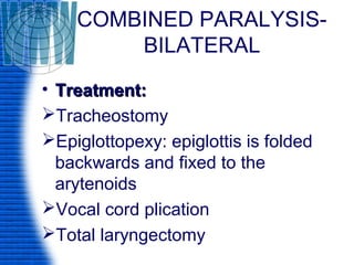


This document discusses laryngeal paralysis, including the nerve supply and causes of paralysis of the larynx. It describes recurrent laryngeal nerve paralysis and superior laryngeal nerve paralysis, including their clinical features and treatments. Bilateral paralysis and combined (complete) paralysis are also covered. Congenital vocal cord paralysis and various phonosurgery procedures are summarized.