Juvenile nasopharyngeal angiofibroma
•Download as PPTX, PDF•
12 likes•1,755 views
Google drive:-https://drive.google.com/open?id=1Qcz7DxCMkrEAgdp7VLk-Hu59zPlTK8yj Youtube:-https://www.youtube.com/watch?v=pxIe-XaGQDA
Report
Share
Report
Share

Recommended
Juvenile papillomatosis
Juvenile papillomatosis is the most common benign laryngeal tumor in children, caused by HPV types 6 and 11. It is thought children contract it from their mothers during birth if the mother had HPV. Papillomas typically affect the supraglottic and glottic regions but can spread lower. Children who had tracheostomies are more likely to have tracheal and stoma involvement due to seeding. Symptoms include hoarseness, difficulty breathing, or stridor in children ages 3-5. Diagnosis is via laryngoscopy and biopsy. Treatment involves laser excision to remove papillomas while preserving the voice, but recurrence is common and requires repeated procedures.
JUVENILE NASOPHARYNGEAL ANGIOFIBROMA
This document discusses juvenile nasopharyngeal angiofibroma (JNA), a rare benign but invasive tumor that arises in adolescent males near the sphenopalatine foramen. JNA presents with nasal obstruction and epistaxis. Diagnosis involves imaging like CT and MRI to determine the extent of involvement. Treatment depends on staging and may include preoperative embolization, surgery such as endoscopic resection, or radiation for advanced cases. Complete resection aims to prevent recurrence while minimizing complications like bleeding, infection, and nerve damage.
JNA
The document discusses juvenile nasopharyngeal angiofibroma (JNA), a benign but locally invasive vascular tumor that primarily affects adolescent males. It arises near the sphenopalatine foramen and can spread medially into the nasopharynx and laterally into surrounding structures. Presentation varies from nasal obstruction to cranial nerve palsies. Imaging shows a hypervascular mass often with bone erosion. Diagnosis is confirmed histologically. Staging guides surgical approach, with endoscopic resection increasingly used for early-stage tumors. Complete resection while preserving function is the goal.
Tympanosclerosis
Tympanosclerosis is characterized by hyaline deposits in the tympanic membrane and middle ear space caused by chronic infection or inflammation. It results in the degeneration of connective tissue and deposition of calcium and phosphate. Common symptoms include conductive hearing loss and occasional tinnitus. Diagnosis is made by otoscopy showing white plaques and audiometry showing a conductive hearing loss. Treatment depends on the size and location of plaques, with small plaques sometimes removed before grafting but large plaques usually just addressed with hearing aids.
Juvenile nasopharyngeal angiofibroma
Juvenile nasopharyngeal angiofibroma is a rare, benign tumor that occurs almost exclusively in adolescent males. It originates from the area around the sphenopalatine foramen and grows locally in an infiltrative manner, often extending into nearby structures like the nasal cavity, paranasal sinuses, and infratemporal fossa. While typically non-cancerous, it can be locally aggressive and in rare cases extend intracranially. Complete surgical removal is the primary treatment, though pre-operative embolization may help control blood loss.
Reinke's oedema
Benign polypoid degeneration of one or both the vocal folds leading to dysphonia.Smoking is the commonest predisposing factor.
Lateral sinus thrombophlebitis
Lateral sinus thrombophlebitis is an inflammation of the inner wall of the lateral venous sinus caused by infection from acute or chronic ear diseases. Bacteria enter the sinus and cause a thrombus formation within the sinus, obstructing drainage. Common symptoms include fever, headache, and papilledema. Diagnosis involves blood tests and imaging like CT or MRI. Treatment requires antibiotics, surgery to drain abscesses and remove clots, and sometimes anticoagulants or jugular vein ligation. Complications can include sepsis, meningitis, abscesses if not treated promptly.
Endoscopic (DCR) Dacryocystorhinostomy
This presentation describes the causes and presentation of Epiphora and describe the technique of Endoscopic DCR surgery.
Glomus Tumour
Glomus tumours are benign, slow-growing, hypervascular tumours that originate from glomus bodies in the middle ear. They are most commonly seen in the 5th decade of life and affect females more often than males. On histology, they appear as clusters of cells arranged in a nested pattern surrounded by a vascular stroma. Surgical excision is the primary treatment, though pre-operative embolization of feeding vessels can help reduce blood loss. Glomus tumours can spread locally within the temporal bone and occasionally metastasize to distant sites like the lungs. Advanced cases may require a combined surgical and radiotherapy approach.
Perichondritis of the external ear
Perichondritis refers to inflammation of the perichondrium of the external ear. It is usually caused by trauma such as piercing or burns and the most common organisms involved are Pseudomonas aeruginosa and Staphylococcus aureus. The diagnosis is clinical based on signs of inflammation and pain in the cartilaginous ear. If left untreated, it can lead to abscess, avascular necrosis of cartilage, and deformity of the pinna. Treatment involves early use of broad-spectrum antibiotics, drainage of any abscesses, and conservative surgery if resistant including irrigation and excision of necrotic cartilage to preserve the structure of the ear.
Atrophic rhinitis
This document discusses three chronic nasal diseases: atrophic rhinitis, rhinosporidiosis, and rhinoscleroma. Atrophic rhinitis causes nasal atrophy and foul odors. It can be primary from infection or nutritional factors, or secondary from other conditions like sinusitis. Rhinosporidiosis is caused by Rhinosporidium seeberi and presents as red nasal lesions. Rhinoscleroma is caused by Klebsiella rhinoscleromatis and presents in stages from rhinitis to fibrosis, commonly affecting the nose and spreading to other areas. Treatment involves antibiotics, surgery, or other measures depending on the specific condition.
Septal perforation
ENT Nasal septal perforation..... for best rhinoplasty and nose reshape surgery contact
Dr Junaid Ahmad (MBBS FCPS) is the best plastic surgeon in Lahore. He is a well known, trained and expert in his field. He is MBBS and FCPS in Plastic and Recosntructive Surgery. He is a post graduate of the College of Physicians and Surgeons Pakistan which is oldest and best institute for post graduation in this area of the world. He is doing his practice in Lahore, Pakistan. He is always kind to the patients and listens them carefully as it is part of modern clinical skill and training. He is expert in both cosmetic as well as reconstructive surgery. He is also skin cancer and burn expert. A few of Dr Junaid Ahmad expertise are listed here..... call 03104037071
Myringoplasty ppt
Myringoplasty is a procedure to repair a perforated eardrum using a graft without examining the middle ear. It aims to replace the missing fibrous layer and allow regeneration of skin and mucosa over the graft. The document discusses the indications, contraindications, surgical approaches, techniques, post-op care, complications and advantages/disadvantages of the underlay and overlay techniques. The key steps of underlay involve freshening the perforation edges, elevating the tympanomeatal flap, placing the graft under the flap and reposing the flap. Overlay places the graft over the remaining eardrum and covers it with the elevated skin.
Laryngeal tuberculosis
About 15-25 percent casesof pulmonary tuberculosis can have laryngeal tuberculosis however primary laryngeal TB is not rare.
Keratosis obturans
Keratosis obturans is a condition characterized by an accumulation of desquamated keratin in the external auditory meatus. It occurs when the normal migration of epithelium from the tympanic membrane to the posterior meatal wall is obstructed by wax or a foreign body, causing an abnormal separation of keratin. There are two main types - a silent type caused by this abnormal keratin separation, and an inflammatory type caused by acute ear canal inflammation. Clinically, it presents as a white keratin plug occluding the ear canal, sometimes with accompanying granulations or canal widening. Treatment involves removing the cause if present, using keratolytic agents, and potentially surgical removal under general anesthesia for recurrent cases
Functional endoscopic sinus surgery
The document discusses Functional Endoscopic Sinus Surgery (FESS). FESS is a minimally invasive procedure that uses an endoscope to access and treat the paranasal sinuses. It aims to restore sinus function by re-establishing ventilation and mucociliary clearance. Key steps in FESS include uncinectomy to remove the uncinate process, maxillary antrostomy to access the maxillary sinus, and ethmoidectomy to access the ethmoid sinuses. Proper identification of anatomical landmarks like the middle turbinate, uncinate process, and bulla ethmoidalis is important for successful FESS.
Nasal polyposis
Nasal polyps are non-cancerous growths that can develop in the nose or sinuses. They are most common in people over 50 years of age and affect males more than females. Histologically, nasal polyps contain edema fluid and high numbers of eosinophil inflammatory cells. The most common sites for nasal polyps are the ethmoid and maxillary sinuses. Nasal polyps can cause symptoms like nasal obstruction, loss of smell, sneezing and headaches. Treatment involves steroid nasal sprays, oral steroids or surgery to remove the polyps.
Maxillary sinus carcinoma
The maxillary sinus is the largest and most commonly involved sinus in malignancies. Maxillary sinus carcinoma arises from the sinus lining and spreads locally through bone destruction and invasion of surrounding structures. Distant metastases occasionally occur in the lungs. Diagnosis involves radiography, CT scan, and biopsy. Treatment depends on tumor stage and may involve surgery, radiation therapy, or chemoradiation. Prognosis diminishes with increased stage, with a 5-year survival rate of 40-50% even with advances in multimodal therapy.
Symposium Vocal Nodules And Polyp
The document discusses vocal nodules and polyps, which are benign growths on the vocal folds caused by vocal abuse or misuse. Vocal nodules are small lesions less than 3mm located at the front of the vocal folds, while polyps are larger lesions. Symptoms include hoarseness, vocal fatigue, and difficulty speaking. Treatment involves voice therapy, medical management, and surgery to remove the growths if they are large or not improving. Surgical complications can include scarring and loss of voice if the layers of the vocal folds are damaged during removal of the nodules or polyps.
Juvenile nasopharyngeal angiofibroma
This document provides information on juvenile nasopharyngeal angiofibroma (JNA), a rare benign tumor that occurs mostly in adolescent males. JNAs originate from sex steroid-stimulated hamartomatous tissue in the nasal cavity. They are highly vascular tumors that can locally invade structures in the nasal cavity, paranasal sinuses, and skull base. Imaging like CT and MRI are used to determine the extent of disease. The Fisch staging system, which classifies JNAs into 4 types based on their extent, is commonly used to guide treatment planning.
Rhinolith
A rhinolith forms around a small foreign body introduced into the nose, causing inflammation and pus secretion high in calcium and magnesium. This allows mineral salts to precipitate over time, forming a stony mass. Rhinoliths typically cause unilateral nasal obstruction, foul discharge, pain, and ulceration. They appear on examination as an irregular, stony hard mass and can be seen on imaging tests. Rhinoliths are usually removed surgically under general anesthesia.
Atrophic rhinitis.pptx
Atrophic rhinitis, also known as ozaena, is a chronic inflammation of the nasal mucosa that results in atrophy, squamous metaplasia, and crust formation. It is characterized by the atrophy of the nasal mucosa and turbinates, scanty viscid secretions, loss of ciliated columnar epithelium, and crust formation. The pathophysiology involves periarteritis and endarteritis of the nasal mucosa, decreasing blood supply and resulting in atrophy of mucous glands, epithelium, and metaplasia of the ciliated columnar epithelium. Treatment involves antibiotics, estrogen therapy, surgical procedures to narrow the nasal cavity and increase lubrication, and sal
Atrophic Rhinitis
Atrophic rhinitis or rhinitis sicca, clinical features, diagnosis and treatment with recent advances.
Smr and septoplasty
This document describes the indications, techniques, and postoperative care for septoplasty surgery. It indicates that septoplasty is performed to correct a deviated nasal septum causing obstruction or other issues. The key steps described are making an incision, raising mucoperichondrial flaps, removing deviated cartilage and bone, and re-approximating the flaps. Potential complications are also outlined.
Diseases of external ear
This document discusses diseases of the external ear. It begins by describing the anatomy of the external ear canal. It then categorizes conditions affecting the external ear into congenital, inflammatory, reactive, traumatic, and tumors. Under congenital conditions it discusses preauricular sinus, congenital ear swellings, fistulas and anomalies. It provides details on preauricular sinus including embryology, clinical features, management and associated syndromes. It also discusses other congenital conditions such as ear swellings, fistulas and atresia. The document further describes inflammatory conditions including erysipelas, perichondritis and malignant otitis externa. It also covers reactive, traumatic, and neoplastic conditions of the external
Rhinomanometry
This document discusses various methods for objectively measuring nasal patency and airflow, which is important for accurately assessing complaints of nasal obstruction. It describes rhinomanometry, which measures nasal resistance, and acoustic rhinomanometry, which provides anatomical data on nasal cross-sectional area. Several other tests are also mentioned, including peak nasal inspiratory flow, body plethysmography, and questionnaires. Overall, the document provides an overview of existing objective methods for evaluating nasal function and structure to help diagnose the cause of a blocked nose.
Nasopharyngeal Carcinoma
The document discusses the anatomy, histology, epidemiology, clinical features, diagnosis, staging, and treatment of nasopharyngeal carcinoma (NPC). Key points include:
- NPC originates from the epithelial lining of the nasopharynx.
- It has a strong association with Epstein-Barr virus.
- Risk factors include genetic predisposition and environmental exposures like salted fish consumption.
- Common symptoms are cervical lymphadenopathy, epistaxis, ear symptoms, and neurological deficits.
- Diagnosis involves biopsy and serological testing for EBV markers.
- Staging systems consider tumor size, node involvement, and serological factors.
- Primary treatment is radiotherapy,
Endoscopic dcr
1) Endoscopic DCR is a minimally invasive procedure to treat nasolacrimal duct obstruction that avoids external incisions and scars.
2) Key steps include identifying bony landmarks to locate the lacrimal sac, making a bone window, inserting a silicone tube, and correcting any associated nasal pathology.
3) Advantages over external DCR include avoiding external scars, direct visualization allowing precise surgery and management of concurrent nasal issues, and lower risks of complications.
Tumours of nasopharynx (2) itp class dr.davis - 03.06.16
This document provides information on tumours of the nasopharynx, including a classification system dividing them into epithelial tumours, soft tissue tumours, bone/cartilage tumours, malignant lymphomas, and miscellaneous tumours. It also discusses juvenile nasopharyngeal angiofibroma, the most common benign nasopharyngeal tumour seen in young males. Nasopharyngeal carcinoma is also summarized, noting its association with Epstein-Barr virus and environmental/genetic risk factors in certain populations. The WHO pathological classification system for NPC divides it into three main types.
Nasopharngeal angiofibroma
1. Nasopharyngeal angiofibroma is a rare, benign tumor that occurs mostly in adolescent males and arises from the posterior nasal cavity.
2. It is locally invasive and can extend into surrounding areas like the nasal cavity, paranasal sinuses, and cranial cavity, causing symptoms like nasal obstruction, epistaxis, and cranial nerve palsies.
3. Diagnosis involves imaging like CT and MRI to determine the extent of the tumor. Surgical excision is the primary treatment but carries a risk of heavy bleeding, so preoperative embolization of feeding vessels is often used to reduce bleeding during surgery.
More Related Content
What's hot
Glomus Tumour
Glomus tumours are benign, slow-growing, hypervascular tumours that originate from glomus bodies in the middle ear. They are most commonly seen in the 5th decade of life and affect females more often than males. On histology, they appear as clusters of cells arranged in a nested pattern surrounded by a vascular stroma. Surgical excision is the primary treatment, though pre-operative embolization of feeding vessels can help reduce blood loss. Glomus tumours can spread locally within the temporal bone and occasionally metastasize to distant sites like the lungs. Advanced cases may require a combined surgical and radiotherapy approach.
Perichondritis of the external ear
Perichondritis refers to inflammation of the perichondrium of the external ear. It is usually caused by trauma such as piercing or burns and the most common organisms involved are Pseudomonas aeruginosa and Staphylococcus aureus. The diagnosis is clinical based on signs of inflammation and pain in the cartilaginous ear. If left untreated, it can lead to abscess, avascular necrosis of cartilage, and deformity of the pinna. Treatment involves early use of broad-spectrum antibiotics, drainage of any abscesses, and conservative surgery if resistant including irrigation and excision of necrotic cartilage to preserve the structure of the ear.
Atrophic rhinitis
This document discusses three chronic nasal diseases: atrophic rhinitis, rhinosporidiosis, and rhinoscleroma. Atrophic rhinitis causes nasal atrophy and foul odors. It can be primary from infection or nutritional factors, or secondary from other conditions like sinusitis. Rhinosporidiosis is caused by Rhinosporidium seeberi and presents as red nasal lesions. Rhinoscleroma is caused by Klebsiella rhinoscleromatis and presents in stages from rhinitis to fibrosis, commonly affecting the nose and spreading to other areas. Treatment involves antibiotics, surgery, or other measures depending on the specific condition.
Septal perforation
ENT Nasal septal perforation..... for best rhinoplasty and nose reshape surgery contact
Dr Junaid Ahmad (MBBS FCPS) is the best plastic surgeon in Lahore. He is a well known, trained and expert in his field. He is MBBS and FCPS in Plastic and Recosntructive Surgery. He is a post graduate of the College of Physicians and Surgeons Pakistan which is oldest and best institute for post graduation in this area of the world. He is doing his practice in Lahore, Pakistan. He is always kind to the patients and listens them carefully as it is part of modern clinical skill and training. He is expert in both cosmetic as well as reconstructive surgery. He is also skin cancer and burn expert. A few of Dr Junaid Ahmad expertise are listed here..... call 03104037071
Myringoplasty ppt
Myringoplasty is a procedure to repair a perforated eardrum using a graft without examining the middle ear. It aims to replace the missing fibrous layer and allow regeneration of skin and mucosa over the graft. The document discusses the indications, contraindications, surgical approaches, techniques, post-op care, complications and advantages/disadvantages of the underlay and overlay techniques. The key steps of underlay involve freshening the perforation edges, elevating the tympanomeatal flap, placing the graft under the flap and reposing the flap. Overlay places the graft over the remaining eardrum and covers it with the elevated skin.
Laryngeal tuberculosis
About 15-25 percent casesof pulmonary tuberculosis can have laryngeal tuberculosis however primary laryngeal TB is not rare.
Keratosis obturans
Keratosis obturans is a condition characterized by an accumulation of desquamated keratin in the external auditory meatus. It occurs when the normal migration of epithelium from the tympanic membrane to the posterior meatal wall is obstructed by wax or a foreign body, causing an abnormal separation of keratin. There are two main types - a silent type caused by this abnormal keratin separation, and an inflammatory type caused by acute ear canal inflammation. Clinically, it presents as a white keratin plug occluding the ear canal, sometimes with accompanying granulations or canal widening. Treatment involves removing the cause if present, using keratolytic agents, and potentially surgical removal under general anesthesia for recurrent cases
Functional endoscopic sinus surgery
The document discusses Functional Endoscopic Sinus Surgery (FESS). FESS is a minimally invasive procedure that uses an endoscope to access and treat the paranasal sinuses. It aims to restore sinus function by re-establishing ventilation and mucociliary clearance. Key steps in FESS include uncinectomy to remove the uncinate process, maxillary antrostomy to access the maxillary sinus, and ethmoidectomy to access the ethmoid sinuses. Proper identification of anatomical landmarks like the middle turbinate, uncinate process, and bulla ethmoidalis is important for successful FESS.
Nasal polyposis
Nasal polyps are non-cancerous growths that can develop in the nose or sinuses. They are most common in people over 50 years of age and affect males more than females. Histologically, nasal polyps contain edema fluid and high numbers of eosinophil inflammatory cells. The most common sites for nasal polyps are the ethmoid and maxillary sinuses. Nasal polyps can cause symptoms like nasal obstruction, loss of smell, sneezing and headaches. Treatment involves steroid nasal sprays, oral steroids or surgery to remove the polyps.
Maxillary sinus carcinoma
The maxillary sinus is the largest and most commonly involved sinus in malignancies. Maxillary sinus carcinoma arises from the sinus lining and spreads locally through bone destruction and invasion of surrounding structures. Distant metastases occasionally occur in the lungs. Diagnosis involves radiography, CT scan, and biopsy. Treatment depends on tumor stage and may involve surgery, radiation therapy, or chemoradiation. Prognosis diminishes with increased stage, with a 5-year survival rate of 40-50% even with advances in multimodal therapy.
Symposium Vocal Nodules And Polyp
The document discusses vocal nodules and polyps, which are benign growths on the vocal folds caused by vocal abuse or misuse. Vocal nodules are small lesions less than 3mm located at the front of the vocal folds, while polyps are larger lesions. Symptoms include hoarseness, vocal fatigue, and difficulty speaking. Treatment involves voice therapy, medical management, and surgery to remove the growths if they are large or not improving. Surgical complications can include scarring and loss of voice if the layers of the vocal folds are damaged during removal of the nodules or polyps.
Juvenile nasopharyngeal angiofibroma
This document provides information on juvenile nasopharyngeal angiofibroma (JNA), a rare benign tumor that occurs mostly in adolescent males. JNAs originate from sex steroid-stimulated hamartomatous tissue in the nasal cavity. They are highly vascular tumors that can locally invade structures in the nasal cavity, paranasal sinuses, and skull base. Imaging like CT and MRI are used to determine the extent of disease. The Fisch staging system, which classifies JNAs into 4 types based on their extent, is commonly used to guide treatment planning.
Rhinolith
A rhinolith forms around a small foreign body introduced into the nose, causing inflammation and pus secretion high in calcium and magnesium. This allows mineral salts to precipitate over time, forming a stony mass. Rhinoliths typically cause unilateral nasal obstruction, foul discharge, pain, and ulceration. They appear on examination as an irregular, stony hard mass and can be seen on imaging tests. Rhinoliths are usually removed surgically under general anesthesia.
Atrophic rhinitis.pptx
Atrophic rhinitis, also known as ozaena, is a chronic inflammation of the nasal mucosa that results in atrophy, squamous metaplasia, and crust formation. It is characterized by the atrophy of the nasal mucosa and turbinates, scanty viscid secretions, loss of ciliated columnar epithelium, and crust formation. The pathophysiology involves periarteritis and endarteritis of the nasal mucosa, decreasing blood supply and resulting in atrophy of mucous glands, epithelium, and metaplasia of the ciliated columnar epithelium. Treatment involves antibiotics, estrogen therapy, surgical procedures to narrow the nasal cavity and increase lubrication, and sal
Atrophic Rhinitis
Atrophic rhinitis or rhinitis sicca, clinical features, diagnosis and treatment with recent advances.
Smr and septoplasty
This document describes the indications, techniques, and postoperative care for septoplasty surgery. It indicates that septoplasty is performed to correct a deviated nasal septum causing obstruction or other issues. The key steps described are making an incision, raising mucoperichondrial flaps, removing deviated cartilage and bone, and re-approximating the flaps. Potential complications are also outlined.
Diseases of external ear
This document discusses diseases of the external ear. It begins by describing the anatomy of the external ear canal. It then categorizes conditions affecting the external ear into congenital, inflammatory, reactive, traumatic, and tumors. Under congenital conditions it discusses preauricular sinus, congenital ear swellings, fistulas and anomalies. It provides details on preauricular sinus including embryology, clinical features, management and associated syndromes. It also discusses other congenital conditions such as ear swellings, fistulas and atresia. The document further describes inflammatory conditions including erysipelas, perichondritis and malignant otitis externa. It also covers reactive, traumatic, and neoplastic conditions of the external
Rhinomanometry
This document discusses various methods for objectively measuring nasal patency and airflow, which is important for accurately assessing complaints of nasal obstruction. It describes rhinomanometry, which measures nasal resistance, and acoustic rhinomanometry, which provides anatomical data on nasal cross-sectional area. Several other tests are also mentioned, including peak nasal inspiratory flow, body plethysmography, and questionnaires. Overall, the document provides an overview of existing objective methods for evaluating nasal function and structure to help diagnose the cause of a blocked nose.
Nasopharyngeal Carcinoma
The document discusses the anatomy, histology, epidemiology, clinical features, diagnosis, staging, and treatment of nasopharyngeal carcinoma (NPC). Key points include:
- NPC originates from the epithelial lining of the nasopharynx.
- It has a strong association with Epstein-Barr virus.
- Risk factors include genetic predisposition and environmental exposures like salted fish consumption.
- Common symptoms are cervical lymphadenopathy, epistaxis, ear symptoms, and neurological deficits.
- Diagnosis involves biopsy and serological testing for EBV markers.
- Staging systems consider tumor size, node involvement, and serological factors.
- Primary treatment is radiotherapy,
Endoscopic dcr
1) Endoscopic DCR is a minimally invasive procedure to treat nasolacrimal duct obstruction that avoids external incisions and scars.
2) Key steps include identifying bony landmarks to locate the lacrimal sac, making a bone window, inserting a silicone tube, and correcting any associated nasal pathology.
3) Advantages over external DCR include avoiding external scars, direct visualization allowing precise surgery and management of concurrent nasal issues, and lower risks of complications.
What's hot (20)
Similar to Juvenile nasopharyngeal angiofibroma
Tumours of nasopharynx (2) itp class dr.davis - 03.06.16
This document provides information on tumours of the nasopharynx, including a classification system dividing them into epithelial tumours, soft tissue tumours, bone/cartilage tumours, malignant lymphomas, and miscellaneous tumours. It also discusses juvenile nasopharyngeal angiofibroma, the most common benign nasopharyngeal tumour seen in young males. Nasopharyngeal carcinoma is also summarized, noting its association with Epstein-Barr virus and environmental/genetic risk factors in certain populations. The WHO pathological classification system for NPC divides it into three main types.
Nasopharngeal angiofibroma
1. Nasopharyngeal angiofibroma is a rare, benign tumor that occurs mostly in adolescent males and arises from the posterior nasal cavity.
2. It is locally invasive and can extend into surrounding areas like the nasal cavity, paranasal sinuses, and cranial cavity, causing symptoms like nasal obstruction, epistaxis, and cranial nerve palsies.
3. Diagnosis involves imaging like CT and MRI to determine the extent of the tumor. Surgical excision is the primary treatment but carries a risk of heavy bleeding, so preoperative embolization of feeding vessels is often used to reduce bleeding during surgery.
angiofibroma.pptx
Juvenile nasopharyngeal angiofibroma is a benign tumor that occurs mainly in males during puberty, most commonly in Southeast Asia, the Middle East, and India. It arises from the base of the medial pterygoid plate and can extend into the nasal cavity, nasopharynx, maxillary sinus, pterygomaxillary fossa, infratemporal fossa, orbit, and occasionally the intracranial cavity. Symptoms include profuse epistaxis, nasal obstruction, and deformities of the face with orbital or cheek involvement. Treatment is generally surgical excision via an endoscopic or open approach, with preoperative embolization or hormone therapy sometimes used to reduce tumor vascular
Juvenile nasopharyngeal angiofibroma
This document provides information about juvenile angiofibroma (JA), a benign tumor that predominantly affects adolescent males. It discusses the anatomy and location of JA tumors, which typically arise near the sphenopalatine foramen. The document covers various theories about the etiology and pathogenesis of JA and describes the clinical presentation, investigations, and treatment approaches for this tumor. The main treatment is surgical resection, which can be challenging due to the tumor's high vascularity and potential for significant bleeding. Preoperative embolization is often used to reduce blood flow and make surgery safer.
Nasopharyngeal angiofribroma
Nasopharyngeal angiofibroma is a benign, vascular tumor that occurs almost exclusively in adolescent males. It arises from the nasopharynx near the sphenopalatine foramen. Clinical features include recurrent epistaxis, nasal obstruction, and conductive hearing loss. While benign, it can be locally invasive and extend into nearby structures like the nasal cavity, paranasal sinuses, orbits, and middle cranial fossa. Treatment involves surgical resection through various approaches like transpalatine or endoscopic. Radiotherapy, hormonal therapy, and chemotherapy may also be used for advanced or recurrent tumors.
Pharynx Lecture_3.doc
The fossa of Rosenmueller is a lateral extension of the nasopharynx located above and behind the Eustachian tube. Nasopharyngeal carcinoma commonly spreads from the fossa of Rosenmueller, which can result in paralysis of cranial nerves and lymph node metastasis. Risk factors for nasopharyngeal carcinoma include genetic susceptibility in Cantonese populations, Epstein-Barr virus infection, and a diet high in salted fish. The primary treatment is radiation therapy, which may be combined with chemotherapy and has improved survival rates compared to radiation alone.
Jna(juvenile nasopharyngeal angiofibroma) current treatment modalities
The document provides information on Juvenile Nasopharyngeal Angiofibroma (JNA), a rare benign tumor that most commonly affects adolescent males. It discusses the tumor's etiology, clinical presentation, diagnostic evaluation including imaging, and various treatment modalities. The current primary treatments are surgical resection via various open or endoscopic approaches, with the goal of complete removal while minimizing complications and risk of recurrence. Future areas of research include investigating the role of hormones and growth factors in tumor development to identify potential non-surgical therapies.
Sino-nasal malignancy
This document provides an overview of sinonasal malignancy including:
- The complex surgical anatomy of the sinonasal region and proximity to vital structures.
- The histopathological classification and TNM staging of sinonasal cancers.
- Presentation, diagnosis, and multidisciplinary management approaches including endoscopic surgery, radiotherapy, and imaging.
- Specific details are provided on surgical procedures like craniofacial resection and midfacial degloving for advanced tumors.
Juvenile Nasopharyngeal Angiofibroma.pptx
Dr. Gladson M. Robin discusses juvenile nasopharyngeal angiofibroma, a rare benign tumor that most commonly affects the posterior nasopharynx near the sphenopalatine foramen. The tumor is composed of blood vessels and fibrous tissue in varying ratios, leading to severe bleeding risks from biopsies. While usually benign, the tumor can invade local structures like the cranial cavity or spread indirectly through nearby sinuses. Treatment involves surgical resection, with radiation used for intracranial extensions or inaccessible recurrences due to its ability to reduce blood supply over time. Recurrence rates for juvenile angiofibroma are reported in up to 35% of cases.
Juvenile angiofibroma (sbo 2)
Juvenile angiofibroma is a rare, benign and highly vascular tumor that develops almost exclusively in adolescent males within the sphenopalatine foramen. It presents with recurrent severe nosebleeds and progressive nasal obstruction. Imaging such as CT and MRI are used to determine the extent of the tumor. While surgery is the primary treatment, preoperative embolization and endoscopic resection are often used for early-stage tumors to reduce bleeding and complications. Advanced tumors may require open approaches like mid-facial degloving. Recurrence rates remain high due to the invasive nature of the tumor.
Pathology of pharynx
1) Rathke's pouch forms the pituitary gland, which must migrate to the hypothalamus during development. In some individuals, pituitary tissue remains in the roof of the pharynx.
2) Down syndrome results from trisomy 21 and is characterized by midface hypoplasia, reducing nasopharynx and oropharynx volume.
3) Crouzon syndrome is an autosomal dominant craniosynostosis with midface hypoplasia and relative mandibular prognathism, potentially causing airway obstruction.
juvenile nasopharyngeal angiofibroma.pptx
Juvenile nasopharyngeal angiofibroma is a rare benign tumor that affects adolescent males. It is locally aggressive and grows in the nasal cavity and nasopharynx. The tumor is highly vascular and can extend to surrounding areas like the pterygopalatine fossa, infratemporal fossa, and intracranially. Treatment involves preoperative embolization followed by surgical resection through an endoscopic or open approach depending on tumor extent. Close postoperative monitoring is needed due to the risk of recurrence.
Angiofibroma
1. Juvenile nasopharyngeal angiofibroma is a rare, benign tumour that occurs primarily in adolescent males and is highly vascular.
2. It originates from the posterior nasal cavity near the sphenopalatine foramen and can extend into local structures like the sinuses, orbit and cranium.
3. Treatment involves preoperative embolization followed by surgical excision via various approaches depending on tumour extent. Endoscopic removal is used for smaller tumours while more extensive approaches are needed for larger or invasive tumours.
ORAL MALIGNANCIES M.pptx
1. Carcinoma of the oral cavity most commonly arises from the lips, tongue, floor of mouth, cheek and retromolar trigone. Major risk factors include smoking, alcohol, betel nut chewing and HPV infection.
2. Early lesions are typically treated with wide local excision or radiotherapy. More advanced tumors involving surrounding bone or lymph nodes may require surgery such as partial glossectomy or mandibulectomy combined with neck dissection followed by postoperative radiotherapy.
3. Prognosis depends on tumor size, location and extent of spread - tumors of the posterior tongue or those invading surrounding structures carry a worse prognosis.
Hypopharyngeal cancer
This document discusses hypopharyngeal cancer. Some key points:
- Hypopharyngeal cancers arise from the mucosa of the hypopharynx and are often advanced at diagnosis due to few symptoms. They have an unfavorable prognosis.
- Risk factors include smoking, alcohol use, poor nutrition. Over 90% of patients have a history of tobacco use. Genetic factors may also play a role.
- The hypopharynx is located posterior to the larynx and above the esophagus. It contains the pyriform sinuses, postcricoid area, and posterior pharyngeal wall.
- Presentation includes sore throat, dysphag
BENIGN NEOPLASMS OF THE NOSE AND PNS.pptx
The document discusses various tumors of the nose and paranasal sinuses. It begins by classifying tumors as benign, intermediate, or malignant. It then describes specific tumor types like squamous papilloma, inverted papilloma, glioma, dermoid, osteoma, chondroma, schwannoma and others. For each tumor type, it discusses characteristics, clinical features, diagnosis using imaging like CT and MRI, and treatment approaches. The document provides detailed information on tumor pathology and management.
hypopharyngealcancer2-151012175726-lva1-app6892.pptx
1. Hypopharyngeal cancers arise from the mucosa of the hypopharynx and are often advanced at diagnosis due to late onset of symptoms.
2. Risk factors include smoking, alcohol use, and HPV infection. Tumors most commonly arise from the pyriform sinus.
3. Diagnosis involves endoscopy, imaging, and biopsy to assess primary tumor size and spread to lymph nodes. Most are squamous cell carcinomas.
Glomus tumours pakistan
Paragangliomas, also known as glomus tumours, are rare tumours that arise from paraganglionic tissue. They most commonly occur in the middle ear, jugular foramen, and along the vagus nerve. Surgical resection is the primary treatment, but pre-operative embolization and radiotherapy can help reduce tumour size and vascularity. Complete surgical removal is difficult due to the complex anatomy of the skull base and risk of damaging nearby cranial nerves. Adjuvant radiotherapy is often used post-operatively to prevent tumour regrowth. Functionally active tumours can cause issues with blood pressure control and circulatory collapse during and after surgery.
Epistaxis & Angiofibroma.pptx
Blood Supply of Nose
Little’s Area & Importance
Causes & Classification of Epistaxis
Management of Epistaxis
Angiofibroma and its Etiology
Pathology of Angiofibroma
Diagnosis of Angiofibroma
Treatment of Angiofibroma
Epistaxis & Angiofibroma.pptx
Blood Supply of Nose
Little’s Area & Importance
Causes & Classification of Epistaxis
Management of Epistaxis
Angiofibroma and its Etiology
Pathology of Angiofibroma
Diagnosis of Angiofibroma
Treatment of Angiofibroma
Situated in the anterior inferior part of nasal septum.
Four arteries anastomose here to form a vascular plexus “Kiesselbach’s Plexus”
Usual site for Epistaxis in children & young adults.
Another plexus of veins is situated inferior to posterior end of inferior turbinate, called “Woodruff’s Plexus”. It is a site of posterior epistaxis.
Similar to Juvenile nasopharyngeal angiofibroma (20)
Tumours of nasopharynx (2) itp class dr.davis - 03.06.16
Tumours of nasopharynx (2) itp class dr.davis - 03.06.16
Jna(juvenile nasopharyngeal angiofibroma) current treatment modalities
Jna(juvenile nasopharyngeal angiofibroma) current treatment modalities
hypopharyngealcancer2-151012175726-lva1-app6892.pptx
hypopharyngealcancer2-151012175726-lva1-app6892.pptx
More from Raafi Ul Zargar
Kernicterus
Drive:-https://drive.google.com/open?id=1bp4kJTvlyRT11rSXnq8kejoezkUwBpQf
Youtube:-https://www.youtube.com/watch?v=2qL_3QAue2M
Arterial arterial occlusion
Drive:-https://drive.google.com/open?id=1VUORIMNocR7Zrlrig8DT85Vp5zjJVwsN
Youtube:-https://www.youtube.com/watch?v=whCJkRVeOK8&t=85s
Trachoma
Google drive:-https://drive.google.com/open?id=1JrWSv4tdQ2DKqNDwApdAvf6IrREQX-v1
Youtube:-https://www.youtube.com/watch?v=kcEh2Ay2sPg
Rhinoscleroma
Google drive:-https://drive.google.com/open?id=1hd2LAIPo3iA5Z5G9RxCSxUT5LPtrohQO
Youtube:-https://www.youtube.com/watch?v=b1pIsGq1XhI
Posterior uveitis
This document discusses uveal tract diseases, specifically uveitis (inflammation of the uveal tract). It defines posterior uveitis as inflammation of the choroid. Common causes of posterior uveitis include infectious diseases like tuberculosis, fungi, viruses, and parasites, as well as non-infectious conditions like autoimmune diseases. Symptoms include vision defects, flashes of light, and positive or negative scotomas. Signs include vitreous opacities, active or healed patches of choroiditis. Choroiditis is classified based on location and number of lesions, and can lead to complications if not treated properly with corticosteroids, immunosuppressants, or specific treatments for the
Panophthalmitis
Google drive:-https://drive.google.com/open?id=1L-l18Q1F2ZdkARgZQj_v5usGo4mYGR0j
Youtube:-https://www.youtube.com/watch?v=X1ntNKjKK2A
Oesophagoscopy
Google drive:-https://drive.google.com/open?id=1J-oqqPBgk4Or9ysZveY-OvnDyqruKid8
Youtube:-https://www.youtube.com/watch?v=rp53LhXX2XI
Malaria
Google drive:-https://drive.google.com/open?id=17XrHKOFezAR6BjcA_efsfroXcwgxrSgs
Youtube:-https://www.youtube.com/watch?v=Tb4Ho7TYtsQ
Leishemania donovani
Google drive:-https://drive.google.com/open?id=1HAgGLnUWYZdNSkNpHbWMUQJYHY-iXEhj
Youtube:-https://www.youtube.com/watch?v=Az_yZ2x8-yY
Hymenolepis nana
Hymenolepis nana, also known as the dwarf tapeworm, is the smallest and most common tapeworm found in the human intestine. It inhabits the proximal ileum and is most prevalent in warm climates. H. nana has a direct lifecycle through ingestion of eggs or an indirect lifecycle involving rat fleas as intermediate hosts. Most infections are asymptomatic but some people experience abdominal pain, diarrhea, and pruritis. Diagnosis is made by finding the pathogen's distinctive eggs on microscopic examination of feces. Treatment involves niclosamide or praziquantel which act against both adult worms and larvae. Maintaining hygiene and sanitation helps prevent transmission.
Endophthalmitis
Google drive:-https://drive.google.com/open?id=1WinQ_Wdg03t9-imtebjEkJX9ibxqX0I3
Youtube:-https://www.youtube.com/watch?v=lcc8uCeFu60
Direct laryngoscopy
Google drive:-https://www.youtube.com/redirect?q=https%3A%2F%2Fdrive.google.com%2Fopen%3Fid%3D1ZET4JzZalyUfM1KWXemKZsQQXMzrYpcJ&v=WHOggpW5Ee8&event=video_description&redir_token=77oOekaJs8_u0RLfrUH8z68tJFt8MTU2MDY1Njc4N0AxNTYwNTcwMzg3
Youtube:-https://www.youtube.com/watch?v=WHOggpW5Ee8
Conjunctivitis
Google drive:-https://www.youtube.com/redirect?redir_token=wEiMpfXg4PMkK33P24q0If4rY7F8MTU2MDY1NjM4NUAxNTYwNTY5OTg1&v=RfdIJLOAqtY&q=https%3A%2F%2Fdrive.google.com%2Ffile%2Fd%2F1nSTePPFenfflkcC7DafOstjK9xq76c2N%2Fview%3Fusp%3Dsharing&event=video_description
Youtube:-https://www.youtube.com/watch?v=RfdIJLOAqtY
More from Raafi Ul Zargar (13)
Recently uploaded
Light House Retreats: Plant Medicine Retreat Europe
Our aim is to organise conscious gatherings and retreats for open and inquisitive minds and souls, with and without the assistance of sacred plants.
The Best Ayurvedic Antacid Tablets in India
Treat the symptoms of indigestion, heartburn and stomach reflux with the 10 Best Ayurvedic Antacid Tablets in India.
The Electrocardiogram - Physiologic Principles
These lecture slides, by Dr Sidra Arshad, offer a quick overview of the physiological basis of a normal electrocardiogram.
Learning objectives:
1. Define an electrocardiogram (ECG) and electrocardiography
2. Describe how dipoles generated by the heart produce the waveforms of the ECG
3. Describe the components of a normal electrocardiogram of a typical bipolar lead (limb II)
4. Differentiate between intervals and segments
5. Enlist some common indications for obtaining an ECG
6. Describe the flow of current around the heart during the cardiac cycle
7. Discuss the placement and polarity of the leads of electrocardiograph
8. Describe the normal electrocardiograms recorded from the limb leads and explain the physiological basis of the different records that are obtained
9. Define mean electrical vector (axis) of the heart and give the normal range
10. Define the mean QRS vector
11. Describe the axes of leads (hexagonal reference system)
12. Comprehend the vectorial analysis of the normal ECG
13. Determine the mean electrical axis of the ventricular QRS and appreciate the mean axis deviation
14. Explain the concepts of current of injury, J point, and their significance
Study Resources:
1. Chapter 11, Guyton and Hall Textbook of Medical Physiology, 14th edition
2. Chapter 9, Human Physiology - From Cells to Systems, Lauralee Sherwood, 9th edition
3. Chapter 29, Ganong’s Review of Medical Physiology, 26th edition
4. Electrocardiogram, StatPearls - https://www.ncbi.nlm.nih.gov/books/NBK549803/
5. ECG in Medical Practice by ABM Abdullah, 4th edition
6. Chapter 3, Cardiology Explained, https://www.ncbi.nlm.nih.gov/books/NBK2214/
7. ECG Basics, http://www.nataliescasebook.com/tag/e-c-g-basics
Hemodialysis: Chapter 4, Dialysate Circuit - Dr.Gawad
- Video recording of this lecture in English language: https://youtu.be/kqbnxVAZs-0
- Video recording of this lecture in Arabic language: https://youtu.be/SINlygW1Mpc
- Link to download the book free: https://nephrotube.blogspot.com/p/nephrotube-nephrology-books.html
- Link to NephroTube website: www.NephroTube.com
- Link to NephroTube social media accounts: https://nephrotube.blogspot.com/p/join-nephrotube-on-social-media.html
Role of Mukta Pishti in the Management of Hyperthyroidism
Muktapishti is a traditional Ayurvedic preparation made from Shoditha Mukta (Purified Pearl), is believed to help regulate thyroid function and reduce symptoms of hyperthyroidism due to its cooling and balancing properties. Clinical evidence on its efficacy remains limited, necessitating further research to validate its therapeutic benefits.
Osteoporosis - Definition , Evaluation and Management .pdf
Osteoporosis is an increasing cause of morbidity among the elderly.
In this document , a brief outline of osteoporosis is given , including the risk factors of osteoporosis fractures , the indications for testing bone mineral density and the management of osteoporosis
Novas diretrizes da OMS para os cuidados perinatais de mais qualidade
Novas diretrizes da OMS para os cuidados perinatais de mais qualidadeProf. Marcus Renato de Carvalho
Recomendações da OMS sobre cuidados maternos e neonatais para uma experiência pós-natal positiva.
Em consonância com os ODS – Objetivos do Desenvolvimento Sustentável e a Estratégia Global para a Saúde das Mulheres, Crianças e Adolescentes, e aplicando uma abordagem baseada nos direitos humanos, os esforços de cuidados pós-natais devem expandir-se para além da cobertura e da simples sobrevivência, de modo a incluir cuidados de qualidade.
Estas diretrizes visam melhorar a qualidade dos cuidados pós-natais essenciais e de rotina prestados às mulheres e aos recém-nascidos, com o objetivo final de melhorar a saúde e o bem-estar materno e neonatal.
Uma “experiência pós-natal positiva” é um resultado importante para todas as mulheres que dão à luz e para os seus recém-nascidos, estabelecendo as bases para a melhoria da saúde e do bem-estar a curto e longo prazo. Uma experiência pós-natal positiva é definida como aquela em que as mulheres, pessoas que gestam, os recém-nascidos, os casais, os pais, os cuidadores e as famílias recebem informação consistente, garantia e apoio de profissionais de saúde motivados; e onde um sistema de saúde flexível e com recursos reconheça as necessidades das mulheres e dos bebês e respeite o seu contexto cultural.
Estas diretrizes consolidadas apresentam algumas recomendações novas e já bem fundamentadas sobre cuidados pós-natais de rotina para mulheres e neonatos que recebem cuidados no pós-parto em unidades de saúde ou na comunidade, independentemente dos recursos disponíveis.
É fornecido um conjunto abrangente de recomendações para cuidados durante o período puerperal, com ênfase nos cuidados essenciais que todas as mulheres e recém-nascidos devem receber, e com a devida atenção à qualidade dos cuidados; isto é, a entrega e a experiência do cuidado recebido. Estas diretrizes atualizam e ampliam as recomendações da OMS de 2014 sobre cuidados pós-natais da mãe e do recém-nascido e complementam as atuais diretrizes da OMS sobre a gestão de complicações pós-natais.
O estabelecimento da amamentação e o manejo das principais intercorrências é contemplada.
Recomendamos muito.
Vamos discutir essas recomendações no nosso curso de pós-graduação em Aleitamento no Instituto Ciclos.
Esta publicação só está disponível em inglês até o momento.
Prof. Marcus Renato de Carvalho
www.agostodourado.com
Efficacy of Avartana Sneha in Ayurveda
Avartana Sneha is a unique method of Preparation of Sneha Kalpana in Ayurveda, mainly it is indicated for the Vataja rogas.
Local Advanced Lung Cancer: Artificial Intelligence, Synergetics, Complex Sys...
Overall life span (LS) was 1671.7±1721.6 days and cumulative 5YS reached 62.4%, 10 years – 50.4%, 20 years – 44.6%. 94 LCP lived more than 5 years without cancer (LS=2958.6±1723.6 days), 22 – more than 10 years (LS=5571±1841.8 days). 67 LCP died because of LC (LS=471.9±344 days). AT significantly improved 5YS (68% vs. 53.7%) (P=0.028 by log-rank test). Cox modeling displayed that 5YS of LCP significantly depended on: N0-N12, T3-4, blood cell circuit, cell ratio factors (ratio between cancer cells-CC and blood cells subpopulations), LC cell dynamics, recalcification time, heparin tolerance, prothrombin index, protein, AT, procedure type (P=0.000-0.031). Neural networks, genetic algorithm selection and bootstrap simulation revealed relationships between 5YS and N0-12 (rank=1), thrombocytes/CC (rank=2), segmented neutrophils/CC (3), eosinophils/CC (4), erythrocytes/CC (5), healthy cells/CC (6), lymphocytes/CC (7), stick neutrophils/CC (8), leucocytes/CC (9), monocytes/CC (10). Correct prediction of 5YS was 100% by neural networks computing (error=0.000; area under ROC curve=1.0).
Histololgy of Female Reproductive System.pptx
Dive into an in-depth exploration of the histological structure of female reproductive system with this comprehensive lecture. Presented by Dr. Ayesha Irfan, Assistant Professor of Anatomy, this presentation covers the Gross anatomy and functional histology of the female reproductive organs. Ideal for students, educators, and anyone interested in medical science, this lecture provides clear explanations, detailed diagrams, and valuable insights into female reproductive system. Enhance your knowledge and understanding of this essential aspect of human biology.
Journal Article Review on Rasamanikya
Rasamanikya is a excellent preparation in the field of Rasashastra, it is used in various Kushtha Roga, Shwasa, Vicharchika, Bhagandara, Vatarakta, and Phiranga Roga. In this article Preparation& Comparative analytical profile for both Formulationon i.e Rasamanikya prepared by Kushmanda swarasa & Churnodhaka Shodita Haratala. The study aims to provide insights into the comparative efficacy and analytical aspects of these formulations for enhanced therapeutic outcomes.
share - Lions, tigers, AI and health misinformation, oh my!.pptx
• Pitfalls and pivots needed to use AI effectively in public health
• Evidence-based strategies to address health misinformation effectively
• Building trust with communities online and offline
• Equipping health professionals to address questions, concerns and health misinformation
• Assessing risk and mitigating harm from adverse health narratives in communities, health workforce and health system
Top-Vitamin-Supplement-Brands-in-India List
Swisschem Dermacare provides the Top 10 Vitamin Supplement Brands in India. To know more about us give us call at our official number
Recently uploaded (20)
Light House Retreats: Plant Medicine Retreat Europe
Light House Retreats: Plant Medicine Retreat Europe
Thyroid Gland- Gross Anatomy by Dr. Rabia Inam Gandapore.pptx
Thyroid Gland- Gross Anatomy by Dr. Rabia Inam Gandapore.pptx
CHEMOTHERAPY_RDP_CHAPTER 6_Anti Malarial Drugs.pdf
CHEMOTHERAPY_RDP_CHAPTER 6_Anti Malarial Drugs.pdf
Hemodialysis: Chapter 4, Dialysate Circuit - Dr.Gawad
Hemodialysis: Chapter 4, Dialysate Circuit - Dr.Gawad
Role of Mukta Pishti in the Management of Hyperthyroidism
Role of Mukta Pishti in the Management of Hyperthyroidism
Osteoporosis - Definition , Evaluation and Management .pdf
Osteoporosis - Definition , Evaluation and Management .pdf
Novas diretrizes da OMS para os cuidados perinatais de mais qualidade
Novas diretrizes da OMS para os cuidados perinatais de mais qualidade
Local Advanced Lung Cancer: Artificial Intelligence, Synergetics, Complex Sys...
Local Advanced Lung Cancer: Artificial Intelligence, Synergetics, Complex Sys...
Muscles of Mastication by Dr. Rabia Inam Gandapore.pptx
Muscles of Mastication by Dr. Rabia Inam Gandapore.pptx
share - Lions, tigers, AI and health misinformation, oh my!.pptx
share - Lions, tigers, AI and health misinformation, oh my!.pptx
Juvenile nasopharyngeal angiofibroma
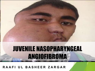
- 1. JUVENILE NASOPHARYNGEAL ANGIOFIBROMA R A A F I U L B A S H E E R Z A R G A R
- 2. INTRODUCTION It is also known as nasopharyngeal fibroma . It is a rare tumor but commonest of all benign tumor of nasopharynx.
- 3. ETIOLOGY Idiopathic Mostly seen in adolescent males in 2nd decade of life. Hormonal Theory • Testosterone dependent • Patients have a hamartomatous nidus of vascular tissue and this is activated to form angiofibroma when male sex hormone appears.
- 4. SITE Posterior part of nasal cavity close to the superior margin of sphenopalatine foramen
- 5. GROWTH Locally invasive benign tumors. From site of origin to nasal cavity, nasopharynx and into the pterygopalatine fossa, running behind the posterior wall of maxillary sinus which is pushed forward as the tumor grows. Laterally it extends into pterygomaxillary fossa then to infratemporal fossa and cheek.
- 6. PATHOLOGY Severe bleeding as the vessels lose ability to contract Made up of vascular and fibrous tissue mostly the vessels are just endothelium lined spaces with no elastic or muscle coat
- 8. EXTENSION Nasal cavity- Causing nasal obstruction, epistaxis and nasal discharge Paranasal sinuses- Maxillary, sphenoid and ethmoid sinuses can all be invaded Pterygomaxillary fossa, infratemporal fossa and cheek Orbits-proptosis, frog face deformity
- 9. EXTENSION CRANIAL CAVITY • ANTERIOR CRANIAL FOSSA through roof of ethmoids or cribriform plate • MIDDLE CRANIAL FOSSA through erosion of floor of middle cranial fossa or indirectly by invading the sphenoid sinus and sella tunica.
- 10. SYMPTOMS SEX-MOSTLY IN MALES AGE-10-20YRS PROFUSE, RECURRENT AND PAINLESS EPISTAXIS LEAD TO ANAEMIC DUE TO REPEATED BLOOD LOSS.
- 11. SYMPTOMS Progressive nasal obstruction and denasal speech Conductive hearing loss and otitis media with effusion Mass in the nasopharynx Broadening of nasal bridge Proptosis Swelling of cheek Conductive hearing loss and serous otitis media due to obstruction of Eustachian tube
- 12. SIGNS Anterior Rhinoscopy – 1. Pink or purplish nasopharyngeal mass 2. sessile, lobulated or smooth 3. obstructs one or both choanae 4. Consistency is firm but digital palpation is never done because it can result in profuse bleeding.
- 13. INVESTIGATIONS CT Scan • Investigation of choice • The extent of tumour, bony destruction or displacements can be seen. • Hollman-Miller Sign Anterior bowing of maxilla and Posterior bowing of ptyerigoid. Contrast CT scan juvenile nasopharyngeal angiofibroma. Note the pterygopalatine fossa and infratemporal fossa extension
- 14. INVESTIGATIONS MRI Carotid angiography shows the extent of tumors
- 15. STAGING Sessions’s classification, modified by Radkowski reflects IA Tumor limited to nose and nasopharyngeal vault IB Extension to paranasal sinuses IIA Minimal extension to pterygomaxillary fissure (PMF) IIB Full extension to PMF and/or erosion of orbital bones IIC Extension to infratemporal fossa and/or cheek or posterior to pterygoid plates IIIA Erosion of skull base: minimal intracranial IIIB Extensive intracranial and/or cavernous sinus extension
- 16. MEASURES TO REDUCE THE VASCULARITY OF THE TUMORREDUCE THE VASCULARITY OF TUMOR LARITY OF TUMOR • . • Embolization of the feeding vessels. • Estrogen therapy: Stilboestrol 2.5 mg three times a day for 3 weeks. Not preferred currently. • Preoperative radiation: Generally not favored. • Cryotherapy.
- 17. TREATMENT(OPEN SURGICAL EXCISION) Surgical approaches Transpalatine: Tumours confined to nasopharynx, nasal cavity and sphenoid sinus. Le Fort 1 osteotomy approach: For extension to paranasal sinuses, pterygopalatine fossa and infratemporal fossa. Medial maxillectomy: It provides access to orbit, ethmoid and sphenoid sinuses and anterior skull base. Sardana’s approach: Transpalatine + Sublabial. Extended lateral rhinotomy Extended Denker’s approach Intracranial-extracranial Infratemporal fossa
- 18. TREATMENT
- 19. TREATMENT RADIATION THERAPY • For advanced tumors with intracranial extension
- 20. COMPLICATIONS Secondary malignancy Abnormal craniofacial development Cataracts Optic atrophy Osteoradionecrosis.
- 21. MANAGEMENT OF RECURRENCE Radiotherapy • 3000-3500 cGy of radiation given in 15 to 20 sittings. Chemotherapy • Drugs like Vincristine, Doxorubicin , Dacarbazine The above methods can arrest the growth and cause tumor regression but not total tumor eradication
- 22. PROGNOSIS Excellent prognosis on complete surgical removal.
- 23. THANK YOU