Dr. Mamoon Ameen
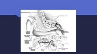
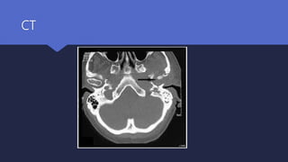
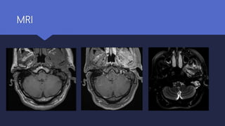
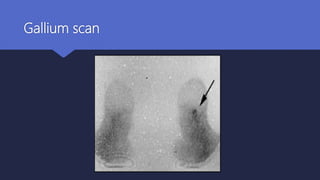
 Malignant otitis externa is an aggressive and potentially life-threatening infection of the soft
tissues of the external ear and surrounding structures, quickly spreading to involve the
periosteum and bone of the skull base.
DEFINITION
NOMENCLATURE
 Malignant otitis externa is a misnomer as it is not a neoplastic process
 In 1968, Chandler described this otitis externa as malignant because he observed an aggressive
clinical behavior, poor treatment outcome, and a high mortality rate for the patients affected by
this disease.
 SYNONAMES :
- Necrotizing otitis externa
-Skull base osteomyelitis
Microbiology
 Bacterial
- Pseudomonas aeruginosa (95%)
- Staphylococcus aureus , S epidermidis .
 Fungal organism
- Aspergillus fumigatus
Predisposing factors
 Old age
 Diabetes mellitus
 Immuno-compromised status
Pathophysiology
Infection from the EAC
spreads
Through the fissures of Santorini
Infection spreads medially to the tympanomastoid suture, and
along venous canals and fascial planes
The compact bone of the skull base becomes replaced with
granulation tissue,
Bone destruction
Progressive spread of infection to skull base foramina causes
cranial neuropathies.
Clinical features
 Long-standing severe otalgia (worst at night)
aggravated by chewing
 Otorrhea
 Hearing loss
Clinical features
 purulent otorrhea with a swollen, tender external
auditory canal
 Hallmark finding: granulation tissue
on floor of the ear canal at the bony-
cartilaginous junction
Clinical features
 Cranial nerve palsy
 Headache
 Neck stiffness
 Altered levels of consciousness
Differential diagnosis
 Carcinoma of the ear canal
 Granulomatous diseases
 Paget's disease
 Nasopharyngeal malignances
 Clival lesions
 Fibrous dysplasia
Diagnosis
 There is no single pathognomonic criterion that defines
malignant otitis externa
 History :(Age ,diabetic ,may give history of trauma to ear by irrigation or
cleaning )
 Complete head and neck examination: (signs of otitis externa with or
without cranial nerve palsy)
Diagnosis
Investigations
 ESR ,CRP raised but not specific
 Ear swab culture/sensitivity ______ Pseudomonas
 Tissues biopsy __________ Rule out malignancy
 HbA1C _________ Diabetic control
Diagnosis
Radiological investigations
 Ct scan :
Defines the anatomical extent of the disease
Remains the initial investigation of choice
 MRI scan
Useful for assessing the initial severity of the disease
Excellent at delineating the extent of soft tissue disease
Intracranial complications
Diagnosis
Radiological investigations
Radioisotope scans
 TC 99 bone scan ------ Bone involvement
 Gallium 67 ------------- Monitoring
 Indium In 111-labeled leukocyte scans
CT
CT
MRI
Gallium scan
TC99
Staging and classification
Stage
1 Clinical evidence of malignant otitis externa with infection of soft tissues beyond the external auditory
canal, but negative Tc-99 bone scan
2 Soft tissue infection beyond external auditory canal with positive Tc-99 bone scan
3 As above, but with cranial nerve paralysis
3a Single
3b Multiple
4 Meningitis, empyema, sinus thrombosis or brain abscess
Management
 Successful management of MOE frequently requires collaboration with an
endocrinologist, neurologist, radiologist, and infectious disease specialist.
 Important principles of treatment include aggressive control of diabetes, reversal of
acidosis, and improvement of immunocompetency where possible
Management
Medical Management
 Meticulous glucose control
 Aural toilet
 systemic anti-Pseudomonas antibiotics (treatment of choice )
Management
Medical Management
 Fluoroquinolones are active against P. aeruginosa.
 For at least 6 to 8 weeks- oral and intravenous ciprofloxacin
 Ceftazidime provide an alternative to ciprofloxacin with or without Aminoglycoside
 Amphotericin B is the most commonly used antifungal agent for fungal
Management
Hyperbaric Oxygen (HBO):
 HBO increases the partial pressure of oxygen, improving hypoxia and allowing
greater oxidative killing of bacteria.
 Used only as an adjunct to antimicrobial therapy
Management
Surgical:
 Removal of sequestra, collections of pus & debridement of
necrotized & granulations
 Facial nerve decompression is not indicated for patients with facial paralysis.
Prognosis
 Disease recurrence
 9-27%
 Due to inadequate length of therapy and manifests as recurrent headache and otalgia
 Can recur as long as one year after treatment is completed
Prognosis
 Mortality
 Decreased to 20% with the introduction of appropriate antibiotics, improved
imaging modalities, and increased awareness of the disease
 Mortality remains high for patients with cranial neuropathies (other than VII),
intracranial complications, or with irreversible systemic immunosuppression.
Thank you

Malignant Otitis Externa

  • 1.
  • 2.
     Malignant otitisexterna is an aggressive and potentially life-threatening infection of the soft tissues of the external ear and surrounding structures, quickly spreading to involve the periosteum and bone of the skull base. DEFINITION
  • 3.
    NOMENCLATURE  Malignant otitisexterna is a misnomer as it is not a neoplastic process  In 1968, Chandler described this otitis externa as malignant because he observed an aggressive clinical behavior, poor treatment outcome, and a high mortality rate for the patients affected by this disease.  SYNONAMES : - Necrotizing otitis externa -Skull base osteomyelitis
  • 4.
    Microbiology  Bacterial - Pseudomonasaeruginosa (95%) - Staphylococcus aureus , S epidermidis .  Fungal organism - Aspergillus fumigatus
  • 5.
    Predisposing factors  Oldage  Diabetes mellitus  Immuno-compromised status
  • 7.
    Pathophysiology Infection from theEAC spreads Through the fissures of Santorini Infection spreads medially to the tympanomastoid suture, and along venous canals and fascial planes The compact bone of the skull base becomes replaced with granulation tissue, Bone destruction Progressive spread of infection to skull base foramina causes cranial neuropathies.
  • 9.
    Clinical features  Long-standingsevere otalgia (worst at night) aggravated by chewing  Otorrhea  Hearing loss
  • 10.
    Clinical features  purulentotorrhea with a swollen, tender external auditory canal  Hallmark finding: granulation tissue on floor of the ear canal at the bony- cartilaginous junction
  • 11.
    Clinical features  Cranialnerve palsy  Headache  Neck stiffness  Altered levels of consciousness
  • 12.
    Differential diagnosis  Carcinomaof the ear canal  Granulomatous diseases  Paget's disease  Nasopharyngeal malignances  Clival lesions  Fibrous dysplasia
  • 13.
    Diagnosis  There isno single pathognomonic criterion that defines malignant otitis externa  History :(Age ,diabetic ,may give history of trauma to ear by irrigation or cleaning )  Complete head and neck examination: (signs of otitis externa with or without cranial nerve palsy)
  • 14.
    Diagnosis Investigations  ESR ,CRPraised but not specific  Ear swab culture/sensitivity ______ Pseudomonas  Tissues biopsy __________ Rule out malignancy  HbA1C _________ Diabetic control
  • 15.
    Diagnosis Radiological investigations  Ctscan : Defines the anatomical extent of the disease Remains the initial investigation of choice  MRI scan Useful for assessing the initial severity of the disease Excellent at delineating the extent of soft tissue disease Intracranial complications
  • 16.
    Diagnosis Radiological investigations Radioisotope scans TC 99 bone scan ------ Bone involvement  Gallium 67 ------------- Monitoring  Indium In 111-labeled leukocyte scans
  • 17.
  • 18.
  • 19.
  • 20.
  • 21.
  • 22.
    Staging and classification Stage 1Clinical evidence of malignant otitis externa with infection of soft tissues beyond the external auditory canal, but negative Tc-99 bone scan 2 Soft tissue infection beyond external auditory canal with positive Tc-99 bone scan 3 As above, but with cranial nerve paralysis 3a Single 3b Multiple 4 Meningitis, empyema, sinus thrombosis or brain abscess
  • 23.
    Management  Successful managementof MOE frequently requires collaboration with an endocrinologist, neurologist, radiologist, and infectious disease specialist.  Important principles of treatment include aggressive control of diabetes, reversal of acidosis, and improvement of immunocompetency where possible
  • 24.
    Management Medical Management  Meticulousglucose control  Aural toilet  systemic anti-Pseudomonas antibiotics (treatment of choice )
  • 25.
    Management Medical Management  Fluoroquinolonesare active against P. aeruginosa.  For at least 6 to 8 weeks- oral and intravenous ciprofloxacin  Ceftazidime provide an alternative to ciprofloxacin with or without Aminoglycoside  Amphotericin B is the most commonly used antifungal agent for fungal
  • 26.
    Management Hyperbaric Oxygen (HBO): HBO increases the partial pressure of oxygen, improving hypoxia and allowing greater oxidative killing of bacteria.  Used only as an adjunct to antimicrobial therapy
  • 27.
    Management Surgical:  Removal ofsequestra, collections of pus & debridement of necrotized & granulations  Facial nerve decompression is not indicated for patients with facial paralysis.
  • 28.
    Prognosis  Disease recurrence 9-27%  Due to inadequate length of therapy and manifests as recurrent headache and otalgia  Can recur as long as one year after treatment is completed
  • 29.
    Prognosis  Mortality  Decreasedto 20% with the introduction of appropriate antibiotics, improved imaging modalities, and increased awareness of the disease  Mortality remains high for patients with cranial neuropathies (other than VII), intracranial complications, or with irreversible systemic immunosuppression.
  • 30.

Editor's Notes

  • #5 The causative agent - Pseudomonas aeruginosa ( a gram-negative – aerobe) --------95% - Other, including Staphylococcus aureus , S epidermidis , Proteus mirabilis , Klebsiella,
  • #6 The most common and important risk factor for MOE is long standing diabetes. • People with recurrent attacks of severe MOE should be screened for diabetes if they aren’t already aware of suffering from it. • Age especially elderly patients • Any condition causing immunosuppression
  • #12 IN Advance disease
  • #14  The diagnosis is generally made from a range of clinical, laboratory, and radiographical findings In this study, more than two-thirds of our patients with malignant external otitis had a history of ear irrigation (generally for the purpose of disimpacting cerumen)
  • #16 These are important adjuncts for determining the presence of osteomyelitis, the extent of disease, and response to therapy.
  • #17 Technitium -99 can detect early bone involvement But not used to assess the response to treatment as it is absorbed by osteoblast and osteoclast that continue remodeling even after infection is settled –remain positive for up to 9 month Gallium (Ga-67) is absorbed by leukocytes and is a more sensitive monitor of infection. The scan quickly returns to normal after the infection has resolved and as such, is a good measure to ascertain when to terminate treatment
  • #18 CT scan showing the soft tissue obliterating left external auditory canal left mastoid, infra-temporal fossa, skull base and involving the left TMJ.
  • #19 Axial CT scan of a 63-year-old patient who has diabetes and left malignant otitis externa showing bony erosion of the posterior external auditory canal and mastoid cortex
  • #20 MRI of same patient: axial T1WI (A), post contrast (B) and fat saturated T2WI (C) demonstrates extensive inflammation and enhancement of the left EAC, periauricular region, masticator, parotid and parapharyngeal spaces. These areas are hyperintense on T2WI. Note mastoid airspace disease. This can be mistaken for an aggressive tumor.
  • #22 Fig. 2. Anterior and posterior views of technetium Tc 99m MDP bone scan of patient from Fig. 1showing increased uptake in the left mastoid.
  • #23 Staging depending on clinical and radiological features
  • #25  Culture-directed therapy should always be the goal of treatment
  • #26 Early infections may be treated solely with oral ciprofloxacin In more advanced stages, parenteral antibiotics may be indicated initially Ceftazidime –upto 2 g i/v tds
  • #27 Hyperbaric oxygen therapy Used only as an adjunct to antimicrobial therapy; it should not be used alone. May be helpful for patients with complications, experiencing a poor response to therapy, or with recurrent cases