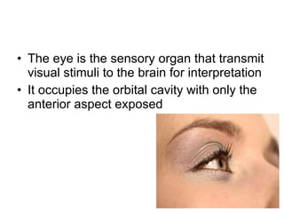
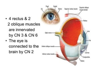
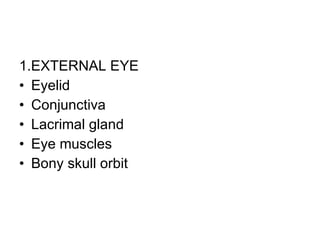
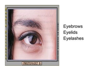
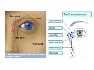
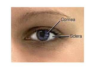
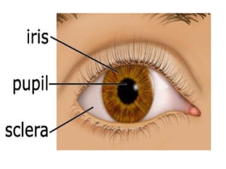
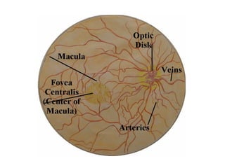
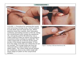
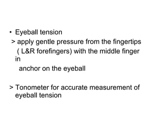
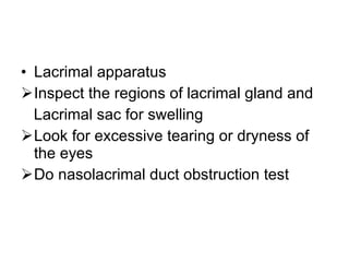
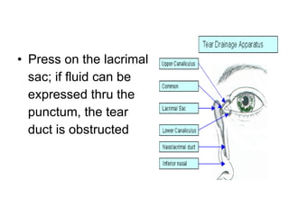
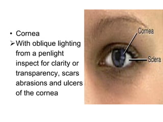
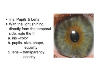
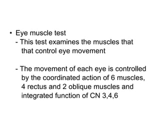
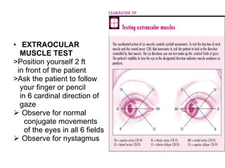
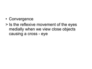
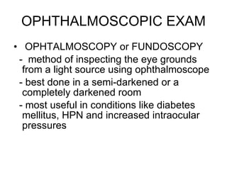
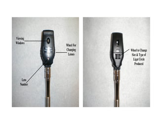
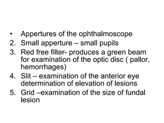
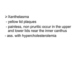
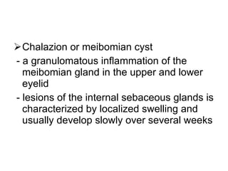
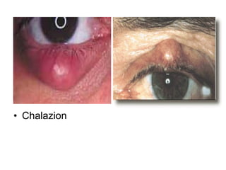
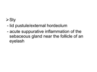
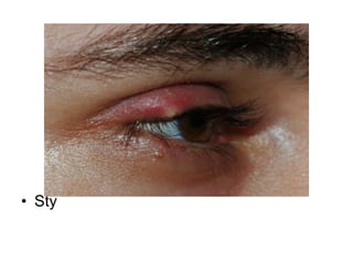
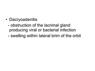
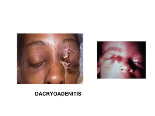
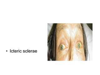
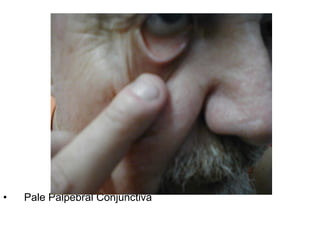
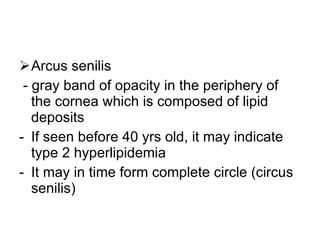
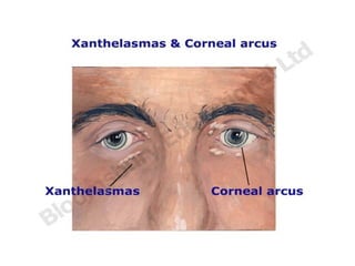
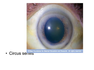
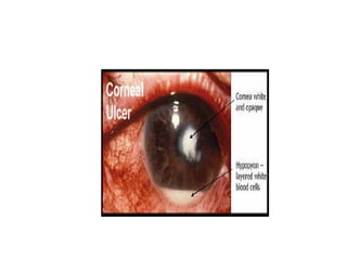
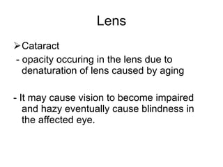
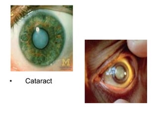
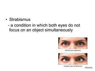
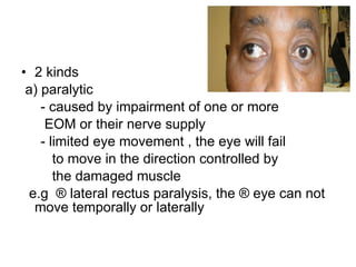
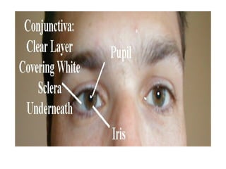
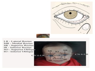
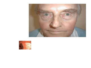
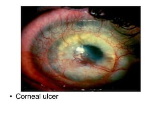
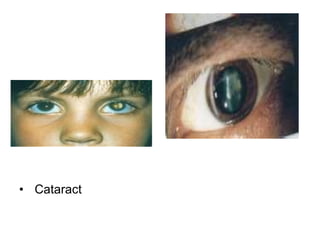
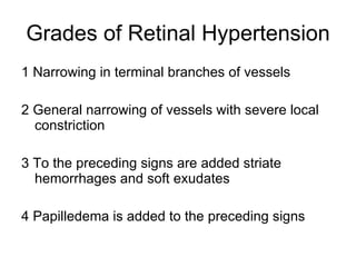
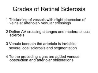
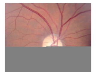
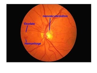
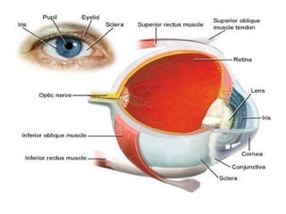
EYES
Anatomy and Landmarks of Eyes
The eye is the sensory organ that transmit visual stimuli to the brain for interpretation It occupies the orbital cavity with only the anterior aspect exposed
2 4 rectus & 2 2 oblique muscles are innervated by CN 3 & CN 6 The eye is connected to the brain by CN 2
1.EXTERNAL EYE Eyelid  Conjunctiva Lacrimal gland Eye muscles Bony skull orbit
Eyebrows Eyelids Eyelashes
 
2.INTERNAL EYE 3 layers  1.Sclerotic coat > this tough layer creates the “white” of the eye except in the front where it forms the transparent cornea
 
2. Choroid coat > this middle layer is deeply pigmented with melanin Reduces reflection of stray light within the eye The choroid coat forms iris and ciliary body in the front of the eye and the choroid posteriorly The size of its opening, the pupil, is variable and under the control of autonomic nervous system
 
3. Retina > forms the inner layer of the eye  > it contains the light receptors, the rods and cones ( thus serve as the “film” of the eye)
 
Eye Examination One of the best ways to protect vision- it can detect eye problems at their earliest stage
Who gives eye exam? Ophthalmologists- or any medical doctors who provide full eye care such as : - complete eye exam - prescribing corrective lenses - diagnosing and treating complex eye diseases - performing surgery
Optometrists  - diagnosing common eye disorders and treating selected eye diseases with drugs - evaluating vision - prescribing corrective lenses - can not perform surgery
Opticians  - fill prescriptions for eyeglasses - some opticians also sell and fit contact lenses
A complete eye exam begins with your medical history: - Are you having any eye problem now? - have you had any eye problems in the past? Do you wear glasses or contacts now? if so, are you satisfied with them?
What health problems have you had in recent years? Are you taking any medications? Do you have any allergies to medications, food or other substances?
Has anyone in the family had eye problems such as cataracts or glaucoma Has anyone in your family had diabetes , high blood pressure, heart disease or any health problems that can affect the whole body?
Eye Examination PROCEDURE…
Eyeball  >Position and alignment of the eyes a. Stand in front of the patient and survey the eyes for position and alignment with  each other .  b. If one or both eyes seem to protrude ,  assess them from the profile and from  above
The eyeball –observe the position within the confines of the bony orbit ( socket)
External examination >Examination of the eyes is carried out in a systematic manner beginning with the appendages that is, the eyebrows and surrounding tissues and moving inward
Surrounding structures Eyebrows  > note for the amount , distribution 2. Inspect the orbital area  > edema > sagging tissues or puffiness > lesion
Eyelids  Assess the palpebral fissure with the patient looking straight ahead and observe the relationship of the lids to the limbus Note for any widening or narrowing of the palpebral fissure
Note for the ability to open and close the eyes completely Observe for ptosis  Note for the eyelashes -Projecting from the border of each lid, should turn outward
Tremors  Redness  Swelling/edema of the lids Note whether there is lid eversion or  lid inversion
Procedure for everting the eyelid and removing a foreign body
 
Eyeball tension > apply gentle pressure from the fingertips ( L&R forefingers) with the middle finger in  anchor on the eyeball > Tonometer for accurate measurement of eyeball tension
Lacrimal apparatus Inspect the regions of lacrimal gland and  Lacrimal sac for swelling Look for excessive tearing or dryness of the eyes Do nasolacrimal duct obstruction test
Press on the lacrimal sac; if fluid can be expressed thru the punctum, the tear duct is obstructed
INTERNAL EXAMINATION
Conjunctivae & Sclerae Ask the patient to look up as you depress both lower lids with your  thumbs Ask the patient to look down as you pull the  upper lids upward
Cornea  With oblique lighting from a penlight inspect for clarity or transparency, scars abrasions and ulcers of the cornea
Iris, Pupils & Lens > With the light shining  directly from the temporal  side, note the ff: a. iris –color b. pupils- size, shape, equality c. lens – transparency, opacity
Pupillary reaction - Pupillary size changes in response to light and to the effort of focusing on a near object
a) The light reaction  Direct light reflex: > From the lateral side of each eye, flash the penlight swiftly into the eye being examined.  >Observe for immediate constriction of the pupil
Indirect light reflex: Ask the patient to rest the radial side of his L or R hand in between the eyes. From the lateral side flash the penlight into one eye Observe for the pupillary constriction of the opposite eye
b) The near reaction - When a person shifts gaze fr. a far object to a near one, the pupils constrict. This response is mediated by the oculomotor nerve. - This is pupillary response to acommodation - This is done if there is defect in the pupillary response to light
 
Eye muscle test - This test examines the muscles that  that control eye movement - The movement of each eye is controlled  by the coordinated action of 6 muscles,  4 rectus and 2 oblique muscles and  integrated function of CN 3,4,6
EXTRAOCULAR  MUSCLE TEST >Position yourself 2 ft in front of the patient >Ask the patient to follow your finger or pencil in 6 cardinal direction of  gaze Observe for normal  conjugate movements of the eyes in all 6 fields Observe for nystagmus
 
Convergence  > Is the reflexive movement of the eyes medially when we view close objects causing a cross - eye
Convergence test - hold the target in the midline and at the eye level about 50 cm (20 inch) from the face gradually moving the target toward the bridge of the nose - convergence is normally maintained until 2-3 inches ( 50 mm) from the bridge of the nose
Lid lag The inability of the upper eyelid to follow the eye’s downward movement A cardinal sign of thyrotoxicosis (Von Graefe’s sign)
Lid lag test - hold the finger or a penlight as a target in the midline above the eye level 20 inches  (50 cm ) away. Move the target rapidly downward in the midline  Watch for the appearance of white sclera between the iris and the upper lid margin
Testing Vision Visual Acuity - This test measures how clearly you can  can see from distance - Gross test for visual acuity can be made  without special equipment. Test a single eye at a time.
Snellen Chart Done when gross visual acuity is fair This is a test for far vision Express the reading as: 20/20 20= distance at which the test is  conducted 20= distance at which line of letters should  be read by a normal eye
SNELLEN CHART  Pinhole test
Jaeger Test > for  near vision  > subject read at one foot or 14” distance Acuity of near vision is expressed as J1,J2,J3 A reading of J1 is  considered normal
Visual field test (perimetry) - visual field is the area in front of you that you can see without moving your eyes - This test determines whether you have difficulty seeing any areas of your peripheral vision- the areas on the side of your visual field
Confrontation test
Tangent screen exam - sit a short distance from a screen and stare at a target at its center.  - tell examiner if you see an object move into your peripheral vision
Automated perimetry - computer program is used that flashes small lights as you look into a special instrument. - press the button when you see the lights
Slit-lamp examination - it uses a microscope that enlarges and illuminate the front of the eye with an intense line of light - this is use to examine the cornea, iris , lens and anterior chamber of the eye
 
OPHTHALMOSCOPIC EXAM OPHTALMOSCOPY or FUNDOSCOPY -  method of inspecting the eye grounds from a light source using ophthalmoscope - best done in a semi-darkened or a completely darkened room - most useful in conditions like diabetes mellitus, HPN and increased intraocular pressures
 
Appertures of the ophthalmoscope Small apperture – small pupils Red free filter- produces a green beam for examination of the optic disc ( pallor, hemorrhages) Slit – examination of the anterior eye determination of elevation of lesions Grid –examination of the size of fundal lesion
 
Optic disc - note for its clarity, disc margin should be sharp and well defined - round or oval vertically - color is yellowish- orange to creamy pink oval
-The physiologic cup if present is normally yellowish white. Its horizontal diameter is usually less than half the horizontal diameter of the disc - Measures about 1.5 mm in diameter and 3 diopters of elevation = 1mm
Identify the arterioles and veins 1) color  A- light red  V- dark red 2) Size A- smaller(2/3 to 4/5 of vein)  V- larger 3) Light reflex A- bright  V- absent
The A:V ratio is generally 3:5 to 2:3
Macula  This is an avascular area, somewhat larger than the disc with no distinct margin To locate the macula, focus on the disc, then move the light temporally about 2 discs diameter
b. To bring it into your vision, ask the patient to look directly at the light c. In the center of the macula the fovea appears as a small darker red area in the retina


Glaucoma test ( tonometry)  - measures intraocular pressure- pressure inside the eyes. - it can detect glaucoma , a disease that causes pressure to build inside the eyes and can cause blindness
SIGNS
Eyeballs Exophthalmos  - increase in the volume of the orbital content causing a protrusion of the globes forward - Due to Grave’s disease if bilateral and retroorbital tumor if unilateral
 
Palpebral fissures Widened palpebral fissures - uncovers the upper border of the limbus to expose white sclera superiorly - fissures maybe widened by retractions of the lids or by protrusion of the  eyeballs
EYELIDS WIDENED PALPEBRAL FISSURE
(+) Lid lag test (Von Graefe sign) - seen in hyperthyroidism
2. Narrowed palpebral fissure: Enophthalmos - the globe is recessed in the orbit - bilateral cause: decrease orbital fat  ( dehydration,inanition) congenital microphthalmos - unilateral cause: trauma or inflammation
Failure of lid closure: Paralysis of orbicularis muscle - seen in Bell’s palsy due to disorder of the facial nerve (CN 7) causing partial or complete paralysis of the orbicularis muscle
BELL’S PALSY
2. Failure of lid opening: Ptosis - indicates a congenital or acquired weakness of the levator muscle or paresis of the 3 rd  CN - Superior eyelids covers more than the iris
Ptosis
Lid swelling: palpebral edema a) inflammatory edema  - redness , warmth, pain b) non inflammatory edema - acute nephritis - myxedema - exophthalmos of grave’s dse. - contact dermatitis
Swollen eyelids
Lid inversion ( Entropion ) a) spastic Occurs only in the lower lid Due to increase tone of the orbicularis oculi usually from inflammation of an eye
b) Cicatricial  - occurs in either lid - due to contracture of scar tissue - eg. Trachoma > Entropion from any cause maybe acc. by blepharospasm from irritation of the inverted eyelashes
LID INVERSION (INTROPION)
Lid eversion ( Ectropion) Lid turns outward Both lids affected by spastic or cicatricial ectropion Paralytic ectropion involves only the lower lid - Senile atrophy of tissues sometimes result in ectropion
LID EVERSION (ECTROPION)
Xanthelasma - yellow lid plaques - painless, non pruritic occur in the upper and lower lids near the inner canthus - ass. with hypercholesterolemia
 
Chalazion or meibomian cyst - a granulomatous inflammation of the meibomian gland in the upper and lower eyelid - lesions of the internal sebaceous glands is characterized by localized swelling and usually develop slowly over several weeks
Chalazion
Sty  - lid pustule/external hordeolum - acute suppurative inflammation of the sebaceous gland near the follicle of an eyelash
Sty
Dacryoadenitis  - obstruction of the lacrimal gland producing viral or bacterial infection - swelling within lateral brim of the orbit
DACRYOADENITIS
Sclera
Icteric sclerae
Conjunctiva
Pale Palpebral Conjunctiva
Pterygium  An abnormal growth of conjunctiva that extends over the cornea from the limbus. More common to people heavily exposed to ultraviolet light Chronic irritation due to wind/dust exposure May interfere vision if it advances over the pupil
 
Subconjunctival Hemorrhage Bright red blood in a sharply defined are surrounded by a normal appearing conjunctiva Bleeding maybe due to coughing, sneezing, weight lifting
Subconjunctival Hemorrhage
Chemosis ( conjunctival edema) - swelling of the eye surface membrane because of accumulation of fluid  -  ass. with grave’s disease
 
Conjunctivitis  - a condition in which the conjunctiva becomes inflamed or infected . - maybe due to viral/bacterial infections - foreign body reaction - allergy
Conjunctivitis
Pupils
Mydriasis  Pupillary dilatation ( > 5 mm diameter) May be due to uremia diabetes mellitus,coma atrophine, alcohol, head trauma, CN 3 damage epilepsy
Miosis  - Pupillary constriction ( < 3 mm in diameter) - maybe due to morphine, pilocarpine, glaucoma
 
Cornea Cloudy cornea - seen in congenital syphilis - Hutchinson triangle : interstitial keratitis deafness notched teeth
 
Arcus senilis - gray band of opacity in the periphery of the cornea which is composed of lipid deposits If seen before 40 yrs old, it may indicate type 2 hyperlipidemia It may in time form complete circle (circus senilis)
 
Circus senilis
Corneal ulcer - infective condition of the cornea involving disruption of its epithelial layer with involvement of the corneal stroma - this involves damage of the cornea as a result of injury, foreign body or excessive or inappropriate wearing of contact lens
 
Lens Cataract  - opacity occuring in the lens due to denaturation of lens caused by aging - It may cause vision to become impaired and hazy eventually cause blindness in the affected eye.
Cataract
EOM muscles
Strabismus  - a condition in which both eyes do not focus on an object simultaneously
2 kinds a) paralytic - caused by impairment of one or more  EOM or their nerve supply - limited eye movement , the eye will fail to move in the direction controlled by the damaged muscle e.g  ® lateral rectus paralysis, the ® eye can not move temporally or laterally
b) Non paralytic strabismus - has no primary muscle weakness - patient can focus with either eye but not with both simultaneously - detected by having a patient observe a near object -
When one eye is covered, the other one will move to  focus on the object if the covered eye is the dominant one This is cover-uncover test
Cranial nerve 3 palsy
Visual Field
 
 
A= total blindness right eye B= nasal hemianopsia of the right eye C= left homonymous hemianopsia  D = bitemporal  heteronymous hemianopsia
Hemianopsia  -involves nerves projecting from both eyes caused by lesion in the optic chiasm, optic tract and brain
Homonymous hemianopsia - defect on the same side of each field e.g.  (L) homonymous hemianopsia can be caused by a lesion in the ® optic tract or the ® side of the brain
(R) Homonymous Hemianopsia (L)
Bitemporal heteronymous hemianopsia - caused by a lesion of the decussating fibers in  the optic chiasm injuring both  nasal retinae - commonly due to pituitary gland tumor
Retina
Papilledema ( choked disc) Due to inc.ICP causes CSF to compress optic nerve -central vision not impaired, there is peripheral loss
Macula  - common site for  diabetes mellitus - microaneurysm occur around the  macula
Retinal spots
Symptoms
Diplopia or double vision - eyes not aligned - perception of 2 visual images due: a) abnormalities of refraction b) impairment of the 3 rd  ,4 th , 6 th  CN or  c) weakness of one of the 6 EOMs that move the eye or of the myoneural junction - ass. With myasthenia gravis, grave’s ophthalmopathy
Blurred vision - loss of sharp focus of light on the retina - may be due to opacities in the cornea, lens or vitreous - Pinhole test is used to determine if blurred vision is optical in origin
Visual loss - injury or impairment to any portion of the visual pathway Acute loss of vision is medical emergency Chronic progressive loss of vision is common with disease of the cornea, lens or retina
Pain in the eye Careful examination of the cornea, anterior chamber, iris, retina are mandatory in patients complaining of eye pain Always assess visual acuity of each eye
Causes: endocrine – thyrotoxicosis idiopathic – cluster headache inflammatory- sty, optic neuritis, chalazion infection- herpes zoster sinusitis – ethmoid, frontal, sphenoid trauma/mechanical - foreign body, corneal abrasion, glaucoma
Thank You


Corneal ulcer
Cataract
Eye Hemorrhage
Grades of Retinal Hypertension 1 Narrowing in terminal branches of vessels 2 General narrowing of vessels with severe local constriction 3 To the preceding signs are added striate hemorrhages and soft exudates 4 Papilledema is added to the preceding signs
Grades of Retinal Sclerosis 1 Thickening of vessels with slight depresion of veins at arteriolar- venular crossings 2 Define AV crossing changes and moderate local sclerosis 3 Venule beneath the arteriole is invisible; severe local sclerosis and segmentation 4 To the preceding signs are added venous obstruction and arteriolar obliterations
 
 
(Lower) Homonymous Hemianopsia (Upper)
Diabetic Retinopathy


Eyes (2)

  • 1.
  • 2.
  • 3.
    The eye isthe sensory organ that transmit visual stimuli to the brain for interpretation It occupies the orbital cavity with only the anterior aspect exposed
  • 4.
    2 4 rectus& 2 2 oblique muscles are innervated by CN 3 & CN 6 The eye is connected to the brain by CN 2
  • 5.
    1.EXTERNAL EYE Eyelid Conjunctiva Lacrimal gland Eye muscles Bony skull orbit
  • 6.
  • 7.
  • 8.
    2.INTERNAL EYE 3layers 1.Sclerotic coat > this tough layer creates the “white” of the eye except in the front where it forms the transparent cornea
  • 9.
  • 10.
    2. Choroid coat> this middle layer is deeply pigmented with melanin Reduces reflection of stray light within the eye The choroid coat forms iris and ciliary body in the front of the eye and the choroid posteriorly The size of its opening, the pupil, is variable and under the control of autonomic nervous system
  • 11.
  • 12.
    3. Retina >forms the inner layer of the eye > it contains the light receptors, the rods and cones ( thus serve as the “film” of the eye)
  • 13.
  • 14.
    Eye Examination Oneof the best ways to protect vision- it can detect eye problems at their earliest stage
  • 15.
    Who gives eyeexam? Ophthalmologists- or any medical doctors who provide full eye care such as : - complete eye exam - prescribing corrective lenses - diagnosing and treating complex eye diseases - performing surgery
  • 16.
    Optometrists -diagnosing common eye disorders and treating selected eye diseases with drugs - evaluating vision - prescribing corrective lenses - can not perform surgery
  • 17.
    Opticians -fill prescriptions for eyeglasses - some opticians also sell and fit contact lenses
  • 18.
    A complete eyeexam begins with your medical history: - Are you having any eye problem now? - have you had any eye problems in the past? Do you wear glasses or contacts now? if so, are you satisfied with them?
  • 19.
    What health problemshave you had in recent years? Are you taking any medications? Do you have any allergies to medications, food or other substances?
  • 20.
    Has anyone inthe family had eye problems such as cataracts or glaucoma Has anyone in your family had diabetes , high blood pressure, heart disease or any health problems that can affect the whole body?
  • 21.
  • 22.
    Eyeball >Positionand alignment of the eyes a. Stand in front of the patient and survey the eyes for position and alignment with each other . b. If one or both eyes seem to protrude , assess them from the profile and from above
  • 23.
    The eyeball –observethe position within the confines of the bony orbit ( socket)
  • 24.
    External examination >Examinationof the eyes is carried out in a systematic manner beginning with the appendages that is, the eyebrows and surrounding tissues and moving inward
  • 25.
    Surrounding structures Eyebrows > note for the amount , distribution 2. Inspect the orbital area > edema > sagging tissues or puffiness > lesion
  • 26.
    Eyelids Assessthe palpebral fissure with the patient looking straight ahead and observe the relationship of the lids to the limbus Note for any widening or narrowing of the palpebral fissure
  • 27.
    Note for theability to open and close the eyes completely Observe for ptosis Note for the eyelashes -Projecting from the border of each lid, should turn outward
  • 28.
    Tremors Redness Swelling/edema of the lids Note whether there is lid eversion or lid inversion
  • 29.
    Procedure for evertingthe eyelid and removing a foreign body
  • 30.
  • 31.
    Eyeball tension >apply gentle pressure from the fingertips ( L&R forefingers) with the middle finger in anchor on the eyeball > Tonometer for accurate measurement of eyeball tension
  • 32.
    Lacrimal apparatus Inspectthe regions of lacrimal gland and Lacrimal sac for swelling Look for excessive tearing or dryness of the eyes Do nasolacrimal duct obstruction test
  • 33.
    Press on thelacrimal sac; if fluid can be expressed thru the punctum, the tear duct is obstructed
  • 34.
  • 35.
    Conjunctivae & ScleraeAsk the patient to look up as you depress both lower lids with your thumbs Ask the patient to look down as you pull the upper lids upward
  • 36.
    Cornea Withoblique lighting from a penlight inspect for clarity or transparency, scars abrasions and ulcers of the cornea
  • 37.
    Iris, Pupils &Lens > With the light shining directly from the temporal side, note the ff: a. iris –color b. pupils- size, shape, equality c. lens – transparency, opacity
  • 38.
    Pupillary reaction -Pupillary size changes in response to light and to the effort of focusing on a near object
  • 39.
    a) The lightreaction Direct light reflex: > From the lateral side of each eye, flash the penlight swiftly into the eye being examined. >Observe for immediate constriction of the pupil
  • 40.
    Indirect light reflex:Ask the patient to rest the radial side of his L or R hand in between the eyes. From the lateral side flash the penlight into one eye Observe for the pupillary constriction of the opposite eye
  • 41.
    b) The nearreaction - When a person shifts gaze fr. a far object to a near one, the pupils constrict. This response is mediated by the oculomotor nerve. - This is pupillary response to acommodation - This is done if there is defect in the pupillary response to light
  • 42.
  • 43.
    Eye muscle test- This test examines the muscles that that control eye movement - The movement of each eye is controlled by the coordinated action of 6 muscles, 4 rectus and 2 oblique muscles and integrated function of CN 3,4,6
  • 44.
    EXTRAOCULAR MUSCLETEST >Position yourself 2 ft in front of the patient >Ask the patient to follow your finger or pencil in 6 cardinal direction of gaze Observe for normal conjugate movements of the eyes in all 6 fields Observe for nystagmus
  • 45.
  • 46.
    Convergence >Is the reflexive movement of the eyes medially when we view close objects causing a cross - eye
  • 47.
    Convergence test -hold the target in the midline and at the eye level about 50 cm (20 inch) from the face gradually moving the target toward the bridge of the nose - convergence is normally maintained until 2-3 inches ( 50 mm) from the bridge of the nose
  • 48.
    Lid lag Theinability of the upper eyelid to follow the eye’s downward movement A cardinal sign of thyrotoxicosis (Von Graefe’s sign)
  • 49.
    Lid lag test- hold the finger or a penlight as a target in the midline above the eye level 20 inches (50 cm ) away. Move the target rapidly downward in the midline Watch for the appearance of white sclera between the iris and the upper lid margin
  • 50.
    Testing Vision VisualAcuity - This test measures how clearly you can can see from distance - Gross test for visual acuity can be made without special equipment. Test a single eye at a time.
  • 51.
    Snellen Chart Donewhen gross visual acuity is fair This is a test for far vision Express the reading as: 20/20 20= distance at which the test is conducted 20= distance at which line of letters should be read by a normal eye
  • 52.
    SNELLEN CHART Pinhole test
  • 53.
    Jaeger Test >for near vision > subject read at one foot or 14” distance Acuity of near vision is expressed as J1,J2,J3 A reading of J1 is considered normal
  • 54.
    Visual field test(perimetry) - visual field is the area in front of you that you can see without moving your eyes - This test determines whether you have difficulty seeing any areas of your peripheral vision- the areas on the side of your visual field
  • 55.
  • 56.
    Tangent screen exam- sit a short distance from a screen and stare at a target at its center. - tell examiner if you see an object move into your peripheral vision
  • 57.
    Automated perimetry -computer program is used that flashes small lights as you look into a special instrument. - press the button when you see the lights
  • 58.
    Slit-lamp examination -it uses a microscope that enlarges and illuminate the front of the eye with an intense line of light - this is use to examine the cornea, iris , lens and anterior chamber of the eye
  • 59.
  • 60.
    OPHTHALMOSCOPIC EXAM OPHTALMOSCOPYor FUNDOSCOPY - method of inspecting the eye grounds from a light source using ophthalmoscope - best done in a semi-darkened or a completely darkened room - most useful in conditions like diabetes mellitus, HPN and increased intraocular pressures
  • 61.
  • 62.
    Appertures of theophthalmoscope Small apperture – small pupils Red free filter- produces a green beam for examination of the optic disc ( pallor, hemorrhages) Slit – examination of the anterior eye determination of elevation of lesions Grid –examination of the size of fundal lesion
  • 63.
  • 64.
    Optic disc -note for its clarity, disc margin should be sharp and well defined - round or oval vertically - color is yellowish- orange to creamy pink oval
  • 65.
    -The physiologic cupif present is normally yellowish white. Its horizontal diameter is usually less than half the horizontal diameter of the disc - Measures about 1.5 mm in diameter and 3 diopters of elevation = 1mm
  • 66.
    Identify the arteriolesand veins 1) color A- light red V- dark red 2) Size A- smaller(2/3 to 4/5 of vein) V- larger 3) Light reflex A- bright V- absent
  • 67.
    The A:V ratiois generally 3:5 to 2:3
  • 68.
    Macula Thisis an avascular area, somewhat larger than the disc with no distinct margin To locate the macula, focus on the disc, then move the light temporally about 2 discs diameter
  • 69.
    b. To bringit into your vision, ask the patient to look directly at the light c. In the center of the macula the fovea appears as a small darker red area in the retina
  • 70.
  • 71.
  • 72.
  • 73.
    Glaucoma test (tonometry) - measures intraocular pressure- pressure inside the eyes. - it can detect glaucoma , a disease that causes pressure to build inside the eyes and can cause blindness
  • 74.
  • 75.
    Eyeballs Exophthalmos - increase in the volume of the orbital content causing a protrusion of the globes forward - Due to Grave’s disease if bilateral and retroorbital tumor if unilateral
  • 76.
  • 77.
    Palpebral fissures Widenedpalpebral fissures - uncovers the upper border of the limbus to expose white sclera superiorly - fissures maybe widened by retractions of the lids or by protrusion of the eyeballs
  • 78.
  • 79.
    (+) Lid lagtest (Von Graefe sign) - seen in hyperthyroidism
  • 80.
    2. Narrowed palpebralfissure: Enophthalmos - the globe is recessed in the orbit - bilateral cause: decrease orbital fat ( dehydration,inanition) congenital microphthalmos - unilateral cause: trauma or inflammation
  • 81.
    Failure of lidclosure: Paralysis of orbicularis muscle - seen in Bell’s palsy due to disorder of the facial nerve (CN 7) causing partial or complete paralysis of the orbicularis muscle
  • 82.
  • 83.
    2. Failure oflid opening: Ptosis - indicates a congenital or acquired weakness of the levator muscle or paresis of the 3 rd CN - Superior eyelids covers more than the iris
  • 84.
  • 85.
    Lid swelling: palpebraledema a) inflammatory edema - redness , warmth, pain b) non inflammatory edema - acute nephritis - myxedema - exophthalmos of grave’s dse. - contact dermatitis
  • 86.
  • 87.
    Lid inversion (Entropion ) a) spastic Occurs only in the lower lid Due to increase tone of the orbicularis oculi usually from inflammation of an eye
  • 88.
    b) Cicatricial - occurs in either lid - due to contracture of scar tissue - eg. Trachoma > Entropion from any cause maybe acc. by blepharospasm from irritation of the inverted eyelashes
  • 89.
  • 90.
    Lid eversion (Ectropion) Lid turns outward Both lids affected by spastic or cicatricial ectropion Paralytic ectropion involves only the lower lid - Senile atrophy of tissues sometimes result in ectropion
  • 91.
  • 92.
    Xanthelasma - yellowlid plaques - painless, non pruritic occur in the upper and lower lids near the inner canthus - ass. with hypercholesterolemia
  • 93.
  • 94.
    Chalazion or meibomiancyst - a granulomatous inflammation of the meibomian gland in the upper and lower eyelid - lesions of the internal sebaceous glands is characterized by localized swelling and usually develop slowly over several weeks
  • 95.
  • 96.
    Sty -lid pustule/external hordeolum - acute suppurative inflammation of the sebaceous gland near the follicle of an eyelash
  • 97.
  • 98.
    Dacryoadenitis -obstruction of the lacrimal gland producing viral or bacterial infection - swelling within lateral brim of the orbit
  • 99.
  • 100.
  • 101.
  • 102.
  • 103.
  • 104.
    Pterygium Anabnormal growth of conjunctiva that extends over the cornea from the limbus. More common to people heavily exposed to ultraviolet light Chronic irritation due to wind/dust exposure May interfere vision if it advances over the pupil
  • 105.
  • 106.
    Subconjunctival Hemorrhage Brightred blood in a sharply defined are surrounded by a normal appearing conjunctiva Bleeding maybe due to coughing, sneezing, weight lifting
  • 107.
  • 108.
    Chemosis ( conjunctivaledema) - swelling of the eye surface membrane because of accumulation of fluid - ass. with grave’s disease
  • 109.
  • 110.
    Conjunctivitis -a condition in which the conjunctiva becomes inflamed or infected . - maybe due to viral/bacterial infections - foreign body reaction - allergy
  • 111.
  • 112.
  • 113.
    Mydriasis Pupillarydilatation ( > 5 mm diameter) May be due to uremia diabetes mellitus,coma atrophine, alcohol, head trauma, CN 3 damage epilepsy
  • 114.
    Miosis -Pupillary constriction ( < 3 mm in diameter) - maybe due to morphine, pilocarpine, glaucoma
  • 115.
  • 116.
    Cornea Cloudy cornea- seen in congenital syphilis - Hutchinson triangle : interstitial keratitis deafness notched teeth
  • 117.
  • 118.
    Arcus senilis -gray band of opacity in the periphery of the cornea which is composed of lipid deposits If seen before 40 yrs old, it may indicate type 2 hyperlipidemia It may in time form complete circle (circus senilis)
  • 119.
  • 120.
  • 121.
    Corneal ulcer -infective condition of the cornea involving disruption of its epithelial layer with involvement of the corneal stroma - this involves damage of the cornea as a result of injury, foreign body or excessive or inappropriate wearing of contact lens
  • 122.
  • 123.
    Lens Cataract - opacity occuring in the lens due to denaturation of lens caused by aging - It may cause vision to become impaired and hazy eventually cause blindness in the affected eye.
  • 124.
  • 125.
  • 126.
    Strabismus -a condition in which both eyes do not focus on an object simultaneously
  • 127.
    2 kinds a)paralytic - caused by impairment of one or more EOM or their nerve supply - limited eye movement , the eye will fail to move in the direction controlled by the damaged muscle e.g ® lateral rectus paralysis, the ® eye can not move temporally or laterally
  • 128.
    b) Non paralyticstrabismus - has no primary muscle weakness - patient can focus with either eye but not with both simultaneously - detected by having a patient observe a near object -
  • 129.
    When one eyeis covered, the other one will move to focus on the object if the covered eye is the dominant one This is cover-uncover test
  • 130.
  • 131.
  • 132.
  • 133.
  • 134.
    A= total blindnessright eye B= nasal hemianopsia of the right eye C= left homonymous hemianopsia D = bitemporal heteronymous hemianopsia
  • 135.
    Hemianopsia -involvesnerves projecting from both eyes caused by lesion in the optic chiasm, optic tract and brain
  • 136.
    Homonymous hemianopsia -defect on the same side of each field e.g. (L) homonymous hemianopsia can be caused by a lesion in the ® optic tract or the ® side of the brain
  • 137.
  • 138.
    Bitemporal heteronymous hemianopsia- caused by a lesion of the decussating fibers in the optic chiasm injuring both nasal retinae - commonly due to pituitary gland tumor
  • 139.
  • 140.
    Papilledema ( chokeddisc) Due to inc.ICP causes CSF to compress optic nerve -central vision not impaired, there is peripheral loss
  • 141.
    Macula -common site for diabetes mellitus - microaneurysm occur around the macula
  • 142.
  • 143.
  • 144.
    Diplopia or doublevision - eyes not aligned - perception of 2 visual images due: a) abnormalities of refraction b) impairment of the 3 rd ,4 th , 6 th CN or c) weakness of one of the 6 EOMs that move the eye or of the myoneural junction - ass. With myasthenia gravis, grave’s ophthalmopathy
  • 145.
    Blurred vision -loss of sharp focus of light on the retina - may be due to opacities in the cornea, lens or vitreous - Pinhole test is used to determine if blurred vision is optical in origin
  • 146.
    Visual loss -injury or impairment to any portion of the visual pathway Acute loss of vision is medical emergency Chronic progressive loss of vision is common with disease of the cornea, lens or retina
  • 147.
    Pain in theeye Careful examination of the cornea, anterior chamber, iris, retina are mandatory in patients complaining of eye pain Always assess visual acuity of each eye
  • 148.
    Causes: endocrine –thyrotoxicosis idiopathic – cluster headache inflammatory- sty, optic neuritis, chalazion infection- herpes zoster sinusitis – ethmoid, frontal, sphenoid trauma/mechanical - foreign body, corneal abrasion, glaucoma
  • 149.
  • 150.
  • 151.
  • 152.
  • 153.
  • 154.
  • 155.
  • 156.
  • 157.
    Grades of RetinalHypertension 1 Narrowing in terminal branches of vessels 2 General narrowing of vessels with severe local constriction 3 To the preceding signs are added striate hemorrhages and soft exudates 4 Papilledema is added to the preceding signs
  • 158.
    Grades of RetinalSclerosis 1 Thickening of vessels with slight depresion of veins at arteriolar- venular crossings 2 Define AV crossing changes and moderate local sclerosis 3 Venule beneath the arteriole is invisible; severe local sclerosis and segmentation 4 To the preceding signs are added venous obstruction and arteriolar obliterations
  • 159.
  • 160.
  • 161.
  • 162.
  • 163.
  • 164.