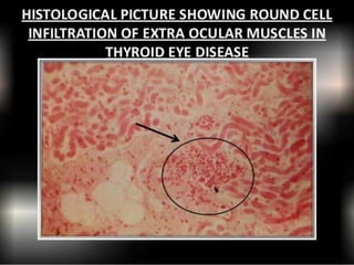
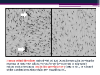
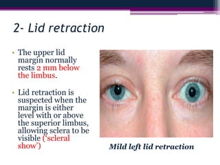
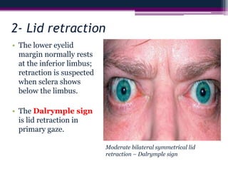
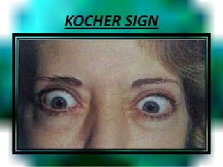
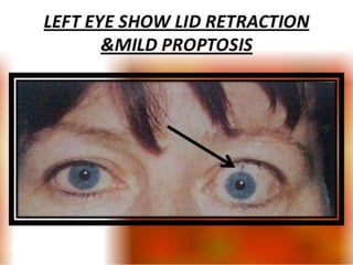
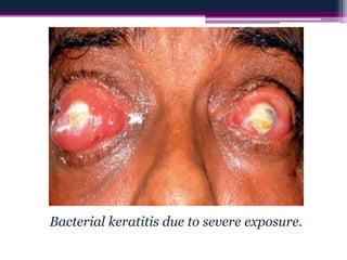
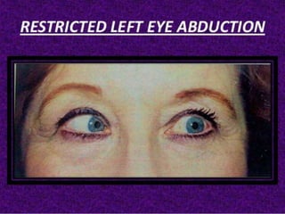
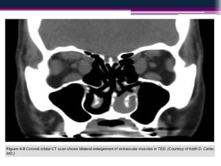
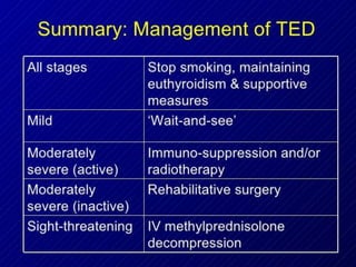
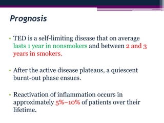
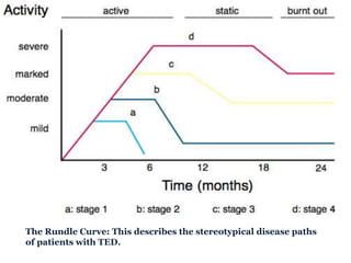
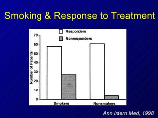
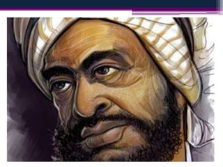
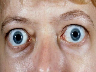
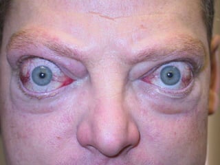
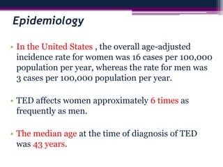
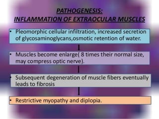
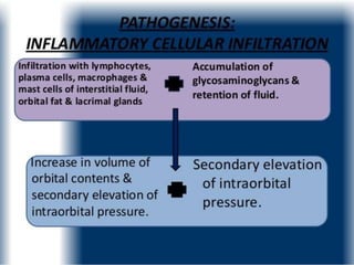
Thyroid eye disease (TED), also known as Graves' ophthalmopathy, is an autoimmune disorder affecting the eyes that is commonly associated with Graves' disease and hyperthyroidism. It causes inflammation and swelling of the muscles and fatty tissues behind the eyes. The document discusses the epidemiology, risk factors, pathogenesis, clinical features including proptosis, lid retraction, optic neuropathy, and restrictive myopathy, as well as treatments such as steroids, radiation, orbital decompression surgery, and eyelid surgery. Management involves treatment of both the eye symptoms and any underlying thyroid abnormalities.


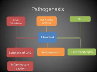
![Potential novel therapeutic targets in
Graves’ ophthalmopathy. [1] Inhibition of T
and B cell activation: costimulation
inhibitors (CTLA4-Ig, alefacept). [2]
Inhibition of B cell maturation,
autoantibody production: rituximab. [3]
Inhibition of autoantibody binding to
insulin-like growth factor-1 receptor (IGF-
1r) and thyroid-stimulating hormone
receptor (TSHr): specific anti-IGF-1r or
TSHr antibodies, inhibitors of IGF-1r
tyrosine kinase, antisense RNA. [4]
Inhibition of adipogenesis: peroxisome
proliferator–activated receptor-
γ antagonists. [5] Decrease inflammation:
(NSAIDs) or anticytokine agents (infliximab,
adalimumab, etanercept, and anakinra).
Pathogenesis of Graves’ Ophthalmopathy: The
Role of Autoantibodies
Teck Kim Khoo and Rebecca S. Bahn
Thyroid. Author manuscript; available in PMC
2014 Jan 25.](https://image.slidesharecdn.com/ted-final-170320165025/85/Thyroid-eye-disease-19-320.jpg)