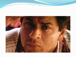
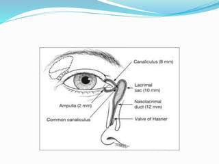
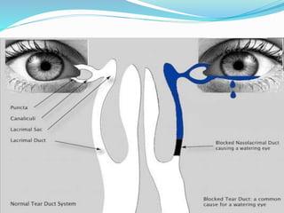
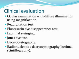
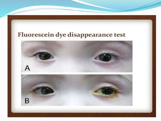
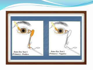
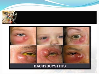
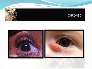
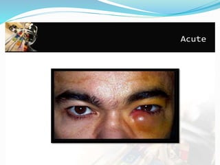
Dr. SOMESH.K.N
watering eye is characterised by
overflow of tears from conjunctival
sac.
Watering eye is mainly due to
hyperlacrimation and epiphora.
Causes of hyperlacrimation
Primary hyperlacrimation
Reflux hyperlacrimation
Central lacrimation
Epiphora(DOWN POUR)
Obstruction to the outflow of
normally secreted tears.
Epiphora may be due to
physiological (lacrimal pump
failure) or anatomical(mechanical
obstruction) cause.
Mechanical obstruction
Punctal causes
Canaliculi
Lacrimal sac
NLD
Clinical evaluation
 Ocular examination with diffuse illumination
using magnifiaction.
 Regurgitation test.
 Fluorescein dye disappearance test.
 Lacrimal syringing.
 Jones dye test.
 Dacryocystography.
 Radionucleotide dacryocystography(lacrimal
scintillography).
Dacryocystitis
 Dacryocystitis is an infection of the lacrimal sac
secondary to obstruction of the nasolacrimal
duct at the junction of lacrimal sac.
 Dacryon= tear, cyst= sac
 Simply dacryocystitis is an inflammation of
lacrimal sac.
 Dacryocystitis may be congenital or
acquired.
Congenital dacryocystitis
 Inflammation of lacrimal sac occuring in new
born infants( dacryocystitis neonatorum)
 Etiology
congenital blockage in NLD
 Clinical picture
1.epiphora
2.positive regurgitation test
3.swelling
Differential diagnosis
 It should be differentiated from ophthalmia
neonatorum and congenital glaucoma
Complications
 Recurrent conjunctivitis
 Lacrimal abscess
 Fistulae formation
Treatment
 Massage over lacrimal sac area and topical
antibiotics in first month(3-4 weeks of age).
 Lacrimal syringing (if not cured up to 2months
of age)
 Probing of NLD with Bowmans probe(if not
cured up to 3-4months)
 Balloon catheter dilatation(if probing fails)
 Intubation with silicone tube(If probings and
catheter dilation fails)
 DCR operation(final approach at the age of 4)
Acquired dacryocystitis
 It may be acute or chronic
Chronic dacryocystitis:
Etiology
 Etiology is of multifactorial.
 Vicious cycle of stasis and mild infection of long
duration
 Predisposing factors.
 Factors responsible for stasis of tears in lacrimal
sac(anatomical factors,foreign body,inflammation of
lacrimal sac, obstruction of NLD)
Clinical picture
1.chronic catarrhal dacryocystitis
 Only symptom is watering eye
 DCG reveals block in NLD, normal sized lacrimal sac
with healthy mucosa.
2.Lacrimal mucocoele
 Distension of lacrimal sac.
 Characterised by constant epiphora associated with
swelling below inner canthus.
 Milky or gelatinous fluid from lower punctum on
pressing swelling.
 DCG reveals distended sac with blockage in NLD
 Some times due to chronic infection, opening of both
canaliculi into sac are blocked leading to negative
regurgitation test. This is called encysted mucocele.
3.Chronic suppurative dacryocystitis
 Due to pyogenic infection, the mucoid discharge becomes
purulent, converting it into pyocoele
 Characterised by epiphora, associated recurrent
conjunctivitis and swelling at the inner canthus with mild
erythema of overlying skin.
 On regurgitation test, frank purulent discharge flows from
lower punctum. If openings of canaliculi are blocked then
encysted pyocole.
4.Chronic fibrotic sac.
 Low grade repeated infections for a
prolonged period ultimately result in a small
fibrotic sac due to thickening of mucosa,
which is often associated with persistant
epiphora and discharge.
 DCG at this stage reveals very small sac with
irregular folds in mucosa.
Treatment
 Conservative treatment with repeated lacrimal
syringing.
 Balloon catheter dilatation(balloon dacryocystoplasty)
 DCR
 DCT
 Conjunctivo dacryocystorhinostomy
Acute dacryocystitis
 It is an acute suppurative inflammation
of lacrimal sac, characterised by
presence of a painful swelling in region
of sac.
 Clinical picture
stage of cellulitis
stage of lacrimal abscess
stage of fistula formation
Treatment
 During cellulitis stage
 During stage of lacrimal abscess
 Treatment of external lacrimal fistula
Surgical technique of DCR:
1.Conventional external approach DCR
2.Endonasal DCR
3.Endocanalicular laser DCR
Watering eye
Watering eye
Watering eye

Watering eye

  • 1.
  • 3.
    watering eye ischaracterised by overflow of tears from conjunctival sac. Watering eye is mainly due to hyperlacrimation and epiphora.
  • 5.
    Causes of hyperlacrimation Primaryhyperlacrimation Reflux hyperlacrimation Central lacrimation
  • 6.
    Epiphora(DOWN POUR) Obstruction tothe outflow of normally secreted tears. Epiphora may be due to physiological (lacrimal pump failure) or anatomical(mechanical obstruction) cause.
  • 7.
  • 9.
    Clinical evaluation  Ocularexamination with diffuse illumination using magnifiaction.  Regurgitation test.  Fluorescein dye disappearance test.  Lacrimal syringing.  Jones dye test.  Dacryocystography.  Radionucleotide dacryocystography(lacrimal scintillography).
  • 14.
    Dacryocystitis  Dacryocystitis isan infection of the lacrimal sac secondary to obstruction of the nasolacrimal duct at the junction of lacrimal sac.  Dacryon= tear, cyst= sac  Simply dacryocystitis is an inflammation of lacrimal sac.  Dacryocystitis may be congenital or acquired.
  • 15.
    Congenital dacryocystitis  Inflammationof lacrimal sac occuring in new born infants( dacryocystitis neonatorum)  Etiology congenital blockage in NLD  Clinical picture 1.epiphora 2.positive regurgitation test 3.swelling
  • 17.
    Differential diagnosis  Itshould be differentiated from ophthalmia neonatorum and congenital glaucoma Complications  Recurrent conjunctivitis  Lacrimal abscess  Fistulae formation
  • 18.
    Treatment  Massage overlacrimal sac area and topical antibiotics in first month(3-4 weeks of age).  Lacrimal syringing (if not cured up to 2months of age)  Probing of NLD with Bowmans probe(if not cured up to 3-4months)  Balloon catheter dilatation(if probing fails)  Intubation with silicone tube(If probings and catheter dilation fails)  DCR operation(final approach at the age of 4)
  • 19.
    Acquired dacryocystitis  Itmay be acute or chronic Chronic dacryocystitis: Etiology  Etiology is of multifactorial.  Vicious cycle of stasis and mild infection of long duration  Predisposing factors.  Factors responsible for stasis of tears in lacrimal sac(anatomical factors,foreign body,inflammation of lacrimal sac, obstruction of NLD)
  • 20.
    Clinical picture 1.chronic catarrhaldacryocystitis  Only symptom is watering eye  DCG reveals block in NLD, normal sized lacrimal sac with healthy mucosa. 2.Lacrimal mucocoele  Distension of lacrimal sac.  Characterised by constant epiphora associated with swelling below inner canthus.  Milky or gelatinous fluid from lower punctum on pressing swelling.  DCG reveals distended sac with blockage in NLD
  • 21.
     Some timesdue to chronic infection, opening of both canaliculi into sac are blocked leading to negative regurgitation test. This is called encysted mucocele. 3.Chronic suppurative dacryocystitis  Due to pyogenic infection, the mucoid discharge becomes purulent, converting it into pyocoele  Characterised by epiphora, associated recurrent conjunctivitis and swelling at the inner canthus with mild erythema of overlying skin.  On regurgitation test, frank purulent discharge flows from lower punctum. If openings of canaliculi are blocked then encysted pyocole.
  • 23.
    4.Chronic fibrotic sac. Low grade repeated infections for a prolonged period ultimately result in a small fibrotic sac due to thickening of mucosa, which is often associated with persistant epiphora and discharge.  DCG at this stage reveals very small sac with irregular folds in mucosa.
  • 24.
    Treatment  Conservative treatmentwith repeated lacrimal syringing.  Balloon catheter dilatation(balloon dacryocystoplasty)  DCR  DCT  Conjunctivo dacryocystorhinostomy
  • 25.
    Acute dacryocystitis  Itis an acute suppurative inflammation of lacrimal sac, characterised by presence of a painful swelling in region of sac.  Clinical picture stage of cellulitis stage of lacrimal abscess stage of fistula formation
  • 27.
    Treatment  During cellulitisstage  During stage of lacrimal abscess  Treatment of external lacrimal fistula Surgical technique of DCR: 1.Conventional external approach DCR 2.Endonasal DCR 3.Endocanalicular laser DCR