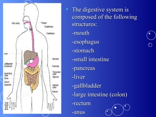
Dietary Management of GI Diseases Jinky Harvey and Mah Asombang
Topics  Diarrhea Atonic constipation Spastic constipation Acute gastritis Chronic gastritis Peptic Ulcer Gastric Ulcer
The Digestive system
The digestive system is composed of the following structures: -mouth -esophagus -stomach -small intestine -pancreas -liver -gallbladder -large intestine (colon) -rectum -anus
What is the function of the digestive system? Digestion begins in the mouth, which grinds the food into smaller particles that are easier to digest. The enzyme, amylase (ptyalin) is  produced by the salivary glands. This enzyme is responsible for the digestion of carbohydrates. The esophagus enables the food to get from the mouth to the stomach via peristalsis
What is the function of the digestive system? Once the stomach receives the food, the mechanoreceptors/strecth receptors (via vagovagal reflex)  stimulate the parietal cells  to secrete hydrochloric acid.  This in turn, enables the chief cells to begin the digestion of proteins via  pepsin.
What is the function of the digestive system? In response to acid entering from the stomach secretin is released from the duodenum. Secretin stimulates the pancreas to secrete bicarbonate. This neutralizes the stomach acid entering the duodenum.  Cholecystokinin(CCK) is also released from the duodenum in response to the partially digested food (i.e. fats, amino acids, peptides)
What is the function of the digestive system? CCK also stimulates the pancreas to release enzymes, such as; amylases, lipases, proteases. This enzyme is also responsible for gallbladder contraction and sphincter of Oddi relaxation. Bile salts are actively secreted by the liver. Once they become concentrated, they form micelles. These water-soluble spheres with a lipid-soluble interior, providing a vehicle to transport lipid-soluble materials in the aqueous medium of the bile fluid and the small intestine.
What is the function of the digestive system? The small intestine functions to reabsorb water and electrolytes with the help of the microvilli on the surface epithelial cells. Most of the water and electrolytes must be reabsorbed in the small intestine, or the colon becomes overwhelmed. Although, the colon may reabsorb water and sodium chloride. It mainly serves as a storage site.
What is the function of the digestive system? The high-amplitude propagated contractions (HAPCS) transports the chyme through the colon.  After meal ingestion, the colonic contractility increases , the initial phase is mediated by the vagus nerve and hormones  in response to mechanical distension of the stomach.
What is the function of the digestive system? Distention of the rectum results in transient relaxation of the internal anal sphincter via intrinsic and reflex sympathetic innervation. As sigmoid and rectal contractions increase the pressure with in the recutm,  the rectosigmoid angle opens by 15 degrees. Voluntary relaxation of the external anal sphincter permits the evacuation of feces in response to the sensation produced by distention.
Diarrhea the passage of abnormally liquid or unformed stools at an increased frequency. The 24-hour stool weighs less than 250g and contains about 150ml of water. When daily stool weight exceeds 200g (approx. 250 ml water).
Four basic pathophysiologic causes of diarrhea Increased secretion of electrolytes and water into the bowel lumen Increased osmotic load within the intestine, leading to water retention in the bowel lumen Inflammation leading to exudation of protein and fluid from the intestinal mucosa Altered intestinal motility leading to rapid transit times
Classifications of diarrhea: organic diarrhea functional diarrhea
Functional diarrhea less severe than organic diarrhea May occur in people who are exposed to irritants compromising the function  of the intestine
Major causes of functinoal diarrhea Overeating Eating the wrong foods Fermentation caused by incomplete digestion of starch Putrefaction in the intestinal tract Overeating Eating the wrong foods Fermentation caused by incomplete digestion of starch Putrefaction in the intestinal tract
Organic diarrhea
Organic diarrhea External poisons Amoebic dysentery Bacillary dysentery or shigellosis Enzyme deficiency (i.e. lactose intolerance) External poisons Amoebic dysentery Bacillary dysentery or shigellosis Enzyme deficiency (i.e. lactose intolerance)
Dietary management Fluid replacement such as; fruit juices that are high in potassium, oral rehydrating solution(ORS), in the absence of Oresol, a solution may be prepared at home using: ¼ tsp crude rock salt + 1 tbsp sugar Dissolved in 1 glass of boiled water or tea
Dietary management cont’d The individual should return to their normal diet gradually. Initially only fruit and vegetable juices should be consumed. Then creamed vegetable soup , and lastly selected whole cooked foods.
Dietary management cont’d If the fluid and electorlyte loss is critical, intravenous fluids may be given. Simple foods may be given such as; broth, gruel, dry toast and tea. Powdered milk or carbohydrates like glucose and lactose may be added to beverages. Emulsified fats like butter and cream may be added to foods as tolerated.
What is constipation? Constipation is a common complaint in clinical practice and usually refers to persistent, difficult, infrequent, or seemingly incomplete defecation. Because of the wide range of normal bowel habits, constipation is difficult to define precisely. Most persons have at least three bowel movements per week; however, low stool frequency alone is not the sole criterion for the diagnosis of constipation. Many constipated patients have a normal frequency of defecation but complain of excessive straining, hard stools, lower abdominal fullness, or a sense of incomplete evacuation
Constipation Constipation occurs when the colon absorbs too much water or if the colon’s muscle contractions are slow or sluggish, causing the stool to move through the colon too slowly. As a result, stools can become hard and dry.
Classifications of constipation Atonic Constipation Spastic Constipation
Atonic constipation also known as “lazy  colon” a lack of normal muscle tone or strength in the colon.
Who is affected? Elderly people or individuals who lack physical activity. Obese individuals. Individuals recovering from a surgical operation. During pregnancy or following childbirth. Individuals with a diet low in fiber and high in fats
Most Important causes Inadequate diet Irregular meals Insufficient liquids and fibers Failure to establish a regular time for defecation
Dietary Management High-fiber diet Regularity of habit through bowel training program Eating regular meals Adequate fluids exercise
Dietary Management
Spastic constipation Also known as irritable colon syndrome. it is secondary to the overstimulationof the intestinal nerve endings resulting in the irregular contractions of the bowel.
Manifestations Uncoordinated sigmoidal mobility Loss of rectal sensibility Abdominal pain Belching Heartburn Heartburn Flatulence  Headache Palpitation nervousness
Management A low fiber diet in order to avoid irritation of the mucous membrane of the intestinal tract.  Diet should consist of non-irritating foods, such as; milk, eggs, refined bread and cereals, butter, oil, finely ground meat, fish, poultry . Vitamin supplements are also recommended during the period of a long restricted diet.
GASTRITIS A term used to describe a group of condition with one thing in common: inflammation of the stomach lining Results from infection w/same bacterium that causes most stomach ulcers Other factors include traumatic injury & regular use of certain pain relievers, drinking too much alcohol can contribute to gastritis
Classification  Acute gastritis:  is an inflammation of the gastric mucosa, occurs suddenly and sometimes violent in onset.  Chronic gastritis:  slowly develop over time and it causes dull pain, feeling of fullness and loss of appetite.
Normal Increased Attack   Hyperacidity Weak defense   Helicobacter pylori   Stress, drugs, smoking
Signs and Symptoms Gnawing or burning pain in upper abdomen  Loss of appetite Bloating Belching Nausea Vomiting  Feeling of fullness after eating a meal Weight loss
Acute Gastritis Acute mucosal inflammatory process of a transient nature Attacks follow after dietary indiscretions: Overeating Eating too quickly Eating while tired or stressed Specific food Other factors: Spoiled foods containing staphylococci Drugs (salicylates, ammonium chloride)
Management Dietary Management Initial Treatment Remove offending substance ASAP by emptying the stomach: induced vomiting and lavage or both NPO for 24-48hrs Initiate IV fluids After fasting period: Low fiber liquid diet can be given as tolerated Milk, toast, cereal and cream soup are fed at an interval of an hour Avoid highly seasoned and spicy foods
Management Cont’d The amount of food and the number of feedings are increased according to the patient’s tolerance until eating a full regular diet Therapeutic diet: soft diet
Chronic Gastritis Presence of chronic mucosal inflammatory changes such as erosions, ulcerations, changes  in the blood vessels, and the destruction of the surface cells Same dietary indiscretions as acute gastritis Illness precedes development of organic gastritis lesions Chief manifestation is pain
Chronic Gastritis Dietary Management Provide adequate calories & nutrients Start patient on soft diet Avoid highly seasoned foods Excess liquids during meals is avoided to reduce discomfort Frequent small meals are recommended  Anti-acid therapy Therapeutic Diet: Soft diet
Peptic Ulcer Disease (PUD)  One of the most common diseases affecting the GIT It causes inflammatory injuries in the gastric or duodenal mucosa with an extension to submucosa and into mucularis mucosa Acidic environment is believed to be the principal cause of PUD
Pathophysiology The normal stomach maintains a balance between protective factors, such as mucus secreted by the glands from the lower esophagus  to the upper duodenum and sodium bicarbonate from the pancreas to neutralize HCL for the protection from its strong acidic environment
Pathophysiology In alkaline environment, pepsin is inactivated and cannot digest the duodenal mucosa Gastric ulcers develop when aggressive factors overcome protective mechanisms
Pathophysiology The two major etiological factors for PUD are  Helicobacter pylori  infection and  nonsteroidal anti-inflammatory drug (NSAID)  consumption. Currently, 70% of all gastric ulcers occurring in the United States can be attributed to H pylori infection. Sanjeeb Shrestha, MD,  Consulting Staff, Division of Gastroenterology, North West Arkansas Gastroenterology Clinic Daryl Lau, MD, MPH, FRCP(C),  Director of Translational Liver Research, Beth Israel Deaconess Medical Center; Associate Professor of Medicine, Harvard Medical School Updated: Aug 24, 2009
Pathophysiology Cigarette smoking can affect gastric mucosal defense adversely. Cigarette smoking is believed to play a facultative role in  H pylori   infection. People who smoke tend to develop more frequent and recurrent ulcers and their ulcers are more resistant to therapy
PUD Most common portion of the GIT affected by PUD: First portion of the duodenum  The antrum of the stomach  The gastroesophageal junction  Ulcer is in the duodenum called duodenal ulcer (DU) Ulcer is found in the stomach called gastric ulcer (GU)
 
H. Pylori A cytotoxin-associated gene ( cag A ) has been isolated in approximately 65% of the bacteria. The products of this gene are associated with more severe gastritis, gastric ulcer, gastric cancer, and lymphoma. 
H. Pylori H pylori  infection also predisposes patients to ulcer disease by disrupting mucosal integrity. The bacterium's spiral shape and flagella facilitate its penetration into the mucous layer and its attachment to the epithelial layer. Subsequently, it releases phospholipase and proteases, which cause further mucosal damage.
Duodenal Ulcer More common than gastric ulcer Usually appear within 3 cm of the pylorus an area where gastric acidity is high  More prevalent in male than in female Usually affect people who are tense, hardworking, and chronic worriers
Duodenal Ulcer Clinical feature: an intense burning epigastric pain 90 minutes to 3 hours after a meal  Aggravated at night and is usually relieved by food  Diagnosis of DU is done by endoscopy or upper GI barium radiography  Increased acid load to the duodenum leads to the development of DU
Gastric Ulcer Majority of gastric ulcers are situated in the antrum or the lesser curvature of the stomach  H. pylori is the primary cause for GU  Gastric acid secretory rates with GU are normal or even decreased  Gastritis and chronic use of salicylate or NSAIDs are other possible causes of GU
 
Predisposing Factors poor dietary habits  excessive smoking  drinking of caffeine (coffee and sodas) and alcohol  Rush through meals  irregular mealtimes Hereditary  Physical and psychological stresses
Objectives Relief pain  healing of the ulcer  prevention of complications and recurrences  Suppression and eradication of H. pylori is indicated for PUD  done by the use of triple or quadruple drug therapy
Medical Treatment antacids can be used to neutralize the overload of acids (Tums, Maalox) Acid secretion may be subdued by the use of anticholinergics like Histamine 2 receptor blockers (cimetidine, ranitidine, and famotidine) w/c block the H2 receptor in the parietal cells Mucosal protectant: Bismuth or Sucralfate can also be effective in healing gastric ulcer
Medical Treatment Proton Pump Inhibitors (PPI): irreversibly inhibit the H/K  ATPase pump effectively inhibiting acid release (omeprazole, lansoprazole, esomeprazole) H. Pylori eradication: triple therapy has consistently been shown to eradicate the organism more than 90% of the time.
Drug therapy Bismuth Subsalicylate (2 tablets QID), plus Metronidazole (250 mg tablet QID) plus tetracycline (500 mg tablet QID).  omeprazole (20 mg tablet OD), bismuth Subsalicylate (2 tablets QID), Metronidazole (250 mg tablet QID) and tetracycline (500 mg tablet QID)
Drug therapy Bismuth, metronidazole, and tetracycline qid with H2 blockers bid Bismuth, metronidazole, and tetracycline bid with a PPI (Helidac) Prevacid, amoxicillin, and clarithromycin bid (PrevPac) Prilosec, metronidazole, and clarithromycin bid Ranitidine, bismuth, and clarithromycin with amoxicillin, metronidazole, or tetracycline bid   The 5 different regimens approved by AmericanCollege of Gastroenterology are as follows (all 5 regimens are given for a total of 2 wk):
Dietary Management The initial dietary management is to consume adequate calories to maintain the desirable body weight of the patient  High protein  To promote healing To buffer acids To replace nitrogen lost from the ulcer
Dietary Management Adequate carbohydrates  High amounts of unsaturated fats To inhibit gastric secretion and motility via Cholecystokinin Small frequent meals:  To rest the organ To maintain constant neutralization of the acid To minimize distention of the stomach, thus minimizing gastrin and HCl secretion
Dietary Management Limit fiber consumption To reduce motility Limit gastric secretagogues such as caffeine and alcohol  Limit Gas-forming foods like cabbage, baked beans, milk, onions, fried foods, spicy foods and orange juice  Avoidance of NSAIDs like salicylates and other drugs known to affect GIT mucosa
References Principles of Diet Therapy Manual http://digestive.niddk.nih.gov/ddiseases/pubs/constipation/ Sanjeeb Shrestha, MD,  Consulting Staff, Division of Gastroenterology, North West Arkansas Gastroenterology Clinic Daryl Lau, MD, MPH, FRCP(C),  Director of Translational Liver Research, Beth Israel Deaconess Medical Center; Associate Professor of Medicine, Harvard Medical School, Updated: Aug 24, 2009
References Soll AH. Consensus conference. Medical treatment of peptic ulcer disease. Practice guidelines. Practice Parameters Committee of the American College of Gastroenterology.  JAMA . Feb 28 1996;275(8):622-9.  [Medline] . Vaira D, Gatta L, Ricci C, et al. Peptic ulcer and Helicobacter pylori: update on testing and treatment.  Postgrad Med . Jun 2005;117(6):17-22, 46.  [Medline]
Thank You!

Git diseases presentn no. 2 applied nutrition

  • 1.
    Dietary Management ofGI Diseases Jinky Harvey and Mah Asombang
  • 2.
    Topics DiarrheaAtonic constipation Spastic constipation Acute gastritis Chronic gastritis Peptic Ulcer Gastric Ulcer
  • 3.
  • 4.
    The digestive systemis composed of the following structures: -mouth -esophagus -stomach -small intestine -pancreas -liver -gallbladder -large intestine (colon) -rectum -anus
  • 5.
    What is thefunction of the digestive system? Digestion begins in the mouth, which grinds the food into smaller particles that are easier to digest. The enzyme, amylase (ptyalin) is produced by the salivary glands. This enzyme is responsible for the digestion of carbohydrates. The esophagus enables the food to get from the mouth to the stomach via peristalsis
  • 6.
    What is thefunction of the digestive system? Once the stomach receives the food, the mechanoreceptors/strecth receptors (via vagovagal reflex) stimulate the parietal cells to secrete hydrochloric acid. This in turn, enables the chief cells to begin the digestion of proteins via pepsin.
  • 7.
    What is thefunction of the digestive system? In response to acid entering from the stomach secretin is released from the duodenum. Secretin stimulates the pancreas to secrete bicarbonate. This neutralizes the stomach acid entering the duodenum. Cholecystokinin(CCK) is also released from the duodenum in response to the partially digested food (i.e. fats, amino acids, peptides)
  • 8.
    What is thefunction of the digestive system? CCK also stimulates the pancreas to release enzymes, such as; amylases, lipases, proteases. This enzyme is also responsible for gallbladder contraction and sphincter of Oddi relaxation. Bile salts are actively secreted by the liver. Once they become concentrated, they form micelles. These water-soluble spheres with a lipid-soluble interior, providing a vehicle to transport lipid-soluble materials in the aqueous medium of the bile fluid and the small intestine.
  • 9.
    What is thefunction of the digestive system? The small intestine functions to reabsorb water and electrolytes with the help of the microvilli on the surface epithelial cells. Most of the water and electrolytes must be reabsorbed in the small intestine, or the colon becomes overwhelmed. Although, the colon may reabsorb water and sodium chloride. It mainly serves as a storage site.
  • 10.
    What is thefunction of the digestive system? The high-amplitude propagated contractions (HAPCS) transports the chyme through the colon. After meal ingestion, the colonic contractility increases , the initial phase is mediated by the vagus nerve and hormones in response to mechanical distension of the stomach.
  • 11.
    What is thefunction of the digestive system? Distention of the rectum results in transient relaxation of the internal anal sphincter via intrinsic and reflex sympathetic innervation. As sigmoid and rectal contractions increase the pressure with in the recutm, the rectosigmoid angle opens by 15 degrees. Voluntary relaxation of the external anal sphincter permits the evacuation of feces in response to the sensation produced by distention.
  • 12.
    Diarrhea the passageof abnormally liquid or unformed stools at an increased frequency. The 24-hour stool weighs less than 250g and contains about 150ml of water. When daily stool weight exceeds 200g (approx. 250 ml water).
  • 13.
    Four basic pathophysiologiccauses of diarrhea Increased secretion of electrolytes and water into the bowel lumen Increased osmotic load within the intestine, leading to water retention in the bowel lumen Inflammation leading to exudation of protein and fluid from the intestinal mucosa Altered intestinal motility leading to rapid transit times
  • 14.
    Classifications of diarrhea:organic diarrhea functional diarrhea
  • 15.
    Functional diarrhea lesssevere than organic diarrhea May occur in people who are exposed to irritants compromising the function of the intestine
  • 16.
    Major causes offunctinoal diarrhea Overeating Eating the wrong foods Fermentation caused by incomplete digestion of starch Putrefaction in the intestinal tract Overeating Eating the wrong foods Fermentation caused by incomplete digestion of starch Putrefaction in the intestinal tract
  • 17.
  • 18.
    Organic diarrhea Externalpoisons Amoebic dysentery Bacillary dysentery or shigellosis Enzyme deficiency (i.e. lactose intolerance) External poisons Amoebic dysentery Bacillary dysentery or shigellosis Enzyme deficiency (i.e. lactose intolerance)
  • 19.
    Dietary management Fluidreplacement such as; fruit juices that are high in potassium, oral rehydrating solution(ORS), in the absence of Oresol, a solution may be prepared at home using: ¼ tsp crude rock salt + 1 tbsp sugar Dissolved in 1 glass of boiled water or tea
  • 20.
    Dietary management cont’dThe individual should return to their normal diet gradually. Initially only fruit and vegetable juices should be consumed. Then creamed vegetable soup , and lastly selected whole cooked foods.
  • 21.
    Dietary management cont’dIf the fluid and electorlyte loss is critical, intravenous fluids may be given. Simple foods may be given such as; broth, gruel, dry toast and tea. Powdered milk or carbohydrates like glucose and lactose may be added to beverages. Emulsified fats like butter and cream may be added to foods as tolerated.
  • 22.
    What is constipation?Constipation is a common complaint in clinical practice and usually refers to persistent, difficult, infrequent, or seemingly incomplete defecation. Because of the wide range of normal bowel habits, constipation is difficult to define precisely. Most persons have at least three bowel movements per week; however, low stool frequency alone is not the sole criterion for the diagnosis of constipation. Many constipated patients have a normal frequency of defecation but complain of excessive straining, hard stools, lower abdominal fullness, or a sense of incomplete evacuation
  • 23.
    Constipation Constipation occurswhen the colon absorbs too much water or if the colon’s muscle contractions are slow or sluggish, causing the stool to move through the colon too slowly. As a result, stools can become hard and dry.
  • 24.
    Classifications of constipationAtonic Constipation Spastic Constipation
  • 25.
    Atonic constipation alsoknown as “lazy colon” a lack of normal muscle tone or strength in the colon.
  • 26.
    Who is affected?Elderly people or individuals who lack physical activity. Obese individuals. Individuals recovering from a surgical operation. During pregnancy or following childbirth. Individuals with a diet low in fiber and high in fats
  • 27.
    Most Important causesInadequate diet Irregular meals Insufficient liquids and fibers Failure to establish a regular time for defecation
  • 28.
    Dietary Management High-fiberdiet Regularity of habit through bowel training program Eating regular meals Adequate fluids exercise
  • 29.
  • 30.
    Spastic constipation Alsoknown as irritable colon syndrome. it is secondary to the overstimulationof the intestinal nerve endings resulting in the irregular contractions of the bowel.
  • 31.
    Manifestations Uncoordinated sigmoidalmobility Loss of rectal sensibility Abdominal pain Belching Heartburn Heartburn Flatulence Headache Palpitation nervousness
  • 32.
    Management A lowfiber diet in order to avoid irritation of the mucous membrane of the intestinal tract. Diet should consist of non-irritating foods, such as; milk, eggs, refined bread and cereals, butter, oil, finely ground meat, fish, poultry . Vitamin supplements are also recommended during the period of a long restricted diet.
  • 33.
    GASTRITIS A termused to describe a group of condition with one thing in common: inflammation of the stomach lining Results from infection w/same bacterium that causes most stomach ulcers Other factors include traumatic injury & regular use of certain pain relievers, drinking too much alcohol can contribute to gastritis
  • 34.
    Classification Acutegastritis: is an inflammation of the gastric mucosa, occurs suddenly and sometimes violent in onset. Chronic gastritis: slowly develop over time and it causes dull pain, feeling of fullness and loss of appetite.
  • 35.
    Normal Increased Attack Hyperacidity Weak defense Helicobacter pylori Stress, drugs, smoking
  • 36.
    Signs and SymptomsGnawing or burning pain in upper abdomen Loss of appetite Bloating Belching Nausea Vomiting Feeling of fullness after eating a meal Weight loss
  • 37.
    Acute Gastritis Acutemucosal inflammatory process of a transient nature Attacks follow after dietary indiscretions: Overeating Eating too quickly Eating while tired or stressed Specific food Other factors: Spoiled foods containing staphylococci Drugs (salicylates, ammonium chloride)
  • 38.
    Management Dietary ManagementInitial Treatment Remove offending substance ASAP by emptying the stomach: induced vomiting and lavage or both NPO for 24-48hrs Initiate IV fluids After fasting period: Low fiber liquid diet can be given as tolerated Milk, toast, cereal and cream soup are fed at an interval of an hour Avoid highly seasoned and spicy foods
  • 39.
    Management Cont’d Theamount of food and the number of feedings are increased according to the patient’s tolerance until eating a full regular diet Therapeutic diet: soft diet
  • 40.
    Chronic Gastritis Presenceof chronic mucosal inflammatory changes such as erosions, ulcerations, changes in the blood vessels, and the destruction of the surface cells Same dietary indiscretions as acute gastritis Illness precedes development of organic gastritis lesions Chief manifestation is pain
  • 41.
    Chronic Gastritis DietaryManagement Provide adequate calories & nutrients Start patient on soft diet Avoid highly seasoned foods Excess liquids during meals is avoided to reduce discomfort Frequent small meals are recommended Anti-acid therapy Therapeutic Diet: Soft diet
  • 42.
    Peptic Ulcer Disease(PUD) One of the most common diseases affecting the GIT It causes inflammatory injuries in the gastric or duodenal mucosa with an extension to submucosa and into mucularis mucosa Acidic environment is believed to be the principal cause of PUD
  • 43.
    Pathophysiology The normalstomach maintains a balance between protective factors, such as mucus secreted by the glands from the lower esophagus to the upper duodenum and sodium bicarbonate from the pancreas to neutralize HCL for the protection from its strong acidic environment
  • 44.
    Pathophysiology In alkalineenvironment, pepsin is inactivated and cannot digest the duodenal mucosa Gastric ulcers develop when aggressive factors overcome protective mechanisms
  • 45.
    Pathophysiology The twomajor etiological factors for PUD are Helicobacter pylori infection and nonsteroidal anti-inflammatory drug (NSAID) consumption. Currently, 70% of all gastric ulcers occurring in the United States can be attributed to H pylori infection. Sanjeeb Shrestha, MD, Consulting Staff, Division of Gastroenterology, North West Arkansas Gastroenterology Clinic Daryl Lau, MD, MPH, FRCP(C), Director of Translational Liver Research, Beth Israel Deaconess Medical Center; Associate Professor of Medicine, Harvard Medical School Updated: Aug 24, 2009
  • 46.
    Pathophysiology Cigarette smokingcan affect gastric mucosal defense adversely. Cigarette smoking is believed to play a facultative role in H pylori infection. People who smoke tend to develop more frequent and recurrent ulcers and their ulcers are more resistant to therapy
  • 47.
    PUD Most commonportion of the GIT affected by PUD: First portion of the duodenum The antrum of the stomach The gastroesophageal junction Ulcer is in the duodenum called duodenal ulcer (DU) Ulcer is found in the stomach called gastric ulcer (GU)
  • 48.
  • 49.
    H. Pylori Acytotoxin-associated gene ( cag A ) has been isolated in approximately 65% of the bacteria. The products of this gene are associated with more severe gastritis, gastric ulcer, gastric cancer, and lymphoma. 
  • 50.
    H. Pylori Hpylori infection also predisposes patients to ulcer disease by disrupting mucosal integrity. The bacterium's spiral shape and flagella facilitate its penetration into the mucous layer and its attachment to the epithelial layer. Subsequently, it releases phospholipase and proteases, which cause further mucosal damage.
  • 51.
    Duodenal Ulcer Morecommon than gastric ulcer Usually appear within 3 cm of the pylorus an area where gastric acidity is high More prevalent in male than in female Usually affect people who are tense, hardworking, and chronic worriers
  • 52.
    Duodenal Ulcer Clinicalfeature: an intense burning epigastric pain 90 minutes to 3 hours after a meal Aggravated at night and is usually relieved by food Diagnosis of DU is done by endoscopy or upper GI barium radiography Increased acid load to the duodenum leads to the development of DU
  • 53.
    Gastric Ulcer Majorityof gastric ulcers are situated in the antrum or the lesser curvature of the stomach H. pylori is the primary cause for GU Gastric acid secretory rates with GU are normal or even decreased Gastritis and chronic use of salicylate or NSAIDs are other possible causes of GU
  • 54.
  • 55.
    Predisposing Factors poordietary habits excessive smoking drinking of caffeine (coffee and sodas) and alcohol Rush through meals irregular mealtimes Hereditary Physical and psychological stresses
  • 56.
    Objectives Relief pain healing of the ulcer prevention of complications and recurrences Suppression and eradication of H. pylori is indicated for PUD done by the use of triple or quadruple drug therapy
  • 57.
    Medical Treatment antacidscan be used to neutralize the overload of acids (Tums, Maalox) Acid secretion may be subdued by the use of anticholinergics like Histamine 2 receptor blockers (cimetidine, ranitidine, and famotidine) w/c block the H2 receptor in the parietal cells Mucosal protectant: Bismuth or Sucralfate can also be effective in healing gastric ulcer
  • 58.
    Medical Treatment ProtonPump Inhibitors (PPI): irreversibly inhibit the H/K ATPase pump effectively inhibiting acid release (omeprazole, lansoprazole, esomeprazole) H. Pylori eradication: triple therapy has consistently been shown to eradicate the organism more than 90% of the time.
  • 59.
    Drug therapy BismuthSubsalicylate (2 tablets QID), plus Metronidazole (250 mg tablet QID) plus tetracycline (500 mg tablet QID). omeprazole (20 mg tablet OD), bismuth Subsalicylate (2 tablets QID), Metronidazole (250 mg tablet QID) and tetracycline (500 mg tablet QID)
  • 60.
    Drug therapy Bismuth,metronidazole, and tetracycline qid with H2 blockers bid Bismuth, metronidazole, and tetracycline bid with a PPI (Helidac) Prevacid, amoxicillin, and clarithromycin bid (PrevPac) Prilosec, metronidazole, and clarithromycin bid Ranitidine, bismuth, and clarithromycin with amoxicillin, metronidazole, or tetracycline bid   The 5 different regimens approved by AmericanCollege of Gastroenterology are as follows (all 5 regimens are given for a total of 2 wk):
  • 61.
    Dietary Management Theinitial dietary management is to consume adequate calories to maintain the desirable body weight of the patient High protein To promote healing To buffer acids To replace nitrogen lost from the ulcer
  • 62.
    Dietary Management Adequatecarbohydrates High amounts of unsaturated fats To inhibit gastric secretion and motility via Cholecystokinin Small frequent meals: To rest the organ To maintain constant neutralization of the acid To minimize distention of the stomach, thus minimizing gastrin and HCl secretion
  • 63.
    Dietary Management Limitfiber consumption To reduce motility Limit gastric secretagogues such as caffeine and alcohol Limit Gas-forming foods like cabbage, baked beans, milk, onions, fried foods, spicy foods and orange juice Avoidance of NSAIDs like salicylates and other drugs known to affect GIT mucosa
  • 64.
    References Principles ofDiet Therapy Manual http://digestive.niddk.nih.gov/ddiseases/pubs/constipation/ Sanjeeb Shrestha, MD, Consulting Staff, Division of Gastroenterology, North West Arkansas Gastroenterology Clinic Daryl Lau, MD, MPH, FRCP(C), Director of Translational Liver Research, Beth Israel Deaconess Medical Center; Associate Professor of Medicine, Harvard Medical School, Updated: Aug 24, 2009
  • 65.
    References Soll AH. Consensusconference. Medical treatment of peptic ulcer disease. Practice guidelines. Practice Parameters Committee of the American College of Gastroenterology.  JAMA . Feb 28 1996;275(8):622-9.  [Medline] . Vaira D, Gatta L, Ricci C, et al. Peptic ulcer and Helicobacter pylori: update on testing and treatment.  Postgrad Med . Jun 2005;117(6):17-22, 46.  [Medline]
  • 66.