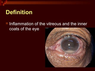
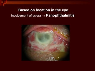
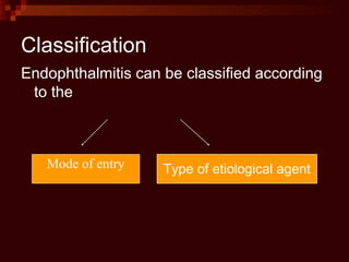
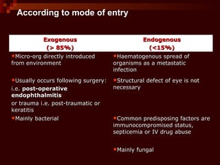
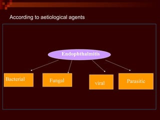
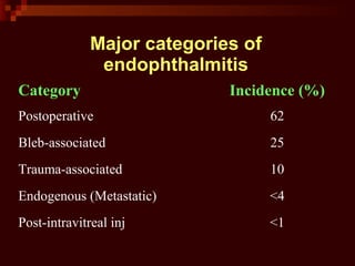
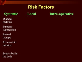
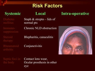
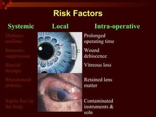
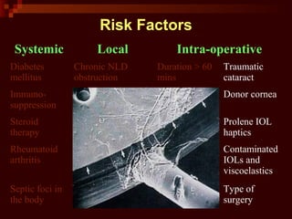
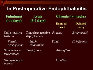
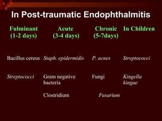
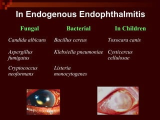
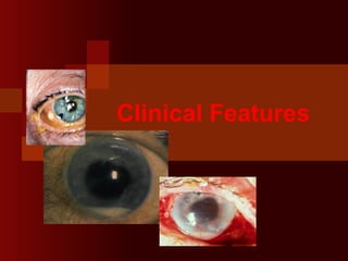
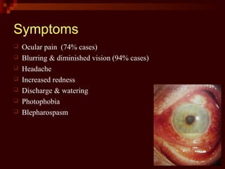
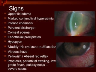
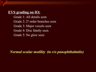
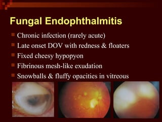
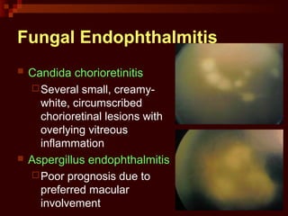
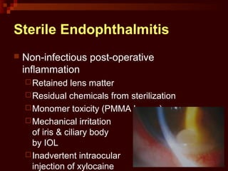
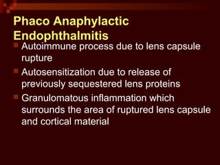
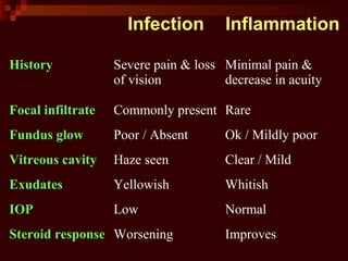
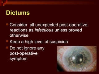
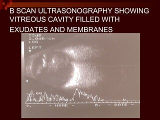
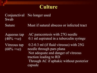
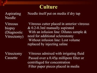
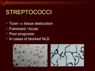
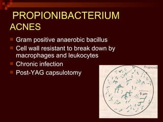
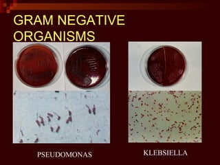
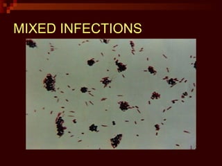
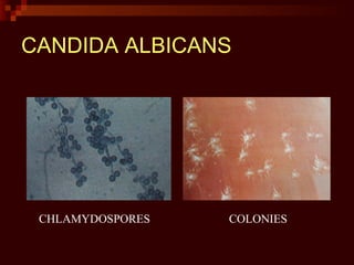
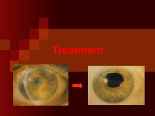
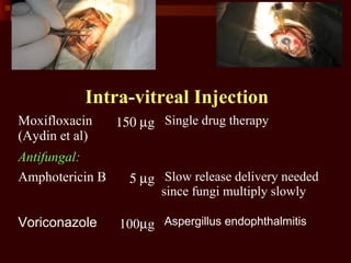
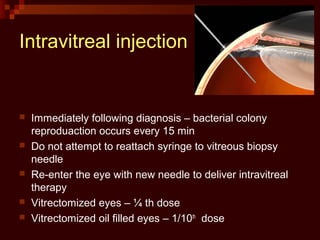
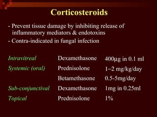
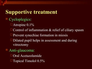
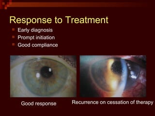
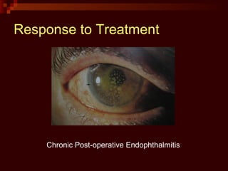
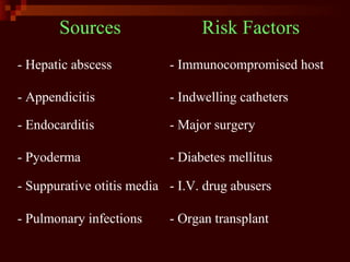
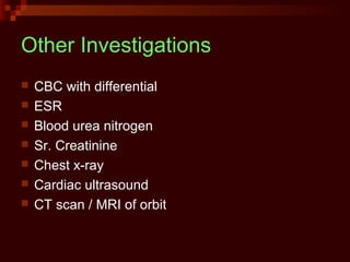
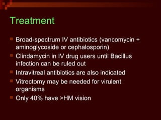
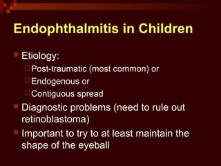
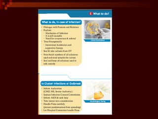
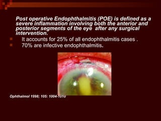
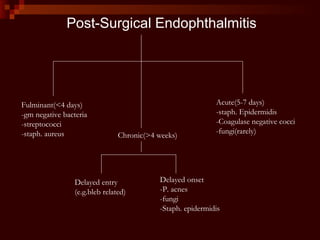
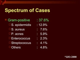
EndophthalmitisEndophthalmitis
Dr.Shah-Noor HassanDr.Shah-Noor Hassan
Vitreo-Retina ConsultantVitreo-Retina Consultant
Bangladesh Eye Hospital & InstituteBangladesh Eye Hospital & Institute
Definition
 Inflammation of the vitreous and the inner
coats of the eye
Based on location in the eye
Involvement of sclera → Panophthalmitis
Classification
Endophthalmitis can be classified according
to the
Mode of entry Type of etiological agent
According to mode of entry
ExogenousExogenous
(> 85%)(> 85%)
EndogenousEndogenous
(<15%)(<15%)
Micro-org directly introducedMicro-org directly introduced
from environmentfrom environment
Haematogenous spread ofHaematogenous spread of
organisms as a metastaticorganisms as a metastatic
infectioninfection
Usually occurs following surgery:Usually occurs following surgery:
i.e.i.e. post-operativepost-operative
endophthalmitisendophthalmitis
or trauma i.e. post-traumatic oror trauma i.e. post-traumatic or
keratitiskeratitis
Structural defect of eye is notStructural defect of eye is not
necessarynecessary
Mainly bacterialMainly bacterial Common predisposing factors areCommon predisposing factors are
immunocompromised status,immunocompromised status,
septicemia or IV drug abusesepticemia or IV drug abuse
Mainly fungalMainly fungal
According to aetiological agents
Bacterial Fungal
viral Parasitic
Endophthalmitis
Exogenous
Endophthalmitis
Major categories of
endophthalmitis
Category Incidence (%)
Postoperative 62
Bleb-associated 25
Trauma-associated 10
Endogenous (Metastatic) <4
Post-intravitreal inj <1
Risk Factors
Systemic Local Intra-operative
Diabetes
mellitus
Immuno-
suppression
Steroid
therapy
Rheumatoid
arthritis
Septic foci in
the body
Risk Factors
Systemic Local Intra-operative
Diabetes
mellitus
Staph & strepto – lids of
normal pts
Immuno-
suppression
Chronic NLD obstruction
Steroid
therapy
Blepharitis, canaculitis
Rheumatoid
arthritis
Conjunctivitis
Septic foci in
the body
Contact lens wear,
Ocular prosthesis in other
eye
Risk Factors
Systemic Local Intra-operative
Diabetes
mellitus
Chronic NLD
obstruction
Prolonged
operating time
Immuno-
suppression
Blepharitis,
canaculitis
Wound
dehiscence
Steroid
therapy
Conjunctivitis Vitreous loss
Rheumatoid
arthritis
Contact lens wear Retained lens
matter
Septic foci in
the body
Ocular prosthesis
in other eye
Contaminated
instruments &
soln
Risk Factors
Systemic Local Intra-operative
Diabetes
mellitus
Chronic NLD
obstruction
Duration > 60
mins
Traumatic
cataract
Immuno-
suppression
Blepharitis,
canaculitis
Poor wound
integrity
Donor cornea
Steroid
therapy
Conjunctivitis Suture
dehiscence
Prolene IOL
haptics
Rheumatoid
arthritis
Contact lens wear Retained lens
matter
Contaminated
IOLs and
viscoelastics
Septic foci in
the body
Ocular prosthesis
in other eye
Use of many
instruments
Type of
surgery
Microbial
Spectrum
Organism Postoperativ
e
(n=63) (%)
Bleb
associated
(n=30) (%)
Trauma
(n=30)
(%)
Staph
epidermidis
38 0 20
S. Aureus 21 7 0
Strepto. Spp 11 57 13
Bacillus spp 0 0 27
Haemophilus
influenzae
3 23 0
Other Gram –ve 13 7 20
Fungi 8 3 17
Other 6 3 3
Mixed flora 2 0 11
In Post-operative Endophthalmitis
Fulminant
(< 4 days)
Acute
(5-7 days)
Chronic (>4 weeks)
Delayed
onset
Delayed
entry
Gram negative
bacteria
Coagulase negative
staphylococci
P. acnes Streptococci
Pseudo.
aeuruginosa
Staph.
epidermidis
Fungi H. influenza
Streptococcus
pneumoniae
Fungi (rare) Aspergillus
Staphylococcus
aureus
Candida
Day of presentation of infection
0
10
20
30
40
50
60
70
80
% infection
1-7 days 8-14 days >15 days >1 month
In most cases, infection occurs in immediate post-op period,
In Post-traumatic Endophthalmitis
Fulminant
(1-2 days)
Acute
(3-4 days)
Chronic
(5-7days)
In Children
Bacillus cereus Staph. epidermidis P. acnes Streptococci
Streptococci Gram negative
bacteria
Fungi Kingella
kingae
Clostridium Fusarium
In Endogenous Endophthalmitis
Fungal Bacterial In Children
Candida albicans Bacillus cereus Toxocara canis
Aspergillus
fumigatus
Klebsiella pneumoniae Cysticercus
cellulosae
Cryptococcus
neoformans
Listeria
monocytogenes
Clinical Features
 Ocular pain (74% cases)
 Blurring & diminished vision (94% cases)
 Headache
 Increased redness
 Discharge & watering
 Photophobia
 Blepharospasm
Symptoms
Signs
 Upper lid edema
 Marked conjunctival hyperaemia
 Intense chemosis
 Purulent discharge
 Corneal edema
 Endothelial precipitates
 Hypopyon
 Muddy iris resistant to dilatation
 Vitreous haze
 Yellowish / Absent red reflex
 Proptosis, periorbital swelling, low-
grade fever, leukocyotosis –
severe cases
-EVS grading on IO:
-Grade 1: All details seen
-Grade 2: 2nd
order branches seen
-Grade 3: Major vessels seen
-Grade 4: Disc faintly seen
-Grade 5: No glow seen
-Normal ocular motility (to r/o panophthalmitis)
Fungal Endophthalmitis
 Chronic infection (rarely acute)
 Late onset DOV with redness & floaters
 Fixed cheesy hypopyon
 Fibrinous mesh-like exudation
 Snowballs & fluffy opacities in vitreous
Fungal Endophthalmitis
 Candida chorioretinitis
Several small, creamy-
white, circumscribed
chorioretinal lesions with
overlying vitreous
inflammation
 Aspergillus endophthalmitis
Poor prognosis due to
preferred macular
involvement
Differential
Diagnosis
Differential Diagnosis
Post-Surgical
Non-Surgical
 Retained IOFB (iron / copper)
 Severe pars planitis
 Old vitreous hemorrhage
 Toxoplasma / Toxocara infestation
 Necrotic retinoblastoma
Sterile Endophthalmitis
 Non-infectious post-operative
inflammation
Retained lens matter
Residual chemicals from sterilization
Monomer toxicity (PMMA lenses)
Mechanical irritation
of iris & ciliary body
by IOL
Inadvertent intraocular
injection of xylocaine
Phaco Anaphylactic
Endophthalmitis
 Autoimmune process due to lens capsule
rupture
 Autosensitization due to release of
previously sequestered lens proteins
 Granulomatous inflammation which
surrounds the area of ruptured lens capsule
and cortical material
Infection Inflammation
History Severe pain & loss
of vision
Minimal pain &
decrease in acuity
Focal infiltrate Commonly present Rare
Fundus glow Poor / Absent Ok / Mildly poor
Vitreous cavity Haze seen Clear / Mild
Exudates Yellowish Whitish
IOP Low Normal
Steroid response Worsening Improves
Dictums
 Consider all unexpected post-operative
reactions as infectious unless proved
otherwise
 Keep a high level of suspicion
 Do not ignore any
post-operative
symptom
Investigations
USG B-scan
Must be done before
invasive, diagnostic or therapeutic intervention
To look for: Retinal or Choroidal detachment
Dislocated lens / nucleus
Retained Intra-Ocular Foreign Body (IOFB)
Parasite infestation
B SCAN ULTRASONOGRAPHY SHOWING
VITREOUS CAVITY FILLED WITH
EXUDATES AND MEMBRANES
Culture
Conjunctival
Swab
No longer used
Suture Must if sutural abscess or infected tract
Aqueous tap
(40% +ve)
-AC paracentesis with 27G needle
-0.1 ml aspirated in a tuberculin syringe
Vitreous tap
(60% +ve)
-0.2-0.3 ml (if fluid vitreous) with 25G
needle through pars plana
-Not adequate and danger of vitreous
traction leading to RD
-Through AC if aphakic without posterior
capsule
Culture
Aspirating
Needle
Needle itself put on media if dry tap
Vitreous
biopsy
(Diagnostic
Vitrectomy)
-Vitreous cutter placed in anterior vitreous
& 0.2-0.3ml manually aspirated
-With an infusion line: Dilutes sample &
need for additional sclerostomy
-Without infusion line: Lost volume
replaced by injecting saline
Vitrectomy
Cassette
-Vitreous admixed with irrigating fluid
-Passed over a 0.45µ millipore filter or
centrifuged for concentration
-Filter paper pieces placed in media
 Nested PCR – used to detect &
differentiate between species of Gram +ve
& -ve organisms
 Improved sensitivity
 PCR-RFLP has sensitivity of 1 organism
Vitreous Biopsy
Stains Cultures
(kept for at least 2 weeks)
Gram stain Blood agar Bacteria + Fungi
Giemsa stain Chocolate agar (with
CO2
)
Aerobic organisms
KOH mount Sabouraud’s agar Fungi
Air dried & fixed with
absolute methanol
Thioglycolate broth Anaerobic organisms
Centrifugal cytology is
superior but not a must
Brain-heart infusion
media
Fastidious fungi
Culture Media
STAPHYLOCOCCI
 Coagulase -ve cocci
Staph. epidermidis
 Coagulase +ve cocci
Staph. aureus
STREPTOCOCCI
 Toxin ⇒ tissue destruction
 Fulminant / Acute
 Poor prognosis
 In cases of blocked NLD
PROPIONIBACTERIUM
ACNES
 Gram positive anaerobic bacillus
 Cell wall resistant to break down by
macrophages and leukocytes
 Chronic infection
 Post-YAG capsulotomy
GRAM NEGATIVE
ORGANISMS
PSEUDOMONAS KLEBSIELLA
MIXED INFECTIONS
CHLAMYDOSPORES
CANDIDA ALBICANS
COLONIES
Laboratory confirmation of
diagnosis
Laboratory confirmed growth is defined as :
1. At least semi confluent growth on solid media
2. Any growth on more than or equal to 2 media
3. Growth on one media supported by a positive
gram stain
www.aios.org
No culture should be considered negative until two
weeks of observation
Treatment
Primary Objectives
1. Control and eradicate infection
2. Manage complications
3. Restore vision
Secondary Objectives
1. Symptomatic relief
2. Remove pupillary membrane in miosis
3. Maintain globe integrity
Determinants of Outcome
 Duration between onset & treatment
(Poor if >48hrs)
 Virulence & organism load
(Poor if Gram negative bacteria)
 Pharmacokinetics & Spectrum of activity of
antibiotics used
Treatment Modalities
 Antimicrobial treatment
 Corticosteroids
 Supportive treatment
 Vitrectomy
Intra-vitreal Injection
Gram-positive organisms:
Vancomycin 1 mg -Effective against MRSA
-Precipitates with ceftazidime
Gram-negative organisms:
Ceftazidime 2.25 mg -Effective against Pseudomonas
Amikacin 375µg -Least toxic & Most effective
aminoglycoside
Gentamicin 200µg -Macular toxicity and drug
resistance
Imipenem 25µg -Less toxic
Intra-vitreal Injection
Moxifloxacin
(Aydin et al)
150 µg -Single drug therapy
Antifungal:
Amphotericin B 5 µg -Slow release delivery needed
since fungi multiply slowly
Voriconazole 100µg -Aspergillus endophthalmitis
Intravitreal injection
 Immediately following diagnosis – bacterial colony
reproduaction occurs every 15 min
 Do not attempt to reattach syringe to vitreous biopsy
needle
 Re-enter the eye with new needle to deliver intravitreal
therapy
 Vitrectomized eyes – ¼ th dose
 Vitrectomized oil filled eyes – 1/10th
dose
 Direct injection of antibiotics into the
capsule may enhance clearing of the
infection and allow the IOL & capsule to
be retained
 Vancomycin + Dexamethsone in same
syringe – precipitate
Complications
 Retinal toxicity (aminoglycosides)
 Macular infarction
Systemic treatment
- Only an adjuvant (poor penetration into vitreous)
- High cost and systemic side-effects
- Antibacterial: Ceftazidime, Cefazolin, Imipenem,
Ciprofloxacin, Pefloxacin
- Antifungal: Amphotericin B (i.v.), Fluconazole or
Ketoconazole (oral)
- Important role in fungal and endogenous
endophthalmitis
Topical treatment
- Immediately following intravitreal inj, & on an hourly
basis
- 2-drug combinations (5% vancomycin with 2% amikacin
or 10% ceftazidime)
- Fortified drops in case of corneal involvement
- 0.3% Gatifloxacin, 0.5% Moxifloxacin: Dual action
- Frequency reduced after healing ensured
Sub-conjunctival injection
-Only if patient not compliant to topical instillation
-More discomfort, conjunctival tearing and SCH
Corticosteroids
- Prevent tissue damage by inhibiting release of
inflammatory mediators & endotoxins
- Contra-indicated in fungal infection
Intravitreal Dexamethasone 400µg in 0.1 ml
Systemic (oral) Prednisolone 1−2 mg/kg/day
Betamethasone 0.5-5mg/day
Sub-conjunctival Dexamethasone 1mg in 0.25ml
Topical Prednisolone 1%
Supportive treatment
 Cycloplegics:
Atropine 0.1%
Control of inflammation & relief of ciliary spasm
Prevent synechiae formation in miosis
Dilated pupil helps in assessment and during
vitrectomy
 Anti-glaucoma:
Oral Acetazolamide
Topical Timolol 0.5%
Response to Treatment
 Early diagnosis
 Prompt initiation
 Good compliance
Good response Recurrence on cessation of therapy
Response to Treatment
Chronic Post-operative Endophthalmitis
Vitrectomy
 Primary vitrectomy
Done for acute infection
 Secondary vitrectomy
Done in resolved phase to clear vitreous
opacification and membranes
Vitrectomy
Advantages Reduces the infectious & inflammatory load
Provides adequate specimen for culture
Increases the efficacy of intravit. antibiotics
Removes the media opacities
Indications Severe Endophthalmitis
Gram-negative organism or fungi identified
No response to intra-vitreal injections
Endophthalmitis Vitrectomy Study
 Multicenter randomized trial at 24 centres in
US (1990-94)
 Purpose:
Role of Immediate vitrectomy
Role of IV antibiotics
Endophthalmitis Vitrectomy Study
 Results:
No role of systemic antibiotics
No role of immediate vitrectomy for HM or better
vision
Immediate Vitrectomy beneficial for only PL
Acuity at presentation is an important prognostic
factor for vitrectomy
Vitreous is the best source for culture
Vitrectomy Considerations
 Obtain an undiluted vitreous sample
 Aggressive removal of all infiltrated vitreous
in the basal area is not necessary
 Discontinue if good visiblity cannot be
obtained
 GA is preferred
Beneficial effect of Silicone oil
Endophthalmitis in Silicone-filled
Eyes
 Anti-microbial activity
 AC & vitreous taps for culture
 Intravitreal injection of antibiotic dose
 Purulent exudate in vitreous cavity may be
removed with the oil using infusion fluid
 Silicone oil can be reinjected immediately or
following resolution of the inflammation
Combined vitrectomy + keratoplasty
Special Cases
P. acnes endophthalmitis
 Most common cause of chronic post-operative
endophthalmitis
 Propionibacterium acnes: Gram positive
anaerobic bacillus with intracellular habitat
 Commensal of conjunctival sac
P. acnes endophthalmitis
 White plaques on capsular bag (colonies + cells)
 Unexpected inflammation after Nd:YAG
posterior capsulotomy
 Relapse after initial response to steroids
 Treatment:
 Intravitreal
Vancomycin
(1mg in 0.1ml)
 Vitrectomy +
IOL explant +
Total capsulectomy
Bleb-related Endophthalmitis
 Delayed trans-conjunctival entry of virulent
organisms after AGS (with anti-mitotic agents)
 Streptococci (50%), H. influenza (25%):
Both penetrate intact conjunctiva
 Risk factors:
 Inferior or nasal or a high bleb
 Late-onset bleb leak
 Use of antimitotic agents
 Preceeded by conjunctivitis,
contact lens use or URTI
 Treatment: Early vitrectomy
Blebitis
 Definition:
 Isolated bleb infection
 Varying anterior segment inflammation
 Without vitreous involvement
 Symptoms:
 Redness,
 Photophobia
 Pain
 Discharge
 Lid swelling
Blebitis
 “White on Red” ⇒
Opalescent bleb against
a background of diffuse
conjunctival injection
 Seidel test is often
positive
 Responds to therapy
within 24 to 48 hours
 May represent an early
stage of endophthalmitis
Post-Traumatic
Endophthalmitis
 Disorganisation of normal anatomy
 Non-metallic IOFB
 Fulminant presentation with chemosis,
proptosis and corneal ring infiltrate
 Treatment:
Prophylactic systemic +/- intravitreal
antibiotics may be considered
Early vitrectomy with IOFB removal and
culture
Endogenous
endophthalmitis
Endogenous Endophthalmitis
 15% of endophthalmitis cases1
 Fungal > Bacterial
 2-8% are bacterial2
 10-40% cases of systemic fungal
infections develop endophthalmitis3
1. Rowsey JJ, Newsom DL, Sexton DJ, et al. Endophthalmitis: current approaches. Ophthalmology 1982;89(9):1055-66.
2. Okada AA, Johnson RP, Liles WC, et al. Endogenous bacterial endophthalmitis. Report of a ten-year retrospective
study. Ophthalmology 1994;101:832-838
3. Donahue SP, Greven CM, Zuravleff JJ, et al. Intraocular candidiasis in patients with candidemia. Ophthalmology
1994;101:1302-1309.
Endogenous Endophthalmitis
 Organisms permeate the blood-ocular
barrier by:
Direct invasion (i.e. septic emboli) or
Changes in vascular endothelium caused by
substrates released during infection
Endogenous Endophthalmitis
 Fungal cases
Candida albicans: 75-80%
 Bacterial cases1
 Gram-positive organisms: 66%
 Streptococcal species: 32%
 Staphylococcus aureus: 25%
 Source identified in 93%1
 Endocarditis, Gastrointestinal tract
1. Okada AA, Johnson RP, Liles WC, et al. Endogenous bacterial endophthalmitis. Report of a ten-year retrospective
study. Ophthalmology 1994;101:832-838
Diffuse scleral
thickening with
preseptal inflammation
and proptosis
Endogenous Endophthalmitis
 DM, decubitus ulcers & nephropathy
 MRSA endophthalmitis
Upper lid erythema &
swelling
Purulent discharge
Sources Risk Factors
- Hepatic abscess - Immunocompromised host
- Appendicitis - Indwelling catheters
- Endocarditis - Major surgery
- Pyoderma - Diabetes mellitus
- Suppurative otitis media - I.V. drug abusers
- Pulmonary infections - Organ transplant
Other Investigations
 CBC with differential
 ESR
 Blood urea nitrogen
 Sr. Creatinine
 Chest x-ray
 Cardiac ultrasound
 CT scan / MRI of orbit
Treatment
 Broad-spectrum IV antibiotics (vancomycin +
aminoglycoside or cephalosporin)
 Clindamycin in IV drug users until Bacillus
infection can be ruled out
 Intravitreal antibiotics are also indicated
 Vitrectomy may be needed for virulent
organisms
 Only 40% have >HM vision
Endophthalmitis in Children
 Etiology:
Post-traumatic (most common) or
Endogenous or
Contiguous spread
 Diagnostic problems (need to rule out
retinoblastoma)
 Important to try to at least maintain the
shape of the eyeball
Prophylaxis
Prophylaxis
Antibiotics in
irrigating fluid
Post-operative
antibiotic drops
Painting with
povidone-iodine
Draping of
eyelashes & lid
margin
Intracameral
antibiotics
Pre-operative
antibiotic drops
Topical 5%
povidone-iodine
Empirical / ControversialProven benefit
Prophylaxis
Antibiotics in
irrigating fluid
Post-operative
antibiotic drops
Painting with
povidone-iodine
Draping of
eyelashes & lid
margin
Intracameral
antibiotics
Pre-operative
antibiotic drops
Topical 5%
povidone-iodine
Empirical / ControversialProven benefit
Post operative Endophthalmitis (POE) is defined as a
severe inflammation involving both the anterior and
posterior segments of the eye after any surgical
intervention.
 It accounts for 25% of all endophthalmitis cases .
 70% are infective endophthalmitis.
Ophthalmol 1998; 105: 1004-1010
Post-Surgical Endophthalmitis
Fulminant(<4 days)
-gm negative bacteria
-streptococci
-staph. aureus
Acute(5-7 days)
-staph. Epidermidis
-Coagulase negative cocci
-fungi(rarely)
Delayed entry
(e.g.bleb related)
Delayed onset
-P. acnes
-fungi
-Staph. epidermidis
Chronic(>4 weeks)
Spectrum of Cases
*IJO-2000
 Gram-positive : 37.6%
S. epidermidis : 12.9%
S. aureus : 7.6%
P. acnes : 5.9%
Enterococcus : 2.3%
Streptococcus : 4.1%
Others : 4.8%
Spectrum of Cases
 Gram-negative : 41.7%
 P. aeruginosa : 7.1%
 Pseudomonas : 8.8%
 Non-fermenters : 10.6%
 Others : 5.8%
 Fungi : 21.8%
*IJO-2000
Other clinical features
Symptoms Signs
-Congestion, chemosis
-Corneal involvement
-Hypotony
Laboratory confirmation of
diagnosis
-Direct plating is better than sending the sample in
transportation media
-If direct plating is not possible, then the sample
should be sent at the earliest for plating in the
laboratory and immediate Gram and Giemsa
staining performed
-Lid margin and conjunctival swab cultures are no
longer recommended
Laboratory confirmation of
diagnosis
-Culture of suture removed in the presence of a
suture abscess or infected suture track is a must
-Samples from both aqueous and vitreous must be
cultured
-Aqueous and vitreous samples must be obtained
with sterile precautions
- Plating should be on all three culture media:
aerobic, anaerobic and fungal
www.aios.org
- The preferred culture media are :
1.Chocolate agar (37° C in C02)
2.Fresh enriched thioglycolate (37° C)
3.Fresh Sabourauds dextrose agar (25° C)
-
www.aios.org
Laboratory confirmation of
diagnosis
Toxoplasma retinochoroiditis
 A form of chronic recurrent
endophthalmitis
 T/t – repeated inj of 1 mg Clindamycin + 1
mg Dexamethasone

Endophthalmitis

  • 1.
    EndophthalmitisEndophthalmitis Dr.Shah-Noor HassanDr.Shah-Noor Hassan Vitreo-RetinaConsultantVitreo-Retina Consultant Bangladesh Eye Hospital & InstituteBangladesh Eye Hospital & Institute
  • 2.
    Definition  Inflammation ofthe vitreous and the inner coats of the eye
  • 3.
    Based on locationin the eye Involvement of sclera → Panophthalmitis
  • 4.
    Classification Endophthalmitis can beclassified according to the Mode of entry Type of etiological agent
  • 5.
    According to modeof entry ExogenousExogenous (> 85%)(> 85%) EndogenousEndogenous (<15%)(<15%) Micro-org directly introducedMicro-org directly introduced from environmentfrom environment Haematogenous spread ofHaematogenous spread of organisms as a metastaticorganisms as a metastatic infectioninfection Usually occurs following surgery:Usually occurs following surgery: i.e.i.e. post-operativepost-operative endophthalmitisendophthalmitis or trauma i.e. post-traumatic oror trauma i.e. post-traumatic or keratitiskeratitis Structural defect of eye is notStructural defect of eye is not necessarynecessary Mainly bacterialMainly bacterial Common predisposing factors areCommon predisposing factors are immunocompromised status,immunocompromised status, septicemia or IV drug abusesepticemia or IV drug abuse Mainly fungalMainly fungal
  • 6.
    According to aetiologicalagents Bacterial Fungal viral Parasitic Endophthalmitis
  • 7.
  • 8.
    Major categories of endophthalmitis CategoryIncidence (%) Postoperative 62 Bleb-associated 25 Trauma-associated 10 Endogenous (Metastatic) <4 Post-intravitreal inj <1
  • 9.
    Risk Factors Systemic LocalIntra-operative Diabetes mellitus Immuno- suppression Steroid therapy Rheumatoid arthritis Septic foci in the body
  • 10.
    Risk Factors Systemic LocalIntra-operative Diabetes mellitus Staph & strepto – lids of normal pts Immuno- suppression Chronic NLD obstruction Steroid therapy Blepharitis, canaculitis Rheumatoid arthritis Conjunctivitis Septic foci in the body Contact lens wear, Ocular prosthesis in other eye
  • 11.
    Risk Factors Systemic LocalIntra-operative Diabetes mellitus Chronic NLD obstruction Prolonged operating time Immuno- suppression Blepharitis, canaculitis Wound dehiscence Steroid therapy Conjunctivitis Vitreous loss Rheumatoid arthritis Contact lens wear Retained lens matter Septic foci in the body Ocular prosthesis in other eye Contaminated instruments & soln
  • 12.
    Risk Factors Systemic LocalIntra-operative Diabetes mellitus Chronic NLD obstruction Duration > 60 mins Traumatic cataract Immuno- suppression Blepharitis, canaculitis Poor wound integrity Donor cornea Steroid therapy Conjunctivitis Suture dehiscence Prolene IOL haptics Rheumatoid arthritis Contact lens wear Retained lens matter Contaminated IOLs and viscoelastics Septic foci in the body Ocular prosthesis in other eye Use of many instruments Type of surgery
  • 13.
  • 14.
    Organism Postoperativ e (n=63) (%) Bleb associated (n=30)(%) Trauma (n=30) (%) Staph epidermidis 38 0 20 S. Aureus 21 7 0 Strepto. Spp 11 57 13 Bacillus spp 0 0 27 Haemophilus influenzae 3 23 0 Other Gram –ve 13 7 20 Fungi 8 3 17 Other 6 3 3 Mixed flora 2 0 11
  • 15.
    In Post-operative Endophthalmitis Fulminant (<4 days) Acute (5-7 days) Chronic (>4 weeks) Delayed onset Delayed entry Gram negative bacteria Coagulase negative staphylococci P. acnes Streptococci Pseudo. aeuruginosa Staph. epidermidis Fungi H. influenza Streptococcus pneumoniae Fungi (rare) Aspergillus Staphylococcus aureus Candida
  • 16.
    Day of presentationof infection 0 10 20 30 40 50 60 70 80 % infection 1-7 days 8-14 days >15 days >1 month In most cases, infection occurs in immediate post-op period,
  • 17.
    In Post-traumatic Endophthalmitis Fulminant (1-2days) Acute (3-4 days) Chronic (5-7days) In Children Bacillus cereus Staph. epidermidis P. acnes Streptococci Streptococci Gram negative bacteria Fungi Kingella kingae Clostridium Fusarium
  • 18.
    In Endogenous Endophthalmitis FungalBacterial In Children Candida albicans Bacillus cereus Toxocara canis Aspergillus fumigatus Klebsiella pneumoniae Cysticercus cellulosae Cryptococcus neoformans Listeria monocytogenes
  • 19.
  • 20.
     Ocular pain(74% cases)  Blurring & diminished vision (94% cases)  Headache  Increased redness  Discharge & watering  Photophobia  Blepharospasm Symptoms
  • 21.
    Signs  Upper lidedema  Marked conjunctival hyperaemia  Intense chemosis  Purulent discharge  Corneal edema  Endothelial precipitates  Hypopyon  Muddy iris resistant to dilatation  Vitreous haze  Yellowish / Absent red reflex  Proptosis, periorbital swelling, low- grade fever, leukocyotosis – severe cases
  • 22.
    -EVS grading onIO: -Grade 1: All details seen -Grade 2: 2nd order branches seen -Grade 3: Major vessels seen -Grade 4: Disc faintly seen -Grade 5: No glow seen -Normal ocular motility (to r/o panophthalmitis)
  • 23.
    Fungal Endophthalmitis  Chronicinfection (rarely acute)  Late onset DOV with redness & floaters  Fixed cheesy hypopyon  Fibrinous mesh-like exudation  Snowballs & fluffy opacities in vitreous
  • 24.
    Fungal Endophthalmitis  Candidachorioretinitis Several small, creamy- white, circumscribed chorioretinal lesions with overlying vitreous inflammation  Aspergillus endophthalmitis Poor prognosis due to preferred macular involvement
  • 25.
  • 26.
    Differential Diagnosis Post-Surgical Non-Surgical  RetainedIOFB (iron / copper)  Severe pars planitis  Old vitreous hemorrhage  Toxoplasma / Toxocara infestation  Necrotic retinoblastoma
  • 27.
    Sterile Endophthalmitis  Non-infectiouspost-operative inflammation Retained lens matter Residual chemicals from sterilization Monomer toxicity (PMMA lenses) Mechanical irritation of iris & ciliary body by IOL Inadvertent intraocular injection of xylocaine
  • 28.
    Phaco Anaphylactic Endophthalmitis  Autoimmuneprocess due to lens capsule rupture  Autosensitization due to release of previously sequestered lens proteins  Granulomatous inflammation which surrounds the area of ruptured lens capsule and cortical material
  • 29.
    Infection Inflammation History Severepain & loss of vision Minimal pain & decrease in acuity Focal infiltrate Commonly present Rare Fundus glow Poor / Absent Ok / Mildly poor Vitreous cavity Haze seen Clear / Mild Exudates Yellowish Whitish IOP Low Normal Steroid response Worsening Improves
  • 30.
    Dictums  Consider allunexpected post-operative reactions as infectious unless proved otherwise  Keep a high level of suspicion  Do not ignore any post-operative symptom
  • 31.
  • 32.
    USG B-scan Must bedone before invasive, diagnostic or therapeutic intervention To look for: Retinal or Choroidal detachment Dislocated lens / nucleus Retained Intra-Ocular Foreign Body (IOFB) Parasite infestation
  • 33.
    B SCAN ULTRASONOGRAPHYSHOWING VITREOUS CAVITY FILLED WITH EXUDATES AND MEMBRANES
  • 34.
    Culture Conjunctival Swab No longer used SutureMust if sutural abscess or infected tract Aqueous tap (40% +ve) -AC paracentesis with 27G needle -0.1 ml aspirated in a tuberculin syringe Vitreous tap (60% +ve) -0.2-0.3 ml (if fluid vitreous) with 25G needle through pars plana -Not adequate and danger of vitreous traction leading to RD -Through AC if aphakic without posterior capsule
  • 35.
    Culture Aspirating Needle Needle itself puton media if dry tap Vitreous biopsy (Diagnostic Vitrectomy) -Vitreous cutter placed in anterior vitreous & 0.2-0.3ml manually aspirated -With an infusion line: Dilutes sample & need for additional sclerostomy -Without infusion line: Lost volume replaced by injecting saline Vitrectomy Cassette -Vitreous admixed with irrigating fluid -Passed over a 0.45µ millipore filter or centrifuged for concentration -Filter paper pieces placed in media
  • 36.
     Nested PCR– used to detect & differentiate between species of Gram +ve & -ve organisms  Improved sensitivity  PCR-RFLP has sensitivity of 1 organism
  • 37.
  • 38.
    Stains Cultures (kept forat least 2 weeks) Gram stain Blood agar Bacteria + Fungi Giemsa stain Chocolate agar (with CO2 ) Aerobic organisms KOH mount Sabouraud’s agar Fungi Air dried & fixed with absolute methanol Thioglycolate broth Anaerobic organisms Centrifugal cytology is superior but not a must Brain-heart infusion media Fastidious fungi Culture Media
  • 39.
    STAPHYLOCOCCI  Coagulase -vecocci Staph. epidermidis  Coagulase +ve cocci Staph. aureus
  • 40.
    STREPTOCOCCI  Toxin ⇒tissue destruction  Fulminant / Acute  Poor prognosis  In cases of blocked NLD
  • 41.
    PROPIONIBACTERIUM ACNES  Gram positiveanaerobic bacillus  Cell wall resistant to break down by macrophages and leukocytes  Chronic infection  Post-YAG capsulotomy
  • 42.
  • 43.
  • 44.
  • 45.
    Laboratory confirmation of diagnosis Laboratoryconfirmed growth is defined as : 1. At least semi confluent growth on solid media 2. Any growth on more than or equal to 2 media 3. Growth on one media supported by a positive gram stain www.aios.org No culture should be considered negative until two weeks of observation
  • 46.
  • 47.
    Primary Objectives 1. Controland eradicate infection 2. Manage complications 3. Restore vision Secondary Objectives 1. Symptomatic relief 2. Remove pupillary membrane in miosis 3. Maintain globe integrity
  • 48.
    Determinants of Outcome Duration between onset & treatment (Poor if >48hrs)  Virulence & organism load (Poor if Gram negative bacteria)  Pharmacokinetics & Spectrum of activity of antibiotics used
  • 49.
    Treatment Modalities  Antimicrobialtreatment  Corticosteroids  Supportive treatment  Vitrectomy
  • 50.
    Intra-vitreal Injection Gram-positive organisms: Vancomycin1 mg -Effective against MRSA -Precipitates with ceftazidime Gram-negative organisms: Ceftazidime 2.25 mg -Effective against Pseudomonas Amikacin 375µg -Least toxic & Most effective aminoglycoside Gentamicin 200µg -Macular toxicity and drug resistance Imipenem 25µg -Less toxic
  • 51.
    Intra-vitreal Injection Moxifloxacin (Aydin etal) 150 µg -Single drug therapy Antifungal: Amphotericin B 5 µg -Slow release delivery needed since fungi multiply slowly Voriconazole 100µg -Aspergillus endophthalmitis
  • 52.
    Intravitreal injection  Immediatelyfollowing diagnosis – bacterial colony reproduaction occurs every 15 min  Do not attempt to reattach syringe to vitreous biopsy needle  Re-enter the eye with new needle to deliver intravitreal therapy  Vitrectomized eyes – ¼ th dose  Vitrectomized oil filled eyes – 1/10th dose
  • 53.
     Direct injectionof antibiotics into the capsule may enhance clearing of the infection and allow the IOL & capsule to be retained  Vancomycin + Dexamethsone in same syringe – precipitate
  • 54.
    Complications  Retinal toxicity(aminoglycosides)  Macular infarction
  • 55.
    Systemic treatment - Onlyan adjuvant (poor penetration into vitreous) - High cost and systemic side-effects - Antibacterial: Ceftazidime, Cefazolin, Imipenem, Ciprofloxacin, Pefloxacin - Antifungal: Amphotericin B (i.v.), Fluconazole or Ketoconazole (oral) - Important role in fungal and endogenous endophthalmitis
  • 56.
    Topical treatment - Immediatelyfollowing intravitreal inj, & on an hourly basis - 2-drug combinations (5% vancomycin with 2% amikacin or 10% ceftazidime) - Fortified drops in case of corneal involvement - 0.3% Gatifloxacin, 0.5% Moxifloxacin: Dual action - Frequency reduced after healing ensured Sub-conjunctival injection -Only if patient not compliant to topical instillation -More discomfort, conjunctival tearing and SCH
  • 57.
    Corticosteroids - Prevent tissuedamage by inhibiting release of inflammatory mediators & endotoxins - Contra-indicated in fungal infection Intravitreal Dexamethasone 400µg in 0.1 ml Systemic (oral) Prednisolone 1−2 mg/kg/day Betamethasone 0.5-5mg/day Sub-conjunctival Dexamethasone 1mg in 0.25ml Topical Prednisolone 1%
  • 58.
    Supportive treatment  Cycloplegics: Atropine0.1% Control of inflammation & relief of ciliary spasm Prevent synechiae formation in miosis Dilated pupil helps in assessment and during vitrectomy  Anti-glaucoma: Oral Acetazolamide Topical Timolol 0.5%
  • 59.
    Response to Treatment Early diagnosis  Prompt initiation  Good compliance Good response Recurrence on cessation of therapy
  • 60.
    Response to Treatment ChronicPost-operative Endophthalmitis
  • 61.
    Vitrectomy  Primary vitrectomy Donefor acute infection  Secondary vitrectomy Done in resolved phase to clear vitreous opacification and membranes
  • 62.
    Vitrectomy Advantages Reduces theinfectious & inflammatory load Provides adequate specimen for culture Increases the efficacy of intravit. antibiotics Removes the media opacities Indications Severe Endophthalmitis Gram-negative organism or fungi identified No response to intra-vitreal injections
  • 63.
    Endophthalmitis Vitrectomy Study Multicenter randomized trial at 24 centres in US (1990-94)  Purpose: Role of Immediate vitrectomy Role of IV antibiotics
  • 64.
    Endophthalmitis Vitrectomy Study Results: No role of systemic antibiotics No role of immediate vitrectomy for HM or better vision Immediate Vitrectomy beneficial for only PL Acuity at presentation is an important prognostic factor for vitrectomy Vitreous is the best source for culture
  • 65.
    Vitrectomy Considerations  Obtainan undiluted vitreous sample  Aggressive removal of all infiltrated vitreous in the basal area is not necessary  Discontinue if good visiblity cannot be obtained  GA is preferred
  • 66.
  • 67.
    Endophthalmitis in Silicone-filled Eyes Anti-microbial activity  AC & vitreous taps for culture  Intravitreal injection of antibiotic dose  Purulent exudate in vitreous cavity may be removed with the oil using infusion fluid  Silicone oil can be reinjected immediately or following resolution of the inflammation
  • 68.
  • 69.
  • 70.
    P. acnes endophthalmitis Most common cause of chronic post-operative endophthalmitis  Propionibacterium acnes: Gram positive anaerobic bacillus with intracellular habitat  Commensal of conjunctival sac
  • 71.
    P. acnes endophthalmitis White plaques on capsular bag (colonies + cells)  Unexpected inflammation after Nd:YAG posterior capsulotomy  Relapse after initial response to steroids  Treatment:  Intravitreal Vancomycin (1mg in 0.1ml)  Vitrectomy + IOL explant + Total capsulectomy
  • 72.
    Bleb-related Endophthalmitis  Delayedtrans-conjunctival entry of virulent organisms after AGS (with anti-mitotic agents)  Streptococci (50%), H. influenza (25%): Both penetrate intact conjunctiva  Risk factors:  Inferior or nasal or a high bleb  Late-onset bleb leak  Use of antimitotic agents  Preceeded by conjunctivitis, contact lens use or URTI  Treatment: Early vitrectomy
  • 73.
    Blebitis  Definition:  Isolatedbleb infection  Varying anterior segment inflammation  Without vitreous involvement  Symptoms:  Redness,  Photophobia  Pain  Discharge  Lid swelling
  • 74.
    Blebitis  “White onRed” ⇒ Opalescent bleb against a background of diffuse conjunctival injection  Seidel test is often positive  Responds to therapy within 24 to 48 hours  May represent an early stage of endophthalmitis
  • 75.
    Post-Traumatic Endophthalmitis  Disorganisation ofnormal anatomy  Non-metallic IOFB  Fulminant presentation with chemosis, proptosis and corneal ring infiltrate  Treatment: Prophylactic systemic +/- intravitreal antibiotics may be considered Early vitrectomy with IOFB removal and culture
  • 76.
  • 77.
    Endogenous Endophthalmitis  15%of endophthalmitis cases1  Fungal > Bacterial  2-8% are bacterial2  10-40% cases of systemic fungal infections develop endophthalmitis3 1. Rowsey JJ, Newsom DL, Sexton DJ, et al. Endophthalmitis: current approaches. Ophthalmology 1982;89(9):1055-66. 2. Okada AA, Johnson RP, Liles WC, et al. Endogenous bacterial endophthalmitis. Report of a ten-year retrospective study. Ophthalmology 1994;101:832-838 3. Donahue SP, Greven CM, Zuravleff JJ, et al. Intraocular candidiasis in patients with candidemia. Ophthalmology 1994;101:1302-1309.
  • 78.
    Endogenous Endophthalmitis  Organismspermeate the blood-ocular barrier by: Direct invasion (i.e. septic emboli) or Changes in vascular endothelium caused by substrates released during infection
  • 79.
    Endogenous Endophthalmitis  Fungalcases Candida albicans: 75-80%  Bacterial cases1  Gram-positive organisms: 66%  Streptococcal species: 32%  Staphylococcus aureus: 25%  Source identified in 93%1  Endocarditis, Gastrointestinal tract 1. Okada AA, Johnson RP, Liles WC, et al. Endogenous bacterial endophthalmitis. Report of a ten-year retrospective study. Ophthalmology 1994;101:832-838
  • 80.
    Diffuse scleral thickening with preseptalinflammation and proptosis Endogenous Endophthalmitis  DM, decubitus ulcers & nephropathy  MRSA endophthalmitis Upper lid erythema & swelling Purulent discharge
  • 81.
    Sources Risk Factors -Hepatic abscess - Immunocompromised host - Appendicitis - Indwelling catheters - Endocarditis - Major surgery - Pyoderma - Diabetes mellitus - Suppurative otitis media - I.V. drug abusers - Pulmonary infections - Organ transplant
  • 82.
    Other Investigations  CBCwith differential  ESR  Blood urea nitrogen  Sr. Creatinine  Chest x-ray  Cardiac ultrasound  CT scan / MRI of orbit
  • 83.
    Treatment  Broad-spectrum IVantibiotics (vancomycin + aminoglycoside or cephalosporin)  Clindamycin in IV drug users until Bacillus infection can be ruled out  Intravitreal antibiotics are also indicated  Vitrectomy may be needed for virulent organisms  Only 40% have >HM vision
  • 84.
    Endophthalmitis in Children Etiology: Post-traumatic (most common) or Endogenous or Contiguous spread  Diagnostic problems (need to rule out retinoblastoma)  Important to try to at least maintain the shape of the eyeball
  • 85.
  • 86.
    Prophylaxis Antibiotics in irrigating fluid Post-operative antibioticdrops Painting with povidone-iodine Draping of eyelashes & lid margin Intracameral antibiotics Pre-operative antibiotic drops Topical 5% povidone-iodine Empirical / ControversialProven benefit
  • 87.
    Prophylaxis Antibiotics in irrigating fluid Post-operative antibioticdrops Painting with povidone-iodine Draping of eyelashes & lid margin Intracameral antibiotics Pre-operative antibiotic drops Topical 5% povidone-iodine Empirical / ControversialProven benefit
  • 96.
    Post operative Endophthalmitis(POE) is defined as a severe inflammation involving both the anterior and posterior segments of the eye after any surgical intervention.  It accounts for 25% of all endophthalmitis cases .  70% are infective endophthalmitis. Ophthalmol 1998; 105: 1004-1010
  • 97.
    Post-Surgical Endophthalmitis Fulminant(<4 days) -gmnegative bacteria -streptococci -staph. aureus Acute(5-7 days) -staph. Epidermidis -Coagulase negative cocci -fungi(rarely) Delayed entry (e.g.bleb related) Delayed onset -P. acnes -fungi -Staph. epidermidis Chronic(>4 weeks)
  • 98.
    Spectrum of Cases *IJO-2000 Gram-positive : 37.6% S. epidermidis : 12.9% S. aureus : 7.6% P. acnes : 5.9% Enterococcus : 2.3% Streptococcus : 4.1% Others : 4.8%
  • 99.
    Spectrum of Cases Gram-negative : 41.7%  P. aeruginosa : 7.1%  Pseudomonas : 8.8%  Non-fermenters : 10.6%  Others : 5.8%  Fungi : 21.8% *IJO-2000
  • 100.
    Other clinical features SymptomsSigns -Congestion, chemosis -Corneal involvement -Hypotony
  • 101.
    Laboratory confirmation of diagnosis -Directplating is better than sending the sample in transportation media -If direct plating is not possible, then the sample should be sent at the earliest for plating in the laboratory and immediate Gram and Giemsa staining performed -Lid margin and conjunctival swab cultures are no longer recommended
  • 102.
    Laboratory confirmation of diagnosis -Cultureof suture removed in the presence of a suture abscess or infected suture track is a must -Samples from both aqueous and vitreous must be cultured -Aqueous and vitreous samples must be obtained with sterile precautions - Plating should be on all three culture media: aerobic, anaerobic and fungal www.aios.org
  • 103.
    - The preferredculture media are : 1.Chocolate agar (37° C in C02) 2.Fresh enriched thioglycolate (37° C) 3.Fresh Sabourauds dextrose agar (25° C) - www.aios.org Laboratory confirmation of diagnosis
  • 104.
    Toxoplasma retinochoroiditis  Aform of chronic recurrent endophthalmitis  T/t – repeated inj of 1 mg Clindamycin + 1 mg Dexamethasone

Editor's Notes

  • #6 AIDS, DM, cancer, septicemia, cardiac anomalies, asplenic pts
  • #18 Micro of ped endoph differ from adults – strepto being mc org. Incidence of gm –ve inf higher following trauma than postop endoph
  • #38 25 gauge vitrector. Convenient air infusion port tp which a syringe may be attached