
Downloaded 470 times


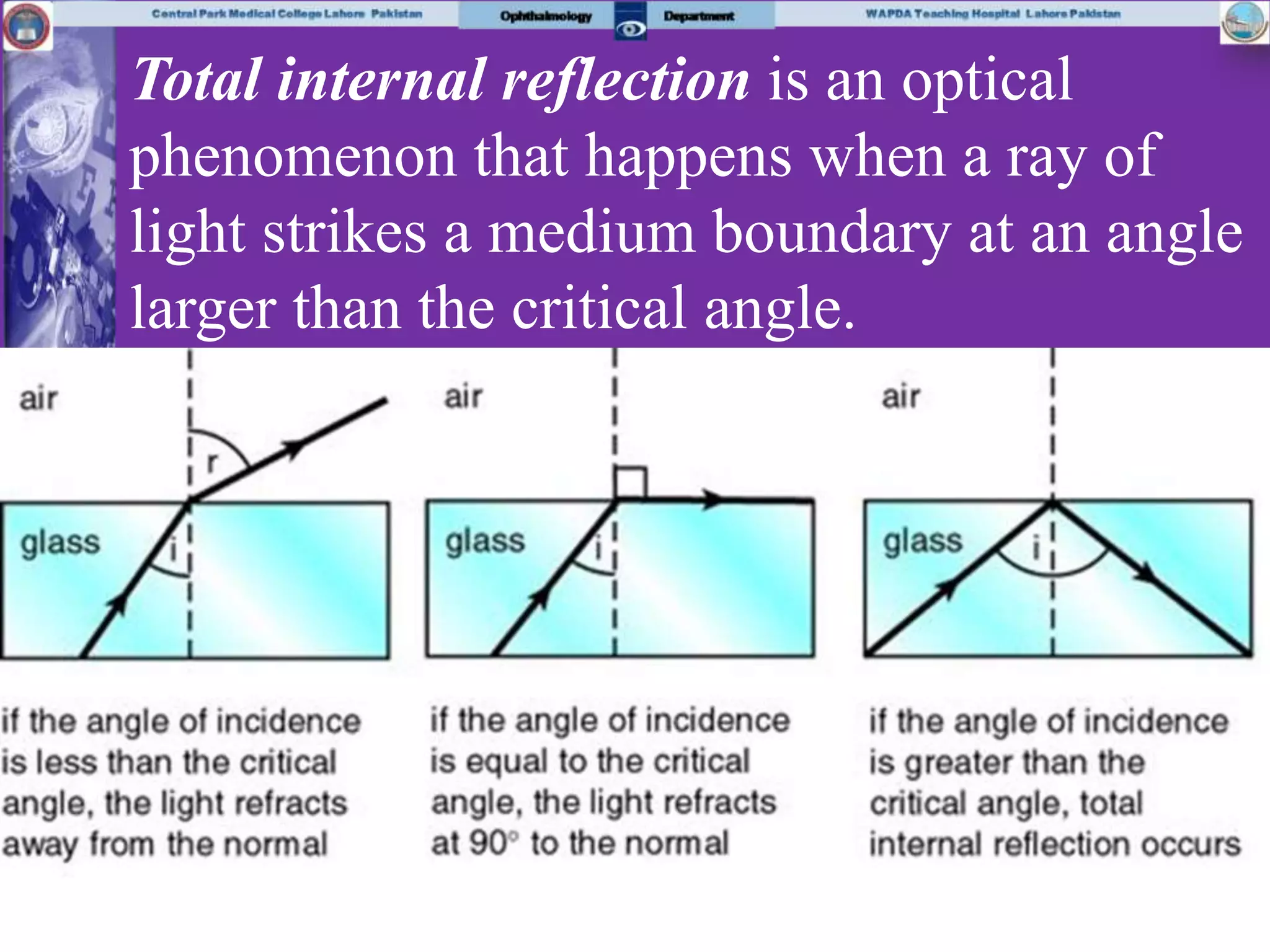


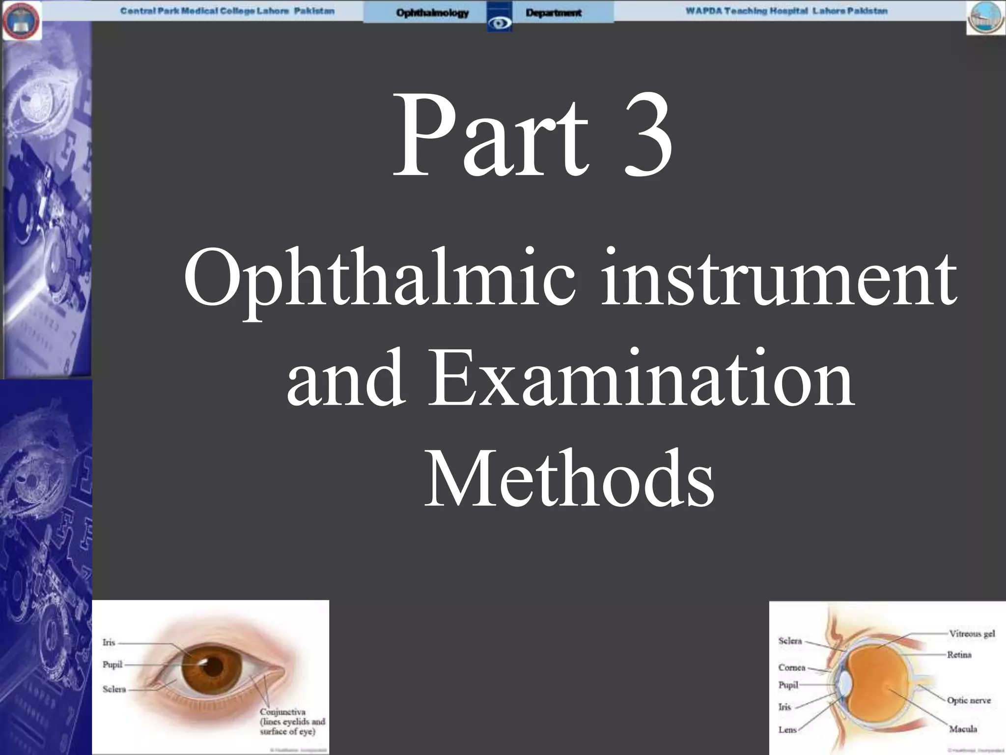


The document provides an extensive overview of clinical optics, ophthalmic instruments, and eye examination techniques, presented in multiple structured parts. It covers the importance of clinical optics in ophthalmology, the use of various instruments for eye examinations, and includes pretests and questions to assess understanding of key concepts. The main objectives are to enable students to understand clinical optics, utilize ophthalmic instruments, and evaluate common ocular conditions effectively.


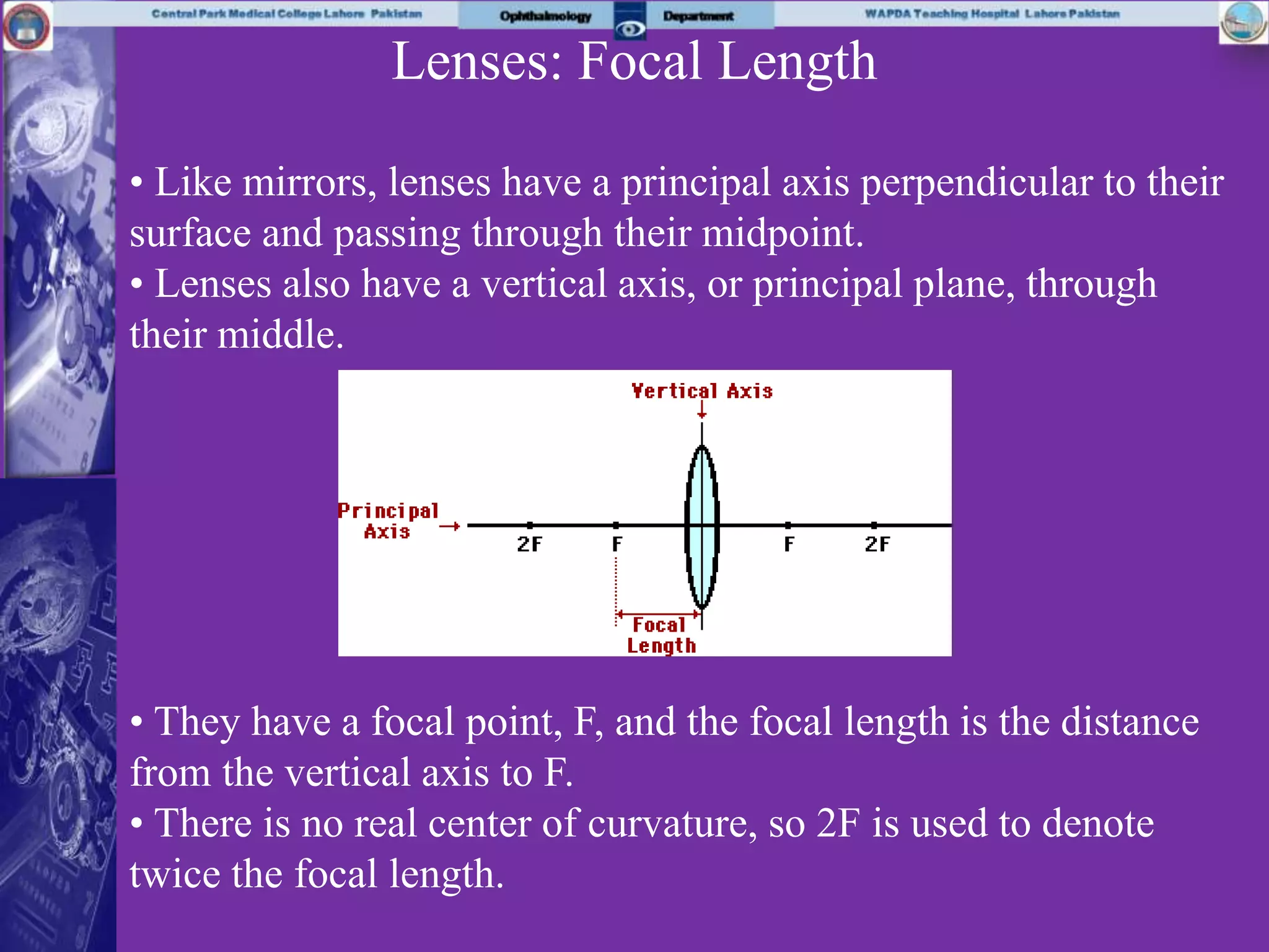
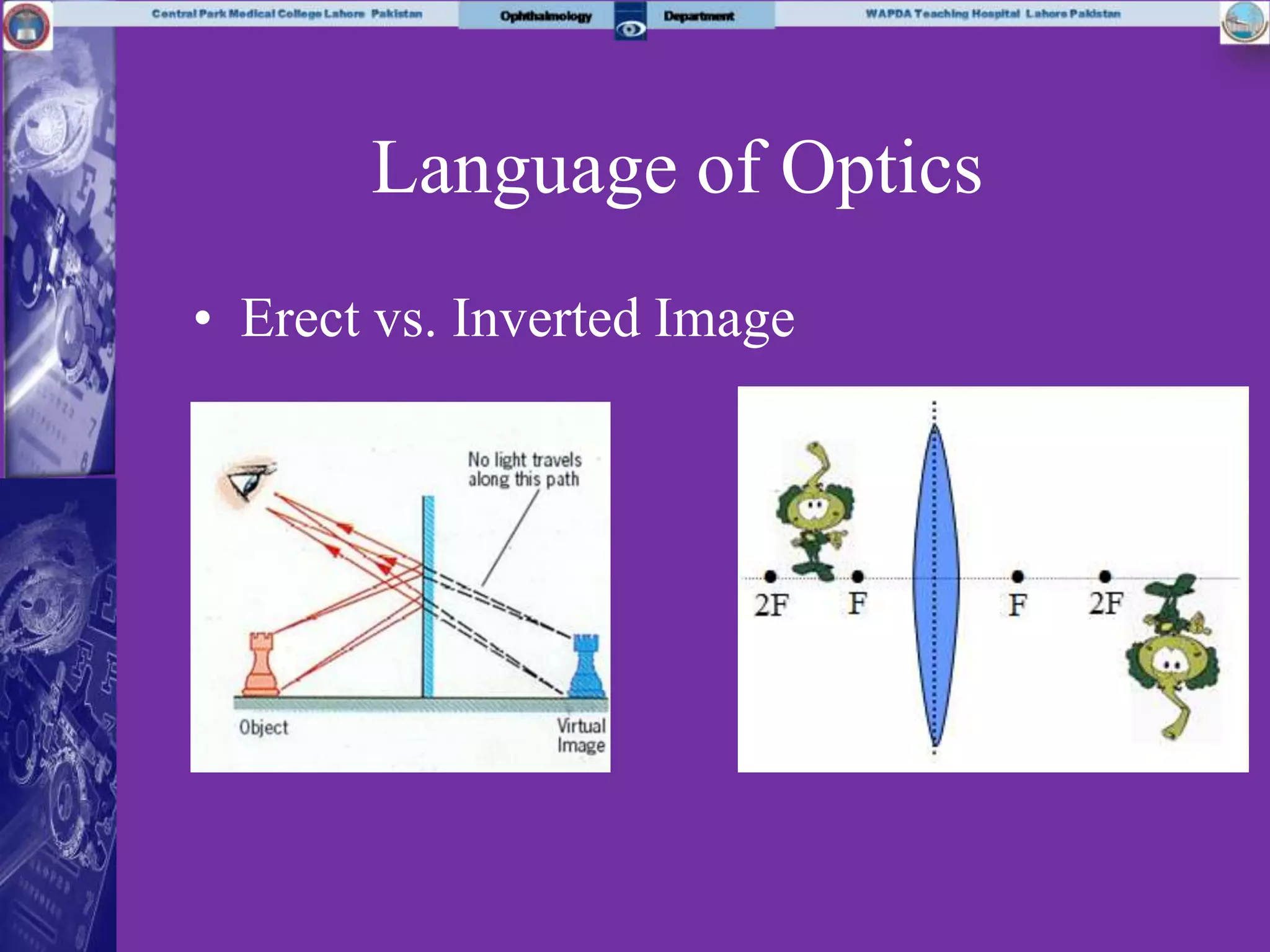
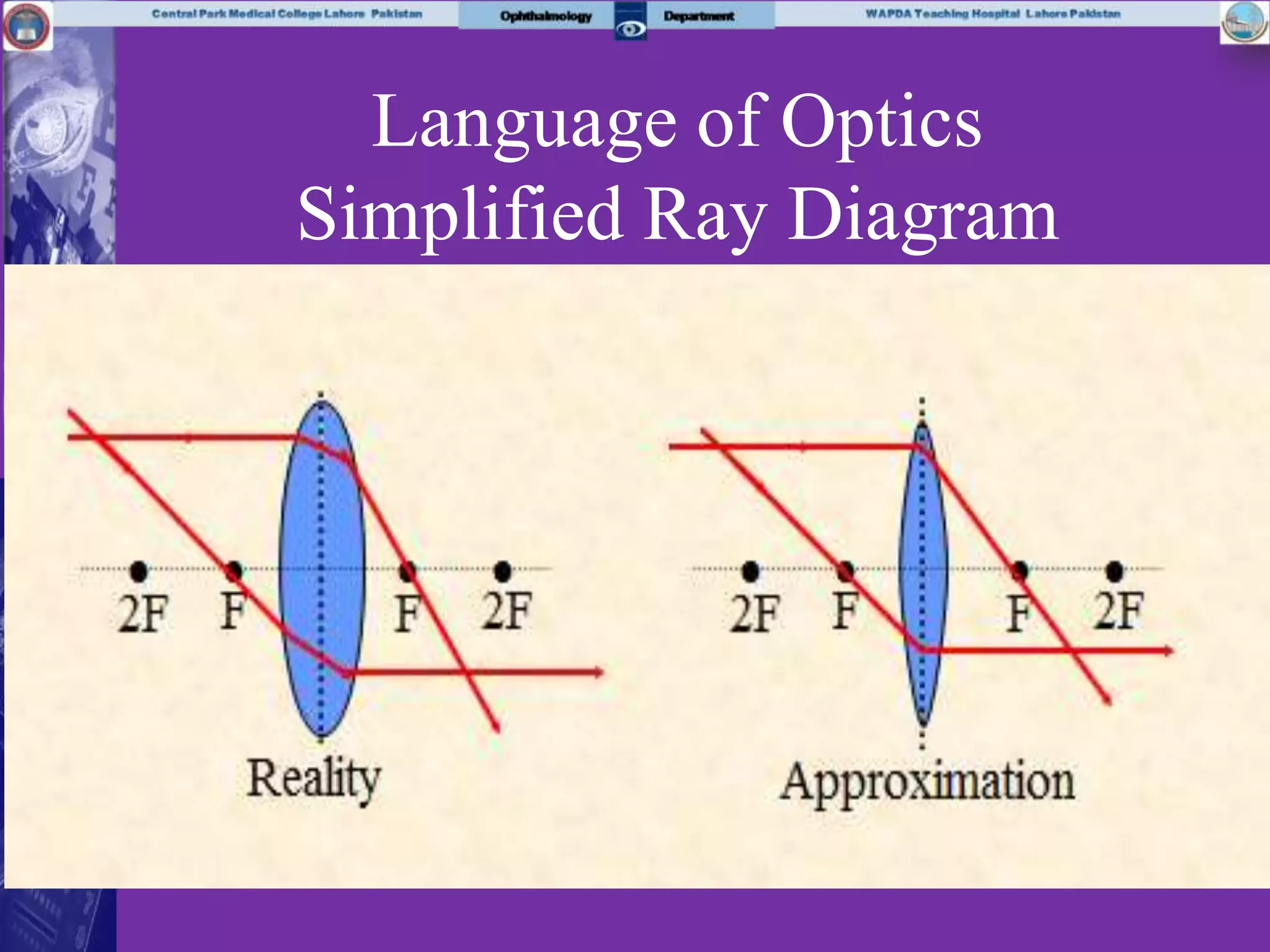
![[Unit 9.03] lens](https://cdn.slidesharecdn.com/ss_thumbnails/unit9-03lens-100916190225-phpapp01-thumbnail.jpg?width=640&height=640&fit=bounds)


![[Unit 12.3] lens](https://cdn.slidesharecdn.com/ss_thumbnails/unit12-3lens-100829070431-phpapp02-thumbnail.jpg?width=640&height=640&fit=bounds)


