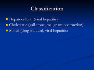
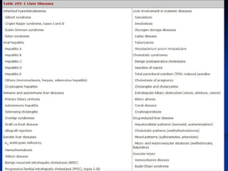
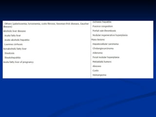
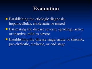
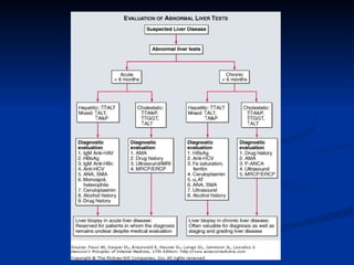
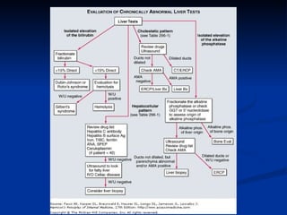
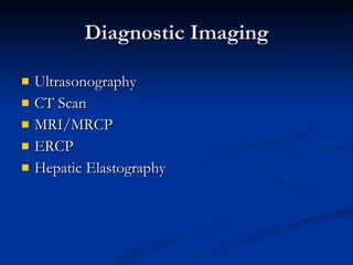
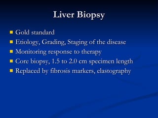
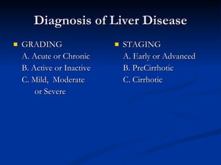
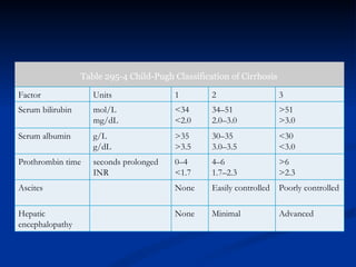
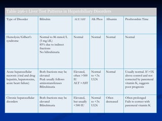
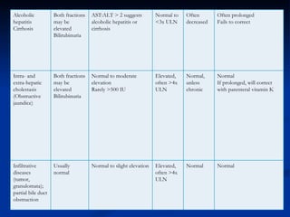
Approach to a Patient  with Liver Disease
Classification Hepatocellular (viral hepatitis) Cholestatic (gall stone, malignant obstruction) Mixed (drug-induced, viral hepatitis)
 
 
Evaluation Establishing the etiologic diagnosis: hepatocellular, cholestatic or mixed Estimating the disease severity (grading): active or inactive, mild to severe Establishing the disease stage: acute or chronic, pre-cirrhotic, cirrhotic, or end stage
 
Clinical Symptomatology Jaundice Fatigue Weakness Pruritus RUQ pain Anorexia Dark urine Light stools Asymptomatic
 
Portal hypertension Ascites Prominent superficial veins Edema Weight loss
Table 295-3 Important Diagnostic Tests in Common Liver Diseases Disease Diagnostic Test Hepatitis A Anti-HAV IgM Hepatitis B      Acute HBsAg and anti-HBc IgM    Chronic HBsAg and HBeAg and/or HBV DNA Hepatitis C Anti-HCV and HCV RNA Hepatitis D (delta) HBsAg and anti-HDV Hepatitis E Anti-HEV Autoimmune hepatitis ANA or SMA, elevated IgG levels, and compatible histology Primary biliary cirrhosis Mitochondrial antibody, elevated IgM levels, and compatible histology Primary sclerosing cholangitis P-ANCA, cholangiography Drug-induced liver disease History of drug ingestion Alcoholic liver disease History of excessive alcohol intake and compatible histology Nonalcoholic steatohepatitis Ultrasound or CT evidence of fatty liver and compatible histology        1  Antitrypsin disease   Reduced         1  antitrypsin levels, phenotypes PiZZ or PiSZ   Wilson disease Decreased serum ceruloplasmin and increased urinary copper; increased hepatic copper level Hemochromatosis Elevated iron saturation and serum ferritin; genetic testing for HFE gene mutations  Hepatocellular cancer Elevated        -fetoprotein level >500; ultrasound or CT image of mass
Clinical History Prodromal symptoms Use of parenteral illicit drugs Maternal history of hepatitis Exposure to jaundiced patients Use of medications (including herbal  meds) Sexual history (multiple partners, same sex) Blood transfusion Family history: Wilson’s disease, hemochromatosis,
Alcohol abuse US: 70% consume alcohol Only 5% have more than 2 drinks/day; 11-15g alcohol/drink Women: 22-30g alcohol/day Men: 33-45g alcohol/day Cirrhosis: at least 10 years of alcohol intake
P.E. Findings Icterus (Bilirubin > 2.5 mg/dL) Hepatomegaly Hepatic tenderness Spider angiomata Palmar erythema Splenomegaly Ascites Edema Skin hyperpigmentation
Hepatic Failure (Encephalopathy) Altered sleeping patterns Mental dullness Altered sensorium – disorientation, confusion, stupor, coma Fetor hepaticus Asterexis
Precipitating Factors GI Bleeding Over diuresis Dehydration Electrolyte imbalance Infection Constipation Use of narcotic analgesics
Diagnostic Evaluation ALT AST Alkaline phosphatase Serum bilirubin (direct, indirect) Albumin Prothrombin time GGT Hepatitis serology Autoimmune markers
Diagnostic Imaging Ultrasonography CT Scan MRI/MRCP ERCP Hepatic Elastography
Liver Biopsy Gold standard Etiology, Grading, Staging of the disease Monitoring response to therapy Core biopsy, 1.5 to 2.0 cm specimen length Replaced by fibrosis markers, elastography
Diagnosis of Liver Disease GRADING A. Acute or Chronic B. Active or Inactive C. Mild,  Moderate or Severe STAGING A. Early or Advanced B. PreCirrhotic C. Cirrhotic
Table 295-4  Child-Pugh Classification of Cirrhosis Factor Units 1 2 3 Serum bilirubin mol/L mg/dL <34 <2.0 34–51 2.0–3.0 >51 >3.0 Serum albumin g/L g/dL >35 >3.5 30–35 3.0–3.5 <30 <3.0 Prothrombin time  seconds prolonged INR 0–4 <1.7 4–6 1.7–2.3 >6 >2.3 Ascites   None Easily controlled Poorly controlled Hepatic encephalopathy   None Minimal Advanced
Table   296-1  Liver Test Patterns in Hepatobiliary Disorders  Type of Disorder Bilirubin  ALT/AST Alk Phos Albumin  Prothrombin Time Hemolysis/Gilbert's syndrome Normal to 86 mmol/L (5 mg/dL) 85% due to indirect fractions No bilirubinuria Normal Normal Normal Normal Acute hepatocellular necrosis (viral and drug hepatitis, hepatotoxins, acute heart failure) Both fractions may be elevated Peak usually follows aminotransferases Bilirubinuria Elevated, often >500 IU ALT >AST Normal to <3x ULN Normal Usually normal. If >5X above control and not corrected by parenteral vitamin K, suggests poor prognosis Chronic hepatocellular disorders Both fractions may be elevated Bilirubinuria Elevated, but usually <300 IU Normal to <3x ULN Often decreased Often prolonged Fails to correct with parenteral vitamin K
Alcoholic hepatitis Cirrhosis Both fractions may be elevated Bilirubinuria AST:ALT > 2 suggests alcoholic hepatitis or cirrhosis Normal to <3x ULN Often decreased Often prolonged Fails to correct  Intra- and extra-hepatic cholestasis (Obstructive jaundice) Both fractions may be elevated Bilirubinuria Normal to moderate elevation Rarely >500 IU Elevated, often >4x ULN Normal, unless chronic Normal If prolonged, will correct with parenteral vitamin K Infiltrative diseases (tumor, granulomata); partial bile duct obstruction Usually normal Normal to slight elevation Elevated, often >4x ULN Normal Normal

Liver disease

  • 1.
    Approach to aPatient with Liver Disease
  • 2.
    Classification Hepatocellular (viralhepatitis) Cholestatic (gall stone, malignant obstruction) Mixed (drug-induced, viral hepatitis)
  • 3.
  • 4.
  • 5.
    Evaluation Establishing theetiologic diagnosis: hepatocellular, cholestatic or mixed Estimating the disease severity (grading): active or inactive, mild to severe Establishing the disease stage: acute or chronic, pre-cirrhotic, cirrhotic, or end stage
  • 6.
  • 7.
    Clinical Symptomatology JaundiceFatigue Weakness Pruritus RUQ pain Anorexia Dark urine Light stools Asymptomatic
  • 8.
  • 9.
    Portal hypertension AscitesProminent superficial veins Edema Weight loss
  • 10.
    Table 295-3 ImportantDiagnostic Tests in Common Liver Diseases Disease Diagnostic Test Hepatitis A Anti-HAV IgM Hepatitis B      Acute HBsAg and anti-HBc IgM    Chronic HBsAg and HBeAg and/or HBV DNA Hepatitis C Anti-HCV and HCV RNA Hepatitis D (delta) HBsAg and anti-HDV Hepatitis E Anti-HEV Autoimmune hepatitis ANA or SMA, elevated IgG levels, and compatible histology Primary biliary cirrhosis Mitochondrial antibody, elevated IgM levels, and compatible histology Primary sclerosing cholangitis P-ANCA, cholangiography Drug-induced liver disease History of drug ingestion Alcoholic liver disease History of excessive alcohol intake and compatible histology Nonalcoholic steatohepatitis Ultrasound or CT evidence of fatty liver and compatible histology        1 Antitrypsin disease   Reduced        1 antitrypsin levels, phenotypes PiZZ or PiSZ   Wilson disease Decreased serum ceruloplasmin and increased urinary copper; increased hepatic copper level Hemochromatosis Elevated iron saturation and serum ferritin; genetic testing for HFE gene mutations  Hepatocellular cancer Elevated       -fetoprotein level >500; ultrasound or CT image of mass
  • 11.
    Clinical History Prodromalsymptoms Use of parenteral illicit drugs Maternal history of hepatitis Exposure to jaundiced patients Use of medications (including herbal meds) Sexual history (multiple partners, same sex) Blood transfusion Family history: Wilson’s disease, hemochromatosis,
  • 12.
    Alcohol abuse US:70% consume alcohol Only 5% have more than 2 drinks/day; 11-15g alcohol/drink Women: 22-30g alcohol/day Men: 33-45g alcohol/day Cirrhosis: at least 10 years of alcohol intake
  • 13.
    P.E. Findings Icterus(Bilirubin > 2.5 mg/dL) Hepatomegaly Hepatic tenderness Spider angiomata Palmar erythema Splenomegaly Ascites Edema Skin hyperpigmentation
  • 14.
    Hepatic Failure (Encephalopathy)Altered sleeping patterns Mental dullness Altered sensorium – disorientation, confusion, stupor, coma Fetor hepaticus Asterexis
  • 15.
    Precipitating Factors GIBleeding Over diuresis Dehydration Electrolyte imbalance Infection Constipation Use of narcotic analgesics
  • 16.
    Diagnostic Evaluation ALTAST Alkaline phosphatase Serum bilirubin (direct, indirect) Albumin Prothrombin time GGT Hepatitis serology Autoimmune markers
  • 17.
    Diagnostic Imaging UltrasonographyCT Scan MRI/MRCP ERCP Hepatic Elastography
  • 18.
    Liver Biopsy Goldstandard Etiology, Grading, Staging of the disease Monitoring response to therapy Core biopsy, 1.5 to 2.0 cm specimen length Replaced by fibrosis markers, elastography
  • 19.
    Diagnosis of LiverDisease GRADING A. Acute or Chronic B. Active or Inactive C. Mild, Moderate or Severe STAGING A. Early or Advanced B. PreCirrhotic C. Cirrhotic
  • 20.
    Table 295-4 Child-Pugh Classification of Cirrhosis Factor Units 1 2 3 Serum bilirubin mol/L mg/dL <34 <2.0 34–51 2.0–3.0 >51 >3.0 Serum albumin g/L g/dL >35 >3.5 30–35 3.0–3.5 <30 <3.0 Prothrombin time seconds prolonged INR 0–4 <1.7 4–6 1.7–2.3 >6 >2.3 Ascites   None Easily controlled Poorly controlled Hepatic encephalopathy   None Minimal Advanced
  • 21.
    Table 296-1 Liver Test Patterns in Hepatobiliary Disorders Type of Disorder Bilirubin ALT/AST Alk Phos Albumin Prothrombin Time Hemolysis/Gilbert's syndrome Normal to 86 mmol/L (5 mg/dL) 85% due to indirect fractions No bilirubinuria Normal Normal Normal Normal Acute hepatocellular necrosis (viral and drug hepatitis, hepatotoxins, acute heart failure) Both fractions may be elevated Peak usually follows aminotransferases Bilirubinuria Elevated, often >500 IU ALT >AST Normal to <3x ULN Normal Usually normal. If >5X above control and not corrected by parenteral vitamin K, suggests poor prognosis Chronic hepatocellular disorders Both fractions may be elevated Bilirubinuria Elevated, but usually <300 IU Normal to <3x ULN Often decreased Often prolonged Fails to correct with parenteral vitamin K
  • 22.
    Alcoholic hepatitis CirrhosisBoth fractions may be elevated Bilirubinuria AST:ALT > 2 suggests alcoholic hepatitis or cirrhosis Normal to <3x ULN Often decreased Often prolonged Fails to correct Intra- and extra-hepatic cholestasis (Obstructive jaundice) Both fractions may be elevated Bilirubinuria Normal to moderate elevation Rarely >500 IU Elevated, often >4x ULN Normal, unless chronic Normal If prolonged, will correct with parenteral vitamin K Infiltrative diseases (tumor, granulomata); partial bile duct obstruction Usually normal Normal to slight elevation Elevated, often >4x ULN Normal Normal