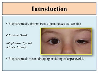
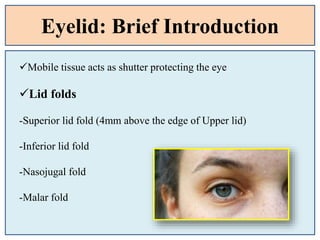
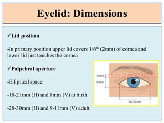
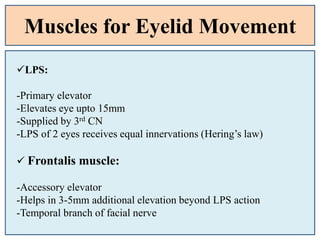
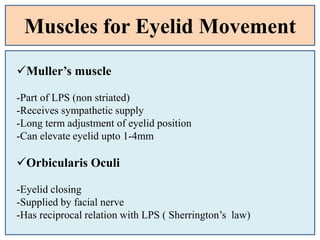
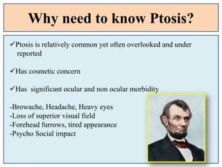
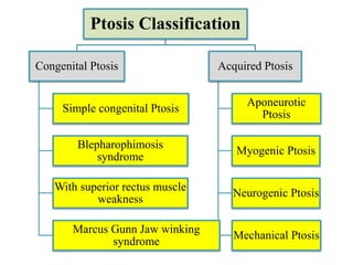
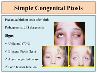
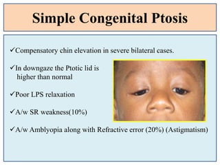
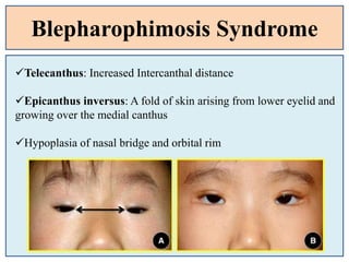
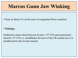
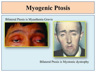
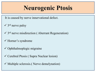
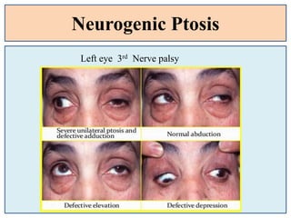
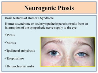
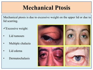
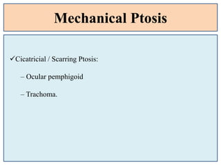
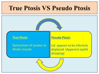
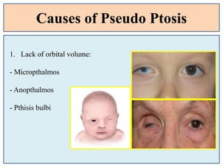
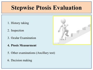
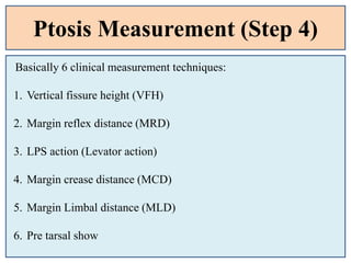
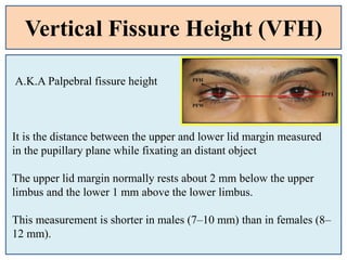
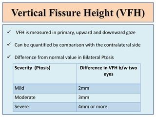
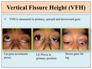
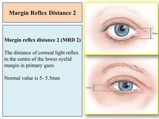
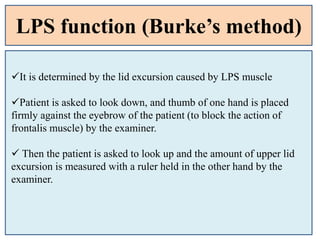
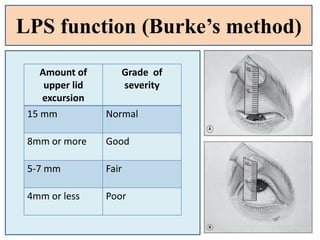
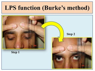
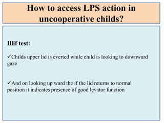
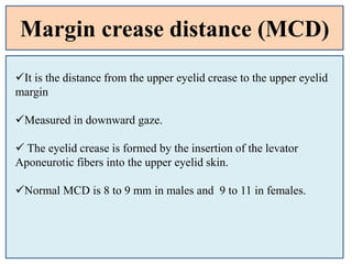
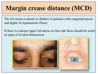
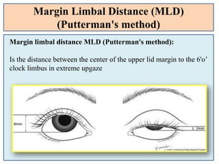
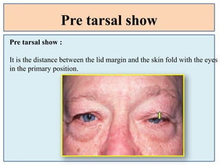
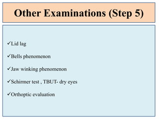
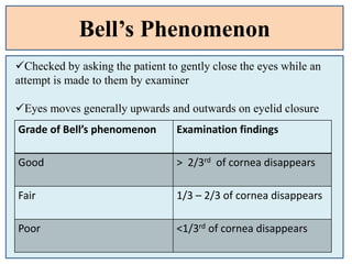
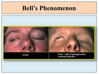
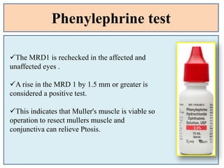
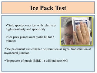
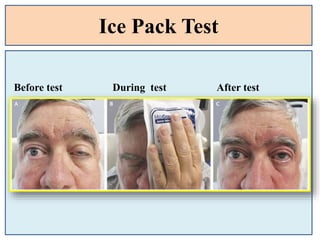
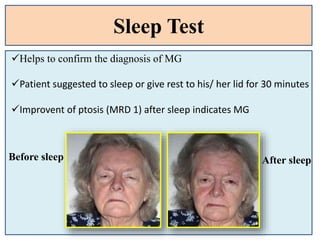
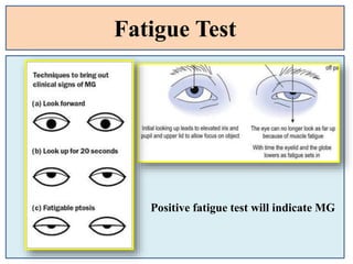
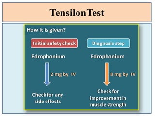
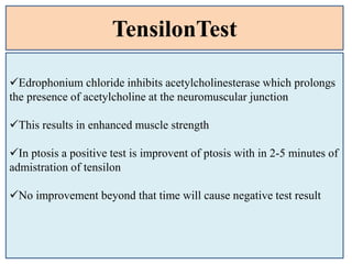
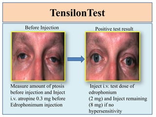
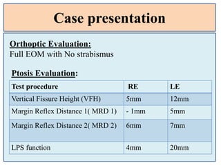
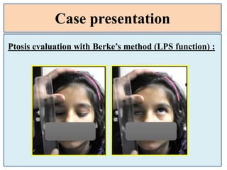
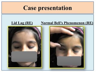
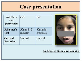
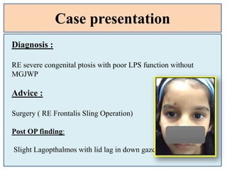
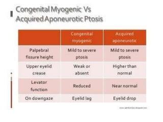
3. Introduction
Blepharoptosis, abbrev. Ptosis (pronounced as “toe-sis)
Ancient Greek:
-Blepharon: Eye lid
-Ptosis: Falling
Blepharoptosis means drooping or falling of upper eyelid.
4. Eyelid: Brief Introduction
Mobile tissue acts as shutter protecting the eye
Lid folds
-Superior lid fold (4mm above the edge of Upper lid)
-Inferior lid fold
-Nasojugal fold
-Malar fold
5. Eyelid: Dimensions
Lid position
-In primary position upper lid covers 1/6th (2mm) of cornea and
lower lid just touches the cornea
Palpebral aperture
-Elliptical space
-18-21mm (H) and 8mm (V) at birth
-28-30mm (H) and 9-11mm (V) adult
6. Muscles for Eyelid Movement
LPS:
-Primary elevator
-Elevates eye upto 15mm
-Supplied by 3rd CN
-LPS of 2 eyes receives equal innervations (Hering’s law)
Frontalis muscle:
-Accessory elevator
-Helps in 3-5mm additional elevation beyond LPS action
-Temporal branch of facial nerve
7. Muscles for Eyelid Movement
Muller’s muscle
-Part of LPS (non striated)
-Receives sympathetic supply
-Long term adjustment of eyelid position
-Can elevate eyelid upto 1-4mm
Orbicularis Oculi
-Eyelid closing
-Supplied by facial nerve
-Has reciprocal relation with LPS ( Sherrington’s law)
8. Why need to know Ptosis?
Ptosis is relatively common yet often overlooked and under
reported
Has cosmetic concern
Has significant ocular and non ocular morbidity
-Browache, Headache, Heavy eyes
-Loss of superior visual field
-Forehead furrows, tired appearance
-Psycho Social impact
9. Ptosis Classification
Congenital Ptosis
Simple congenital Ptosis
Blepharophimosis
syndrome
With superior rectus muscle
weakness
Marcus Gunn Jaw winking
syndrome
Acquired Ptosis
Aponeurotic
Ptosis
Myogenic Ptosis
Neurogenic Ptosis
Mechanical Ptosis
10. Simple Congenital Ptosis
Present at birth or soon after birth
Pathogenesis: LPS dysgenesis
Signs
Unilateral (70%)
Bilateral Ptosis (less)
Absent upper lid crease
Poor levator function.
11. Simple Congenital Ptosis
Compensatory chin elevation in severe bilateral cases.
In downgaze the Ptotic lid is
higher than normal
Poor LPS relaxation
A/w SR weakness(10%)
A/w Amblyopia along with Refractive error (20%) (Astigmatism)
12. Blepharophimosis Syndrome
AD inheritance
Also called BPES( Blepharophimosis Ptosis Epicanthus Syndrome)
Classical features:
Blepharophimosis: narrowing of horizontal palpebral aperture.
Normal 25-30mm. Usually 20-22mm in BPES
Ptosis
-With poor levator function
-Usually bilateral and symmetric and absent lid crease
13. Blepharophimosis Syndrome
Telecanthus: Increased Intercanthal distance
Epicanthus inversus: A fold of skin arising from lower eyelid and
growing over the medial canthus
Hypoplasia of nasal bridge and orbital rim
14. Marcus Gunn Jaw Winking
Seen in about 5% of all cases of congenital Ptosis manifest
Etiology:
Synkinetic innervation between levator (3rd CN) and masticatory
muscle ( 5th CN) i.e. mandibular division of the 5th cranial nerve is
misdirected to the levator muscle.
15. Marcus Gunn Jaw Winking
Common Signs:
Retraction of the ptotic lid in conjunction with stimulation of the
ipsilateral pterygoid muscles by chewing, sucking, opening the
mouth or contralateral jaw movement.
Less common stimuli to winking include jaw protrusion,
smiling, swallowing and clenching of teeth.
Jaw-winking does not improve with age, although patients may
learn to mask it
16. Grading of
Synkinesis
Subjective
(Maximum
Elevation of ptotic
lid)
Objective
(Measurment)
Mild Non ptotic position < 2mm
Moderate Upto superior limbus 2-5 mm
Severe Beyond superior
limbus with scleral
show
> 5mm
Marcus Gunn Jaw Winking
18. Acquired Ptosis
1. Aponeurotic Ptosis
It develops due to defects or dehiscence of the levator
aponeurosis in the presence of a normal functioning muscle.
Senile/ Involutional ptosis
Post traumatic
Post surgical
24. Neurogenic Ptosis
Basic features of Horner’s Syndrome
Ptosis
Miosis
Ipsilateral anhydrosis
Enopthalmos
Heterochromia iridis
Horner’s syndrome or oculosympathetic paresis results from an
interruption of the sympathetic nerve supply to the eye
25. Mechanical Ptosis
Mechanical ptosis is due to excessive weight on the upper lid or due to
lid scarring.
Excessive weight:
• Lid tumours
• Multiple chalazia
• Lid edema
• Dermatochalasis
27. True Ptosis VS Pseudo Ptosis
True Ptosis
Dysfunction of Levator or
Muller muscle
Pseudo Ptosis
Lid appears to be inferiorly
displaced (Apparent eyelid
drooping)
28. Causes of Pseudo Ptosis
1. Lack of orbital volume:
- Micropthalmos
- Anopthalmos
- Pthisis bulbi
29. Causes of Pseudo Ptosis
2. Malposition of eye (Ipsilateral Hypotropia / Contra. Hypertropia
3. Enopthalmos
30. Causes of Pseudo Ptosis
4. Contralateral proptosis/ Contralateral lid
retraction
5. Dermatochalasis
6. Browptosis
7. Blepharospasm
32. Stepwise Ptosis Evaluation
1. History taking
2. Inspection
3. Ocular Examination
4. Ptosis Measurment
5. Other examinations (Ancillary test)
6. Decision making
33. History Taking (Step 1)
Age of onset?
-Since childhood/ Mid teen/
- Late teen/ Adult
Any variability in Ptosis?
- Yes/ No
- If yes then
- Diurnal variation/ Jaw winking/ Fatigueness
34. History Taking (Step 1)
Age of onset?
-Since childhood/ Mid teen/ Late teen/ Adult
Duration/ laterality?
- Recent onset/ long history
- Unilateral / Bilateral ( as reported by patient)
Any variability in Ptosis?
- Yes/ No
- If yes then
- Diurnal variation/ Jaw winking/ Fatigueness/ Rest- sleep
35. History Taking (Step 1)
Any predisposing factors?
-Past trauma/ Eye surgery/ Systemic Diseases/ Steroid Use
-Contact lens use
Any Family history?
-Yes/No
- If yes
- r/o BPES, MG, MD, CPEO, pharyngeal muscular dystrophy
Ask for Old photographs / Old clinical records?
- If available will help to compare the progression also predict the
onset
36. History Taking (Step 1)
Ask for the presence of any other associated features ?
-Diplopia
-Abnormal Head position
-Body muscle weakness
-Eye movement restrictions
37. Inspection (Step 2)
Closely inspect the patients extra ocular features before going to
the ocular examination
Laterality: Unilateral/ bilateral
Change in lid position with eye movement and jaw movement
Compare facial symmetry/ palsy
Abnormal head posture- head tilt/ face turn, chin up/ down
Overaction of Frontalis – Forehead crease
42. Vertical Fissure Height (VFH)
A.K.A Palpebral fissure height
It is the distance between the upper and lower lid margin measured
in the pupillary plane while fixating an distant object
The upper lid margin normally rests about 2 mm below the upper
limbus and the lower 1 mm above the lower limbus.
This measurement is shorter in males (7–10 mm) than in females (8–
12 mm).
43. Vertical Fissure Height (VFH)
VFH is measured in primary, upward and downward gaze
Can be quantified by comparison with the contralateral side
Difference from normal value in Bilateral Ptosis
Severity (Ptosis) Difference in VFH b/w two
eyes
Mild 2mm
Moderate 3mm
Severe 4mm or more
44. Vertical Fissure Height (VFH)
VFH is measured in primary, upward and downward gaze
Up gaze accentuate
ptosis
Down gaze lid
lag
LE Ptosis in
primary position
46. Margin Reflex Distance (MRD)
Total 3 types of margin to reflex distance is measured clinically
during Ptosis evaluation
- Margin reflex distance 1 (MRD 1)
- Margin reflex distance 2 (MRD 2)
- Margin reflex distance 3 (MRD 3)
47. Margin Reflex Distance 1
Margin-to-Reflex distance 1 (MRD1) :
Distance from the central pupillary light reflex to the upper
eyelid margin with the eye in primary gaze.
NORMAL : 4 - 5 mm.
If the margin is above the light reflex the MRD 1 is a +ve value.
If the lid margin is below the corneal reflex in cases of very
severe Ptosis the MRD 1 would be a –ve value.
48. Margin Reflex Distance 1
Severity of
Ptosis
Amount of
Upper eyelid
droop
Mild (B) 2mm
Moderate (C) 3mm
Severe (D) 4mm or more
49. Margin Reflex Distance 2
Margin reflex distance 2 (MRD 2)
The distance of corneal light reflex
to the centre of the lower eyelid
margin in primary gaze.
Normal value is 5- 5.5mm
50. Margin Reflex Distance 3
MRD3 is used to determine how much levator to resect in patients
with congenital Ptosis, who have a vertical strabismus associated
with ptosis and in whom strabismus surgery is not indicated.
MRD3 is less frequently used when compared to MRD 1 or 2.
Margin reflex distance 3(MRD 3)
The distance between the corneal
light reflex and the centre of upper
eyelid margin in extreme up gaze.
51. LPS function (Burke’s method)
It is determined by the lid excursion caused by LPS muscle
Patient is asked to look down, and thumb of one hand is placed
firmly against the eyebrow of the patient (to block the action of
frontalis muscle) by the examiner.
Then the patient is asked to look up and the amount of upper lid
excursion is measured with a ruler held in the other hand by the
examiner.
52. LPS function (Burke’s method)
Amount of
upper lid
excursion
Grade of
severity
15 mm Normal
8mm or more Good
5-7 mm Fair
4mm or less Poor
54. How to access LPS action in
uncooperative childs?
Illif test:
Childs upper lid is everted while child is looking to downward
gaze
And on looking up ward the if the lid returns to normal
position it indicates presence of good levator function
55. Margin crease distance (MCD)
It is the distance from the upper eyelid crease to the upper eyelid
margin
Measured in downward gaze.
The eyelid crease is formed by the insertion of the levator
Aponeurotic fibers into the upper eyelid skin.
Normal MCD is 8 to 9 mm in males and 9 to 11 in females.
56. Margin crease distance (MCD)
The lid crease is absent or shallow in patients with congenital ptosis
and higher in Aponeurotic Ptosis.
If there is a deeper upper lid sulcus on that side these should be noted
as signs of levator disinsertion.
57. Margin Limbal Distance (MLD)
(Putterman's method)
Margin limbal distance MLD (Putterman's method):
Is the distance between the center of the upper lid margin to the 6'o’
clock limbus in extreme upgaze
58. Pre tarsal show
Pre tarsal show :
It is the distance between the lid margin and the skin fold with the eyes
in the primary position.
59. Other Examinations (Step 5)
Lid lag
Bells phenomenon
Jaw winking phenomenon
Schirmer test , TBUT- dry eyes
Orthoptic evaluation
60. Bell’s Phenomenon
Checked by asking the patient to gently close the eyes while an
attempt is made to them by examiner
Eyes moves generally upwards and outwards on eyelid closure
Grade of Bell’s phenomenon Examination findings
Good > 2/3rd of cornea disappears
Fair 1/3 – 2/3 of cornea disappears
Poor <1/3rd of cornea disappears
62. Corneal sensation/TBUT TEST/
lagophthalmos test
The corneal sensation and lagophthalmos should be checked in all
Ptosis patients before planning for surgery.
Normal blink reflex and eyelid closure are essential to prevent dry
eye and exposure keratitis after Ptosis correction surgery.
Schirmer's test, tear film breakup time, and the tear meniscus
should be documented before surgery as the presence of a dry eye
may be a contraindication for Ptosis correction.
63. Ocular Motility Assessment
Orthoptic evaluation and Ocular motility test:
To r/o palsy and DRS
To r/o all causes of
pseudo ptosis
To find associated SR muscle weakness in Double elevator palsy
Strabismus must be corrected prior to the correction of ptosis.
64. Ancillary tests
Phenylephrine test: to see feasibility of Muller’s muscle surgery
Icepack test
Tensilon test
Fatigue/ sleep test
Others :
-Serum acetylcholine receptor assay
-EMG
-T3, T4, TSH
65. Phenylephrine test
Patients with minimal ptosis (2 mm or less)
should have a phenylephrine test performed in the
involved eye or eyes
Either 2.5 or 10% phenylephrine is instilled in
the affected eye or eyes. Usually two drops are
placed and the patient is reexamined 5 minutes
later.
66. Phenylephrine test
The MRD1 is rechecked in the affected and
unaffected eyes .
A rise in the MRD 1 by 1.5 mm or greater is
considered a positive test.
This indicates that Muller's muscle is viable so
operation to resect mullers muscle and
conjunctiva can relieve Ptosis.
67. Ice Pack Test
Safe speedy, easy test with relatively
high sensitivity and specificity
Ice pack placed over ptotic lid for 5
minutes
Ice palcement will enhance neuromuscular signal transmission at
myoneural junction
Improvent of ptosis (MRD 1) will indicate MG
69. Sleep Test
Helps to confirm the diagnosis of MG
Patient suggested to sleep or give rest to his/ her lid for 30 minutes
Improvent of ptosis (MRD 1) after sleep indicates MG
Before sleep After sleep
73. TensilonTest
Edrophonium chloride inhibits acetylcholinesterase which prolongs
the presence of acetylcholine at the neuromuscular junction
This results in enhanced muscle strength
In ptosis a positive test is improvent of ptosis with in 2-5 minutes of
admistration of tensilon
No improvement beyond that time will cause negative test result
74. TensilonTest
Measure amount of ptosis
before injection and Inject
i.v. atropine 0.3 mg before
Edrophonimum injection
Inject i.v. test dose of
edrophonium
(2 mg) and Inject remaining
(8 mg) if no
hypersensitivity
Before Injection Positive test result
75. Final Diagnosis
Do a quick assessment!!!
Unilateral/ bialteral
Congenital/ Acquired
Acute onset/ Chronic disorder
Progressive / Non progresive
Isolated / Systemic association
77. Case presentation
Age : 10/F
C/0 dropping of right eye upper lid since birth
No History of ptosis progression
No history of variation
No any systemic illness
No past history of trauma and surgery
78. Case presentation
Ocular Examination and Inspection:
BCVA: 6/6 OU
IOP: 13mm Hg RE and 15 mm Hg LE
Pupil: R/R/R
Anterior segment : WNL
Posterior Segment : WNL
No abnormal head posture
Brow position: slightly lifted in RE
Lid crease: faintly present in RE
79. Case presentation
Ptosis Evaluation:
Test procedure RE LE
Vertical Fissure Height (VFH) 5mm 12mm
Margin Reflex Distance 1( MRD 1) - 1mm 5mm
Margin Reflex Distance 2( MRD 2) 6mm 7mm
LPS function 4mm 20mm
Orthoptic Evaluation:
Full EOM with No strabismus
82. Case presentation
No Marcus Gunn Jaw Winking
Ancillary
test
performed
OD OS
Schirmer’s
Test
35mm in 3
minutes
35mm in
3minutes
Corneal
Sensation
Normal Normal
83. Case presentation
Diagnosis :
RE severe congenital ptosis with poor LPS function without
MGJWP
Advice :
Surgery ( RE Frontalis Sling Operation)
Post OP finding:
Slight Lagopthalmos with lid lag in down gaze
84. References
1. A.K. Khurana Clinical Ophthalmology
2. Clinical Ophthalmology by J.J kanski
3. Wills Eye Manual
4. Internet
5. Case Courtesy ( Dr. Sabin Shau’s presentation)
Editor's Notes
This is in contrast to acquired ptosis in which the affected lid is either level with or lower than the normal lid on downgaze.
Following surgical correction the lid lag in downgaze may worsen.
Treatment should be carried out during the preschool years once accurate measurements can be obtained, although it may be considered earlier in severe cases to prevent amblyopia. Most cases require levator resection
Ptosis worse on down gaze, normal Eom and pupil
If the onset of Horner syndrome is before two years of age, the colored portions of the eyes (irises) may be different colors (heterochromia iridis)
iris of the affected side lacks color (hypopigmentation).
All to r/o pseudoptosis
Margin reflex distance 3 or MRD3 is an entity described by Putterman, which is the distance from the ocular, not corneal, light reflex to the central upper-eyelid margin when the patient looks in extreme up gaze.[1]
Margin reflex
Margin reflex
Margin reflex
Margin reflex
The skin crease is also used as a guide to the initial incision in some surgical procedures.
Helps to find amount of levator resection required during the ptosis correction
If there is vertical strabismus associated with blepharoptosis (i.e. double elevator palsy), it has been determined that the MLD is not valid because the 6-o’clock limbal level in up gaze is abnormal given vertical restriction
Simpson test also called
Next test is neostigmine test..
Action last for maximum 10 min….starts with in 30 sec