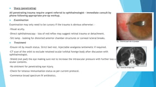
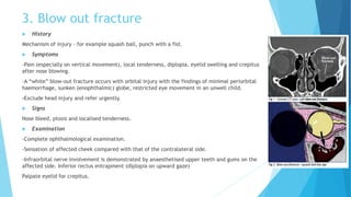
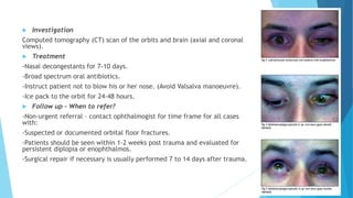
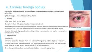
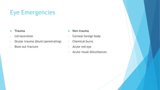
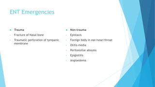
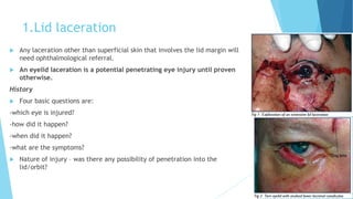
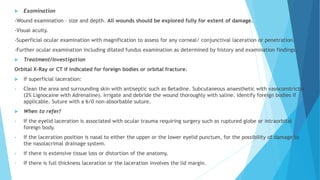
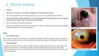
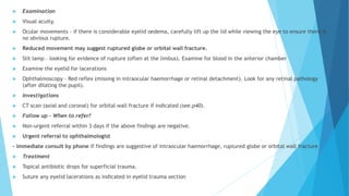
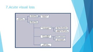
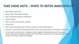
This document provides information on various eye, ear, nose, and throat (EENT) emergencies. It describes trauma-related conditions like lid lacerations, ocular trauma, blowout fractures, and nasal bone fractures. It also covers non-trauma issues such as corneal foreign bodies, chemical burns, acute red eye, acute visual loss, epistaxis, traumatic tympanic membrane perforations, and more. For each condition, it discusses history, examination, treatment, and criteria for referral to a specialist. Immediate referral is indicated for penetrating injuries, suspected globe rupture, intraocular foreign bodies, severe chemical burns, hyphema, uveitis with hypopyon, and recurrent amauro
you will get information about the staphyloma, its types, its etiology, its pathogenesis and its treatment.
you will know about the clinical types of staphyloma in detailed such as anterior staphyloma, posterior staphyloma, equatorial staphyloma, ciliary staphyloma, and Intercalary staphyloma.
Simple eye education for EHW, Ophthalmic eye student, school eye education & first - second year optometry students only .
common eye lid inflammatory conditions .
stye or hordeolum ( external / internal hordeolum ), lid abscess , chalazion or mebomian retention cyst, accessory lacrimal glands , lacrimal gland etc...
Any deviation in the normal nasal septum is called DNS (Deviated Nasal Septum).
Deviated Nasal Septum may be caused by mechanical trauma and may be
associated with some developmental defects.
An ear infection is an inflammation of the middle ear, usually caused
by bacteria, that occurs when fluid builds up behind the eardrum.
Anyone can get an ear infection, but children get them more often than
adults. Three out of four children will have at least one ear infection
by their third birthday. In fact, ear infections are the most common
reason parents bring their child to a doctor. The scientific name for an
ear infection is otitis media (OM).
Glaucoma is caused by a number of different eye diseases that in most cases produce increased pressure within the eye. This elevated pressure is caused by a backup of fluid in the eye. Over time, it causes damage to the optic nerve. Through early detection, diagnosis and treatment, you and your doctor can help to preserve your vision.
Ectropion
It is an outward turning of the eyelid margin . This more frequently affects the lower eyelid.Upper eyelid ectropion is uncommon.Classified in 5 types
1)Congenital 2) Involutional 3) Paralytic 4) Cicatricial 5) Mechanical
Involutional ectropion is more common.Congenital ectropion is very rare.
Symptoms Epiphora :- excessive tearing.Excessive dryness.
Foreign body sensation Irritation.Burning.Redness.Chronic conjunctivitis KeratinizationCorneal exposure
Grading
Lid margin is out rolled and depending on out rolling ectropion can be classified as under:
Grade I –only punctum is everted
Grade II –lid margin is everted and palpebral conjunctiva is visible
Grade III –fornix is also visible
Etiological factors
Horizontal lid laxity:-can be demonstrated by pulling the central part of the lid 8 mm or more from the globe, with a failure to snap back to its normal position on release without the patient first blinking.
Medial canthal tendon laxity
demonstrated by pulling the lower lid laterally and observing the position of the inferior punctum If the lid is normal the punctum should not be displaced more than 1–2 mm
Lateral canthal tendon laxity
characterized by a rounded appearance of the lateral canthus and the ability to pull the lower lid medially more than 2 mm.
>Normally, the displacement should only be 0-2 mm.
Treatment
1 medical therapy
2 surgical therapy
263778731218 Abortion Clinic /Pills In Harare ,sisternakatoto
263778731218 Abortion Clinic /Pills In Harare ,ABORTION WOMEN’S CLINIC +27730423979 IN women clinic we believe that every woman should be able to make choices in her pregnancy. Our job is to provide compassionate care, safety,affordable and confidential services. That’s why we have won the trust from all generations of women all over the world. we use non surgical method(Abortion pills) to terminate…Dr.LISA +27730423979women Clinic is committed to providing the highest quality of obstetrical and gynecological care to women of all ages. Our dedicated staff aim to treat each patient and her health concerns with compassion and respect.Our dedicated group ABORTION WOMEN’S CLINIC +27730423979 IN women clinic we believe that every woman should be able to make choices in her pregnancy. Our job is to provide compassionate care, safety,affordable and confidential services. That’s why we have won the trust from all generations of women all over the world. we use non surgical method(Abortion pills) to terminate…Dr.LISA +27730423979women Clinic is committed to providing the highest quality of obstetrical and gynecological care to women of all ages. Our dedicated staff aim to treat each patient and her health concerns with compassion and respect.Our dedicated group of receptionists, nurses, and physicians have worked together as a teamof receptionists, nurses, and physicians have worked together as a team wwww.lisywomensclinic.co.za/
These simplified slides by Dr. Sidra Arshad present an overview of the non-respiratory functions of the respiratory tract.
Learning objectives:
1. Enlist the non-respiratory functions of the respiratory tract
2. Briefly explain how these functions are carried out
3. Discuss the significance of dead space
4. Differentiate between minute ventilation and alveolar ventilation
5. Describe the cough and sneeze reflexes
Study Resources:
1. Chapter 39, Guyton and Hall Textbook of Medical Physiology, 14th edition
2. Chapter 34, Ganong’s Review of Medical Physiology, 26th edition
3. Chapter 17, Human Physiology by Lauralee Sherwood, 9th edition
4. Non-respiratory functions of the lungs https://academic.oup.com/bjaed/article/13/3/98/278874
Recomendações da OMS sobre cuidados maternos e neonatais para uma experiência pós-natal positiva.
Em consonância com os ODS – Objetivos do Desenvolvimento Sustentável e a Estratégia Global para a Saúde das Mulheres, Crianças e Adolescentes, e aplicando uma abordagem baseada nos direitos humanos, os esforços de cuidados pós-natais devem expandir-se para além da cobertura e da simples sobrevivência, de modo a incluir cuidados de qualidade.
Estas diretrizes visam melhorar a qualidade dos cuidados pós-natais essenciais e de rotina prestados às mulheres e aos recém-nascidos, com o objetivo final de melhorar a saúde e o bem-estar materno e neonatal.
Uma “experiência pós-natal positiva” é um resultado importante para todas as mulheres que dão à luz e para os seus recém-nascidos, estabelecendo as bases para a melhoria da saúde e do bem-estar a curto e longo prazo. Uma experiência pós-natal positiva é definida como aquela em que as mulheres, pessoas que gestam, os recém-nascidos, os casais, os pais, os cuidadores e as famílias recebem informação consistente, garantia e apoio de profissionais de saúde motivados; e onde um sistema de saúde flexível e com recursos reconheça as necessidades das mulheres e dos bebês e respeite o seu contexto cultural.
Estas diretrizes consolidadas apresentam algumas recomendações novas e já bem fundamentadas sobre cuidados pós-natais de rotina para mulheres e neonatos que recebem cuidados no pós-parto em unidades de saúde ou na comunidade, independentemente dos recursos disponíveis.
É fornecido um conjunto abrangente de recomendações para cuidados durante o período puerperal, com ênfase nos cuidados essenciais que todas as mulheres e recém-nascidos devem receber, e com a devida atenção à qualidade dos cuidados; isto é, a entrega e a experiência do cuidado recebido. Estas diretrizes atualizam e ampliam as recomendações da OMS de 2014 sobre cuidados pós-natais da mãe e do recém-nascido e complementam as atuais diretrizes da OMS sobre a gestão de complicações pós-natais.
O estabelecimento da amamentação e o manejo das principais intercorrências é contemplada.
Recomendamos muito.
Vamos discutir essas recomendações no nosso curso de pós-graduação em Aleitamento no Instituto Ciclos.
Esta publicação só está disponível em inglês até o momento.
Prof. Marcus Renato de Carvalho
www.agostodourado.com
These lecture slides, by Dr Sidra Arshad, offer a quick overview of the physiological basis of a normal electrocardiogram.
Learning objectives:
1. Define an electrocardiogram (ECG) and electrocardiography
2. Describe how dipoles generated by the heart produce the waveforms of the ECG
3. Describe the components of a normal electrocardiogram of a typical bipolar lead (limb II)
4. Differentiate between intervals and segments
5. Enlist some common indications for obtaining an ECG
6. Describe the flow of current around the heart during the cardiac cycle
7. Discuss the placement and polarity of the leads of electrocardiograph
8. Describe the normal electrocardiograms recorded from the limb leads and explain the physiological basis of the different records that are obtained
9. Define mean electrical vector (axis) of the heart and give the normal range
10. Define the mean QRS vector
11. Describe the axes of leads (hexagonal reference system)
12. Comprehend the vectorial analysis of the normal ECG
13. Determine the mean electrical axis of the ventricular QRS and appreciate the mean axis deviation
14. Explain the concepts of current of injury, J point, and their significance
Study Resources:
1. Chapter 11, Guyton and Hall Textbook of Medical Physiology, 14th edition
2. Chapter 9, Human Physiology - From Cells to Systems, Lauralee Sherwood, 9th edition
3. Chapter 29, Ganong’s Review of Medical Physiology, 26th edition
4. Electrocardiogram, StatPearls - https://www.ncbi.nlm.nih.gov/books/NBK549803/
5. ECG in Medical Practice by ABM Abdullah, 4th edition
6. Chapter 3, Cardiology Explained, https://www.ncbi.nlm.nih.gov/books/NBK2214/
7. ECG Basics, http://www.nataliescasebook.com/tag/e-c-g-basics
The Gram stain is a fundamental technique in microbiology used to classify bacteria based on their cell wall structure. It provides a quick and simple method to distinguish between Gram-positive and Gram-negative bacteria, which have different susceptibilities to antibiotics
Basavarajeeyam is an important text for ayurvedic physician belonging to andhra pradehs. It is a popular compendium in various parts of our country as well as in andhra pradesh. The content of the text was presented in sanskrit and telugu language (Bilingual). One of the most famous book in ayurvedic pharmaceutics and therapeutics. This book contains 25 chapters called as prakaranas. Many rasaoushadis were explained, pioneer of dhatu druti, nadi pareeksha, mutra pareeksha etc. Belongs to the period of 15-16 century. New diseases like upadamsha, phiranga rogas are explained.
Best Ayurvedic medicine for Gas and IndigestionSwastikAyurveda
Here is the updated list of Top Best Ayurvedic medicine for Gas and Indigestion and those are Gas-O-Go Syp for Dyspepsia | Lavizyme Syrup for Acidity | Yumzyme Hepatoprotective Capsules etc
Muktapishti is a traditional Ayurvedic preparation made from Shoditha Mukta (Purified Pearl), is believed to help regulate thyroid function and reduce symptoms of hyperthyroidism due to its cooling and balancing properties. Clinical evidence on its efficacy remains limited, necessitating further research to validate its therapeutic benefits.
micro teaching on communication m.sc nursing.pdfAnurag Sharma
Microteaching is a unique model of practice teaching. It is a viable instrument for the. desired change in the teaching behavior or the behavior potential which, in specified types of real. classroom situations, tends to facilitate the achievement of specified types of objectives.
2. Eye Emergencies
Trauma
• Lid laceration
• Ocular trauma (blunt/penetrating)
• Blow out fracture
Non trauma
• Corneal foreign body
• Chemical burns
• Acute red eye
• Acute visual disturbances
3. ENT Emergencies
Trauma
• Fracture of Nasal bone
• Traumatic perforation of tympanic
membrane
Non-trauma
• Epistaxis
• Foreign body in ear/nose/throat
• Otitis media
• Peritonsillar abscess
• Epiglotitis
• Angioedema
4. 1.Lid laceration
Any laceration other than superficial skin that involves the lid margin will
need ophthalmological referral.
An eyelid laceration is a potential penetrating eye injury until proven
otherwise.
History
Four basic questions are:
-which eye is injured?
-how did it happen?
-when did it happen?
-what are the symptoms?
Nature of injury – was there any possibility of penetration into the
lid/orbit?
5. Examination
-Wound examination – size and depth. All wounds should be explored fully for extent of damage.
-Visual acuity.
-Superficial ocular examination with magnification to assess for any corneal/ conjunctival laceration or penetration.
-Further ocular examination including dilated fundus examination as determined by history and examination findings.
Treatment/Investigation
Orbital X-Ray or CT if indicated for foreign bodies or orbital fracture.
If superficial laceration:
• Clean the area and surrounding skin with antiseptic such as Betadine. Subcutaneous anaesthetic with vasoconstrictor
(2% Lignocaine with Adrenaline). Irrigate and debride the wound thoroughly with saline. Identify foreign bodies if
applicable. Suture with a 6/0 non-absorbable suture.
When to refer?
• If the eyelid laceration is associated with ocular trauma requiring surgery such as ruptured globe or intraorbital
foreign body.
• If the laceration position is nasal to either the upper or the lower eyelid punctum, for the possibility of damage to
the nasolacrimal drainage system.
• If there is extensive tissue loss or distortion of the anatomy.
• If there is full thickness laceration or the laceration involves the lid margin.
6. 2. Ocular trauma
History
Mechanism of trauma – any history suggestive of a penetrating trauma.
The type of projectile and the likely velocity (e.g. low or high) should be documented.
Small projectiles at high velocities increase the likelihood of penetrating trauma. Symptoms
include loss of vision, pain on movement and diplopia.
Was the patient wearing eye protection?
Any previous history of ocular trauma or previous surgery is to be documented and may suggest
reduced integrity of the wall of the globe.
Blunt
Closed globe injury
Blunt trauma to the eye may result in considerable damage to the intraocular contents. Fracture of
the orbital wall may occur due to the transfer of mechanical energy to relatively thin orbital bone.
Ruptured globe
Trauma of sufficient force may result in globe rupture and typically occurs in the areas where the
scleral wall is thinnest: at the limbus (which would be visible via the slit lamp) or behind the
insertion of the rectus muscle (which would result in reduced ocular motility, loss of red reflex and
vitreous bleeding).
7. Examination
Visual acuity.
Ocular movements – if there is considerable eyelid oedema, carefully lift up the lid while viewing the eye to ensure there is
no obvious rupture.
Reduced movement may suggest ruptured globe or orbital wall fracture.
Slit lamp – looking for evidence of rupture (often at the limbus). Examine for blood in the anterior chamber
Examine the eyelid for lacerations
Ophthalmoscopy – Red reflex (missing in intraocular haemorrhage or retinal detachment). Look for any retinal pathology
(after dilating the pupil).
Investigations
CT scan (axial and coronal) for orbital wall fracture if indicated (see.p40).
Follow up - When to refer?
Non-urgent referral within 3 days if the above findings are negative.
Urgent referral to ophthalmologist
- immediate consult by phone if findings are suggestive of intraocular haemorrhage, ruptured globe or orbital wall fracture
Treatment
Topical antibiotic drops for superficial trauma.
Suture any eyelid lacerations as indicated in eyelid trauma section
8. Sharp (penetrating)
All penetrating trauma require urgent referral to ophthalmologist – immediate consult by
phone following appropriate pre-op workup.
Examination
Examination may only need to be cursory if the trauma is obvious otherwise: –
-Visual acuity.
-Direct ophthalmoscopy – loss of red reflex may suggest retinal trauma or detachment.
-Slit lamp – looking for distorted anterior chamber structures or corneal/scleral breaks.
Treatment
-Ensure nil by mouth status. Strict bed rest. Injectable analgesia/antiemetic if required.
-CT scan of the orbit to exclude retained ocular/orbital foreign body after discussion with
ophthalmologist.
-Shield (not pad) the eye making sure not to increase the intraocular pressure with further loss of
ocular contents.
-No ointment for penetrating eye injury.
-Check for tetanus immunisation status as per current protocol.
-Commence broad spectrum IV antibiotics.
9. 3. Blow out fracture
History
Mechanism of injury - for example squash ball, punch with a fist.
Symptoms
-Pain (especially on vertical movement), local tenderness, diplopia, eyelid swelling and crepitus
after nose blowing.
-A “white” blow-out fracture occurs with orbital injury with the findings of minimal periorbital
haemorrhage, sunken (enophthalmic) globe, restricted eye movement in an unwell child.
-Exclude head injury and refer urgently.
Signs
Nose bleed, ptosis and localised tenderness.
Examination
-Complete ophthalmological examination.
-Sensation of affected cheek compared with that of the contralateral side.
-Infraorbital nerve involvement is demonstrated by anaesthetised upper teeth and gums on the
affected side. Inferior rectus entrapment (diplopia on upward gaze)
Palpate eyelid for crepitus.
10. Investigation
Computed tomography (CT) scan of the orbits and brain (axial and coronal
views).
Treatment
-Nasal decongestants for 7-10 days.
-Broad spectrum oral antibiotics.
-Instruct patient not to blow his or her nose. (Avoid Valsalva manoeuvre).
-Ice pack to the orbit for 24-48 hours.
Follow up - When to refer?
-Non-urgent referral – contact ophthalmogist for time frame for all cases
with:
-Suspected or documented orbital floor fractures.
-Patients should be seen within 1-2 weeks post trauma and evaluated for
persistent diplopia or enophthalmos.
-Surgical repair if necessary is usually performed 7 to 14 days after trauma.
11. 4. Corneal foreign bodies
Any foreign body penetration of the cornea or retained foreign body will require urgent
referral to
ophthalmologist - immediate consult by phone.
History
-What is the likely foreign body?
-Examples include dirt, glass, metal and inorganic material.
-Retained organic material may lead to infection; retained metallic foreign bodies may lead to the
formation of rust rings that produce persistent inflammation and corneal epithelial defect.
-Velocity of impact? High speed motor drilling without eye protection may lead to a penetrating
corneal/scleral injury.
Examination
-Visual acuity
-Slit lamp - assess for the size, site/s and nature of foreign body and the depth of penetration.
-Examine the cornea, anterior chamber, iris, pupil and lens for any distortion that may indicate
ocular penetration and require urgent referral to an ophthalmologist.
-Evert the eyelids to exclude retained foreign bodies – remove if appropriate.
12. 5. Chemical burns
History
-When did it occur?
-What is the chemical? (e.g. acid/alkali; alkalis are more harmful to the eye). Examples of
acids include: toilet cleaner, car battery fluid, pool cleaner. Examples of alkalis include:
lime, mortar & plaster, drain cleaner, oven cleaner, ammonia.
-Any first aid administered and how soon after the incident?
Examination
-Use topical anaesthesia.
-The degree of vascular blanching, particularly at the limbus, is proportional to severity of
chemical burn Immediate treatment as indicated **
Chemical Burns -Measure pH using universal indicator paper to sample the forniceal space
-Test and record visual acuity.
-Contact poisons information or the chemical manufacturer for further information if
required.
13. 1.Instil local anaesthetic drops to affected eye/eyes.
2. Commence irrigation with 1 litre of a neutral solution, eg N/Saline (0.9%),
Hartmann’s.
3. Evert the eyelid and clear the eye of any debris / foreign body that may be
present by
sweeping the conjunctival fornices with a moistened cotton bud.
4. a. Continue to irrigate, aiming for a continuous irrigation with giving set
regulator fully open.
b. If using a Morgan Lens, carefully insert the device now.
5. Review the patient’s pain level every 10 minutes and instil another drop of local
anaesthetic as required.
6. a. After one litre of irrigation, review.
b. If using a Morgan Lens, remove the device prior to review.
7. Wait 5 minutes after ceasing the irrigation luid then check pH. Acceptable pH
range 6.5-8.5.
8. Consult with the senior medical oficer and recommence irrigation if necessary.
9. Severe burns will usually require continuous irrigation for at least 30 minutes.
15. Acute Angle Closure Glaucoma
Eye pain, headache, cloudy vision, colored halos
around lights, conjunctival injection
Fixed, mid-dilated pupil
Increased IOP (40-70 mm Hg)
Normal range is 10 – 20 mm Hg
Nausea, vomiting
16. Scleritis
-Vision may be impaired. Sclera is thickened and discoloured. The globe is tender to palpation.
-Associated history of life-threatening vascular or connective tissue disease
– may require appropriate physician consult (NB look for medications involving systemic
steroids, NSAIDs, antimetabolites).
Urgent referral – see ophthalmologist within 24 hours.
Anterior chamber involvement
Acute Anterior Uveitis (Iritis)
-Pain, photophobia, and red eye.
-Anterior chamber appears cloudy from cells and flare.
Urgent referral – see ophthalmologist within 24 hours.
Hypopyon
-Visible accumulation of white cells inferiorly seen in severe uveitis.
Urgent referral – immediate consult by phone - for investigation of infection, inflammation or
ocular malignancy.
Hyphaema
-Usually trauma related but consider non-accidental injury in children and blood dyscrasias. Bed
rest.
Urgent referral – see ophthalmologist within 24 hours.
18. Amaurosis Fugax
Symptoms
-Monocular visual loss that usually lasts seconds to minutes, but may last 1-2 hours. Vision returns
to normal.
Signs
-Essentially normal fundus exam (an embolus within a retinal arteriole is only occasionally seen
Other neurological signs associated with ischemia of cerebral hemispheres.
Investigation
-As per protocol but usually includes assessment of cardiovascular risk factors:
-Blood count/electrolytes/lipids/fasting blood sugar.
-Thrombophilia screen.
-Echocardiogram.
-Carotid doppler studies.
Management
-Commence aspirin.
Referral to neurology/cardiology or vascular surgery as appropriate.
Patients with recurrent episodes of amaurosis fugax require immediate diagnostic and therapeutic
intervention.
19.
20. TAKE HOME NOTE : WHEN TO REFER IMMEDIATELY??
Decreased visual acuity
Deep, rather than superficial,pain
Pain unrelieved by topical anaesthetics
Corneal edema
Flare or cells in the anterior chamber
Ciliary flush
Pain in the contralateral eye on direct exposure of the unaffected eye
Corneal or conjunctival foreign body which cannot be removed after one attempt in the office
In the case of corrosive burn, implement saline irrigation immediately prior to the patient
being transported to hospital. Irrigate until the effluent is pH neutral or mildly acidic as shown
by the use of blue and pink litmus paper
21. 8.Fracture of nasal bone
Caused by direct trauma to the nose
Features: -distorted nose shape, soft tissue swelling, tenderness over the
nasal bone area
Important to exclude:
-injuries to the other parts of the facial skeleton
-septal hematoma (bluish swelling on both sides of the nasal septum visible from
the front of the nose). If present urgent ENT referral is required for hematoma
aspiration/incision and drainage, in order to prevent development of septal
ischaemia or abscess, which may lead to necrosis, collapse and deformity of the
cartilaginous structure of the nose.
Nasal xray is done for medicolegal reasons. It does not affect mx.
The need for manipulation and reduction (M&R) should be assessed 5 to 7
days post injury when the soft tissue swelling has subsided. M&R is usually
done within 7 to 10 days of injury before the nasal bone become firmly set.
22. 9. Traumatic perforation of tympanic membrane
Usually caused by a slap or punch to the side of the head.
Other causes: foreign body insertion, forceful ear irrigation, barotrauma, and sudden
negative pressure
Features:
- unilateral otalgia, hearing loss, tinnitus, vertigo & bleeding from the affected ear
- Perforation of tympanic membrane seen on otoscopy (often blood stained)
- Variable degree of conductive hearing loss depending on the size of the perforation (the
Weber test lateralizes to the side of the TM perforation, negative rinne test
Management:
-give analgesics. DO NOT prescribe ear drops
-give prophylactic broad spectrum oral antibiotics eg: amoxicillin
-important to instruct patient to keep affected ear dry
23. 10. Epistaxis
Well supplied vasculature underlying mucosa
Dryness: nose is humidifying organ
Anterior vs posterior
-Most anterior bleeding is from the septum, in younger age groups.
Self limited and respond to pressure
-Most posterior nosebleeds are in older population, and are more
serious
Diagnosis
-Laterality: unilateral can be either anterior or posterior, bilateral
usually means posterior
-Response to compression: Anterior bleeding responds to septal
compression
-Severity?
25. Management
Priorities: assess & stabilize hemodynamic status, identify the site & cause of bleeding
and stop the bleeding
Institute stabilization: pinch the nostril btwn a finger and thumb for at least 10 min,
apply ice packs on the bridge of the nose. Have the pt sit up and hold a bowl.
(swallowing which may displace the accumulating clot, must be discouraged)
If pt unstable hemodynamically: transfer to critical care unit. Establish IV lines and
administer crystalloids.
Identify the source of bleeding. Remove clotted blood with Tilley’s forcep or sucker. As
each part of nasal septum comes in view, it may be sprayed with cophenylcaine
Upon cessation of hemorrheage: bleeding points can be cauterized with beads of silver
nitrate on a stick or packed with gauze soaked in adrenaline 1:10000 for 15-30min. If no
further bleed discharge with bed rest and outpatient ENT referral.
26. If bleeding persist, anterior nasal packing is required
-Options: Merocel (8 or 10cm pack for adults) lubricated with tetracycline
ointment or BIPP (bismuth subnitrate and iodoform paste) using a Tilleys nasal
dressing forceps
-Admit pt for observation and start oral antibiotics.
If bleeding persist despite effective anterior nasal pack, posterior nasal
packing is required
-reassess hemodynamic status
-Foley catheter size 12 inserted through nostrils until tip is seen in the oral
pharynx
Always refer ENT for urgent referral:
-if epistaxis is prolonged
-in the event of repeat visits
-if recurrent epistaxis presents
-if the patient is elderly
27. 11. Foreign bodies (ears)
Foreign bodies can be removed using microforceps / a blunt hook (under
otoscopy or syringing)
If uncooperative child, removal under general anaesthesia is advised
-if foreign body is an insect: kill it with a few drops of 1% lignocaine or olive oil
before removing with microforceps.
-organic freign bodies (eg: pea, tissue, paper & sponge). Do not use syringing
technique as they may swell up, rendering removal difficult.
If removal is difficult under otoscopy, refer to ent clinic where a microscope
is available.
One attempt by an emergency dept staff is recommended, falling which the
patient should be referred to the ent clinic.
28. 11. Foreign bodies (nose)
Occurs in children, presenting with unilateral, foul smelling nasal discharge
There is the danger of inhalation and obstruction of the respiratory tract
during removal, esp if the patient is supine during the removal attempts.
If the foreign body is of an irregular shape, use small alligator forceps for
removal.
If it is round or smooth foreign body, use a blunt hook to engage the posterior
end of the foreign body before removing it
A cophenylcaine nasal spray may be used to aid in the removal of the foreign
body by causing mucosal shrinkage.
One attempt at ED, falling which requires referral to ENT officer.
29. 11. Foreign bodies (throat)
Ask about the nature of the foreign body: fish bone, chicken bone
Ask the pt to identify the exact site of pain. Pain over the lower
part of the neck or chest may suggest an oesophageal foreign body
which is not easily visualized clinically/radiologically (lat xray)
Enquire about the presence of haemoptysis or hematemesis &
migratory pain, eg: from the throat to the neck, or neck to chest
Inspect the tonsillar region. Options include:
-indirect laryngoscopy
-fiberoptic nasopharnyngoscopy
-direct laryngoscopy
Inspect closely for foreign bodies at the tonsillar poles, base of the
tongue, vallecular region and pyriform fossae
If no foreign body can be seen, proceed with lateral xray of neck
If xray shows presence of foreign body, inform the ENT officer.
30. If xray and IDL are negative for foreign bodies and the patient is comfortable,
reassure and treat symptomatically: lozenges ang gargle.
-oral antobiotics if theres ulcer or abrasion. Refer to ent as outpatient.
-tca stat if dypnoea, chest pain, fever or hematemesis.
If xray and IDL are negative for forein bodies but patient is still symptomatic
inform ENT officer to review and keep in view the barium swallow test
(especially for lower neck pain and chest pain or rigid oesophagoscopy under
GA.
31. 12. Otitis media
Common childhood illness
Caused by: Strep pneumoniae, Haemophilus influenza & Moraxella catarrhalis
Features: Fever, earache, ear discharge if TM is perforated
Examination: TM appears red and bulging or perforated with mucopurulent
discharge on otoscopy
Treatment:
-Oral antibiotic: amoxicillin
-Topical nasal decongestants
-Oral antihistamine
-Analgesia
-Antibiotic ear drops are indicated ONLY if the TM is ruptured (this differentiates
from treatment of traumatic rupture of tympanic membrane)
32. 13. Peritonsillar abscess
History
–Sore throat, worse despite antiboitics
–Difficulty eating, drinking (swallowing) and pain to swallow
–Difficulty opening mouth (trismus)
Physical findings:
–Soft palate swelling
–“Hot potato voice”
–Asymmetrical tonsil size
–Trismus
–Neck swelling
Incision and drainage under local anaesthesia, refer ENT
33. 14. Epiglottitis
Acute inflammation causing swelling of the
Supraglottic structures of the larynx
Older children & adults
decrease incidence in children secondary to
HIB vaccine
Onset rapid, patients look toxic
prefer to sit, muffled voice, dysphagia, drooling,
restlessness
Management:
-Avoid agitation
-Direct visualization if patient allows soft tissue of neck
-Prepare for emergent airway, best achieved in a
controlled setting
-Unasyn, +/- steroids
34. Soft tissue X-ray of neck
Anterior-posterior view is normal
Lateral view: ***THUMB PRINT***
swelling epiglottis/ary epiglottic folds
fullness of the valleculae
ballooned hypopharyx
assess the retropharyngeal space
37. Management
In Children:
Brought in the operating room
Be ready to Intubate
Have a rigid Bronchoscope ready
Have the Tracheostomy tray opened
***All need to be intubated to secure the airway due to the smaller airway in the
child.***
38. Management
In Adult:
All need to be admitted
ICU or Step-down Unit
Intubation only if compromise airway
Continuous O2 sat monitoring
Daily examination of their larynx