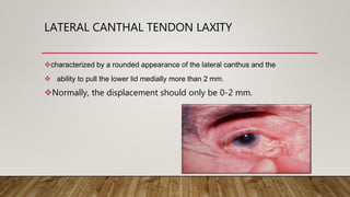
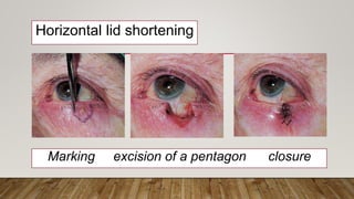
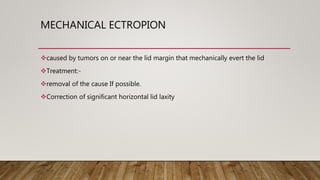
ECTROPION
PRESENTER :- DR RAHUL MAHALA
DNB OPHTHALMOLOGY
BOKARO GENERAL HOSPITAL, BOKARO
JHARKHAND ( INDIA )
ECTROPION
It is an outward turning of the eyelid margin .
This more frequently affects the lower eyelid.
Upper eyelid ectropion is uncommon.
CLASSIFICATION
Classified in 5 types
1)Congenital
2) Involutional
3) Paralytic
4) Cicatricial
5) Mechanical
Involutional ectropion is more common.
Congenital ectropion is very rare.
SYMPTOMS
 Epiphora :- excessive tearing.
 Excessive dryness.
 Foreign body sensation
 Irritation.
 Burning.
 Redness.
 Chronic conjunctivitis
 Keratinization
 Corneal exposure
GRADING
Lid margin is out rolled and depending on out rolling ectropion can be classified
as under:
Grade I –only punctum is everted
Grade II –lid margin is everted and palpebral conjunctiva is visible
Grade III –fornix is also visible
ETIOLOGICAL FACTORS
Horizontal lid laxity:-can be demonstrated by pulling the central part
of the lid 8 mm or more from the globe, with a failure to snap back to its normal
position on release without the patient first blinking.
Classified:-
MILD – takes some time
MODERATE – goes back slowly without blink
SEVERE – does not go back even after a blink
LID LAXITY
MEDIAL CANTHAL TENDON LAXITY
demonstrated by pulling the lower lid laterally and observing the position of the
inferior punctum
If the lid is normal the punctum should not be displaced more than 1–2 mm
Mild:- punctum reaches the limbus
Severe :- may reach the pupil
LATERAL CANTHAL TENDON LAXITY
characterized by a rounded appearance of the lateral canthus and the
 ability to pull the lower lid medially more than 2 mm.
Normally, the displacement should only be 0-2 mm.
PATHOPHYSIOLOGY
1. Senile or involutional :-caused by a horizontal lid laxity
Lengthening of medial and lateral canthal tendons with ageing changes
It is the most common type of ectropion and has a continuous pathological
process that is aggravated by conjunctivitis and epiphora
2. cicatricial ectropion :- when anterior lamella is shortened either postoperatively,
trauma or ulceration.
3) Paralytic ectropion :- supporting of the lower eyelid in its normal
position depending on the orbicularis oculi muscle tone and loss of this
support in case of facial nerve
4) congenital ectropion :- due to shortage of skin as in congenital
ichthyosis or blepharophimosis .
5) mechanical ectropion :- is caused by eversion of lower lid by mass or
tumor
Involution ectropion
Cicatricial ectropion
Paralytic ectropion
Mechanical ectropion
EVALUATION
Proper history:- time, duration, h/o trauma, h/o surgery of eye
Check severity of ectropion
External examination
Slit lamp examination:- for lid, conjunctiva, cornea, pupil
Test for ectropion:- ex eyelid snap test, medial canthus laxity test.
OCT cornea
TREATMENT
1 medical therapy
2 surgical therapy
INVOLUTIONAL ECTROPION
Involutional (age-related) ectropion affects the lower lid of elderly
patients
Cause :- horizontal lid laxity
medial canthal tendon laxity
lateral canthal tendon laxity
Treatment :-
Generalized ectropion treated with repair of horizontal lid laxity.
Lateral tarsal strip procedure :- lower canthal tendon is
tightened by shortening and reattachment to the lateral
orbital rim.
Helpful if the lateral canthus is rounded and lax, with
associated tear overflow
Excision of a tarsoconjunctival pentagon is an
alternative that can be placed to excise an area of
misdirected lashes or keratinized conjunctiva.
Medial ectropion :- if mild, may be treated with a
medial conjunctival diamond excision (medial spindle
procedure)
Medial canthal tendon laxity, if marked, requires
stabilization prior to horizontal shortening to avoid
excessive dragging of the punctum laterally.
Horizontal lid shortening
Marking excision of a pentagon closure
Lateral tarsal strip procedure
Medial diamond excision
CICATRICIAL ECTROPION
caused by scarring or contracture of the skin and underlying tissues
which pulls the eyelid away from the globe
Treatment
Mild localized cases are treated by excision of scar tissue combined with ‘Z’-plasty
Severe cases require transposition flaps or free skin grafts, sources of skin include
the upper lids, posterior auricular, preauricular and supraclavicular areas.
Cicatricial ectropion
PARALYTIC ECTROPION
caused by ipsilateral facial nerve palsy and is associated with retraction of the
upper and lower lids and brow ptosis
Left facial palsy and severe paralytic ectropion
Treatment :-
Temporary :- to protect the cornea,
1)Lubrication with higher viscosity tear substitutes during the
day
2)Taping shut of the lids during sleep, are usually adequate in
mild cases
3)Botulinum toxin injection into the levator to induce temporary
ptosis.
4)Temporary tarsorrhaphy
Permanent treatment:- when irreversible damage to facial
nerve.
Medial canthoplasty may be performed if the medial canthal
tendon is intact. The eyelids are sutured together medial to the
lacrimal puncta.
A lateral canthal sling or tarsal strip may be used to correct
residual ectropion and raise the lateral canthus
Upper eyelid lowering by levator disinsertion.
Gold weight implantation in the upper lid can assist closure.
A small lateral tarsorrhaphy is usually cosmetically acceptable.
Medial canthoplasty
lateral canthal sling –refashioned canthal tendon from the lower lid is passed
through a buttonhole in the tendon from the upper lid
MECHANICAL ECTROPION
caused by tumors on or near the lid margin that mechanically evert the lid
Treatment:-
removal of the cause If possible.
Correction of significant horizontal lid laxity
THANK YOU

Ectropion

  • 1.
    ECTROPION PRESENTER :- DRRAHUL MAHALA DNB OPHTHALMOLOGY BOKARO GENERAL HOSPITAL, BOKARO JHARKHAND ( INDIA )
  • 2.
    ECTROPION It is anoutward turning of the eyelid margin . This more frequently affects the lower eyelid. Upper eyelid ectropion is uncommon.
  • 3.
    CLASSIFICATION Classified in 5types 1)Congenital 2) Involutional 3) Paralytic 4) Cicatricial 5) Mechanical Involutional ectropion is more common. Congenital ectropion is very rare.
  • 4.
    SYMPTOMS  Epiphora :-excessive tearing.  Excessive dryness.  Foreign body sensation  Irritation.  Burning.  Redness.  Chronic conjunctivitis  Keratinization  Corneal exposure
  • 5.
    GRADING Lid margin isout rolled and depending on out rolling ectropion can be classified as under: Grade I –only punctum is everted Grade II –lid margin is everted and palpebral conjunctiva is visible Grade III –fornix is also visible
  • 6.
    ETIOLOGICAL FACTORS Horizontal lidlaxity:-can be demonstrated by pulling the central part of the lid 8 mm or more from the globe, with a failure to snap back to its normal position on release without the patient first blinking. Classified:- MILD – takes some time MODERATE – goes back slowly without blink SEVERE – does not go back even after a blink
  • 7.
  • 8.
    MEDIAL CANTHAL TENDONLAXITY demonstrated by pulling the lower lid laterally and observing the position of the inferior punctum If the lid is normal the punctum should not be displaced more than 1–2 mm Mild:- punctum reaches the limbus Severe :- may reach the pupil
  • 9.
    LATERAL CANTHAL TENDONLAXITY characterized by a rounded appearance of the lateral canthus and the  ability to pull the lower lid medially more than 2 mm. Normally, the displacement should only be 0-2 mm.
  • 10.
    PATHOPHYSIOLOGY 1. Senile orinvolutional :-caused by a horizontal lid laxity Lengthening of medial and lateral canthal tendons with ageing changes It is the most common type of ectropion and has a continuous pathological process that is aggravated by conjunctivitis and epiphora 2. cicatricial ectropion :- when anterior lamella is shortened either postoperatively, trauma or ulceration.
  • 11.
    3) Paralytic ectropion:- supporting of the lower eyelid in its normal position depending on the orbicularis oculi muscle tone and loss of this support in case of facial nerve 4) congenital ectropion :- due to shortage of skin as in congenital ichthyosis or blepharophimosis . 5) mechanical ectropion :- is caused by eversion of lower lid by mass or tumor
  • 12.
  • 13.
    EVALUATION Proper history:- time,duration, h/o trauma, h/o surgery of eye Check severity of ectropion External examination Slit lamp examination:- for lid, conjunctiva, cornea, pupil Test for ectropion:- ex eyelid snap test, medial canthus laxity test. OCT cornea
  • 14.
  • 15.
    INVOLUTIONAL ECTROPION Involutional (age-related)ectropion affects the lower lid of elderly patients Cause :- horizontal lid laxity medial canthal tendon laxity lateral canthal tendon laxity Treatment :- Generalized ectropion treated with repair of horizontal lid laxity.
  • 16.
    Lateral tarsal stripprocedure :- lower canthal tendon is tightened by shortening and reattachment to the lateral orbital rim. Helpful if the lateral canthus is rounded and lax, with associated tear overflow Excision of a tarsoconjunctival pentagon is an alternative that can be placed to excise an area of misdirected lashes or keratinized conjunctiva. Medial ectropion :- if mild, may be treated with a medial conjunctival diamond excision (medial spindle procedure) Medial canthal tendon laxity, if marked, requires stabilization prior to horizontal shortening to avoid excessive dragging of the punctum laterally.
  • 17.
    Horizontal lid shortening Markingexcision of a pentagon closure
  • 18.
  • 19.
  • 20.
    CICATRICIAL ECTROPION caused byscarring or contracture of the skin and underlying tissues which pulls the eyelid away from the globe Treatment Mild localized cases are treated by excision of scar tissue combined with ‘Z’-plasty Severe cases require transposition flaps or free skin grafts, sources of skin include the upper lids, posterior auricular, preauricular and supraclavicular areas.
  • 21.
  • 22.
    PARALYTIC ECTROPION caused byipsilateral facial nerve palsy and is associated with retraction of the upper and lower lids and brow ptosis Left facial palsy and severe paralytic ectropion
  • 23.
    Treatment :- Temporary :-to protect the cornea, 1)Lubrication with higher viscosity tear substitutes during the day 2)Taping shut of the lids during sleep, are usually adequate in mild cases 3)Botulinum toxin injection into the levator to induce temporary ptosis. 4)Temporary tarsorrhaphy
  • 24.
    Permanent treatment:- whenirreversible damage to facial nerve. Medial canthoplasty may be performed if the medial canthal tendon is intact. The eyelids are sutured together medial to the lacrimal puncta. A lateral canthal sling or tarsal strip may be used to correct residual ectropion and raise the lateral canthus Upper eyelid lowering by levator disinsertion. Gold weight implantation in the upper lid can assist closure. A small lateral tarsorrhaphy is usually cosmetically acceptable.
  • 25.
  • 26.
    lateral canthal sling–refashioned canthal tendon from the lower lid is passed through a buttonhole in the tendon from the upper lid
  • 27.
    MECHANICAL ECTROPION caused bytumors on or near the lid margin that mechanically evert the lid Treatment:- removal of the cause If possible. Correction of significant horizontal lid laxity
  • 28.