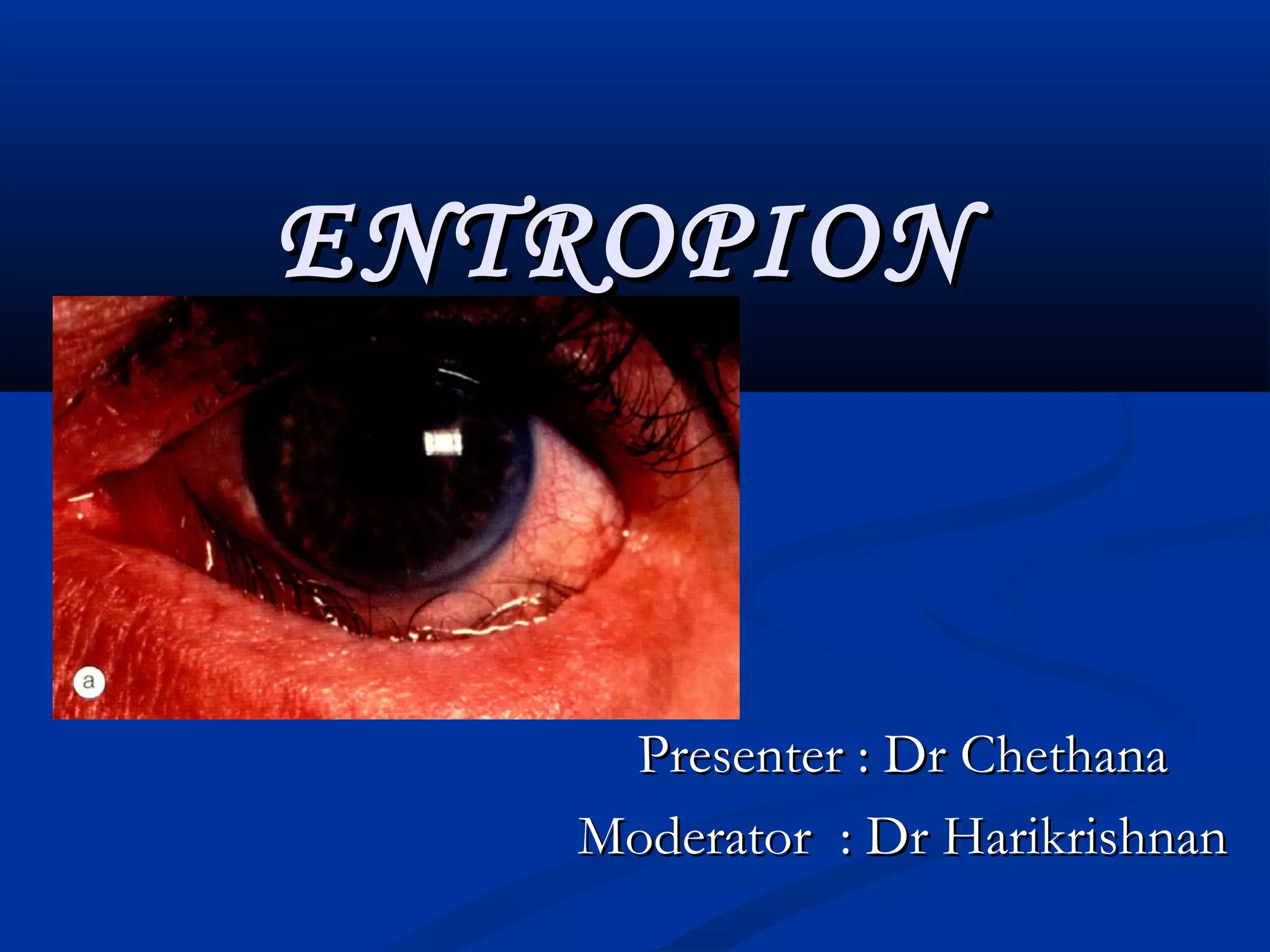
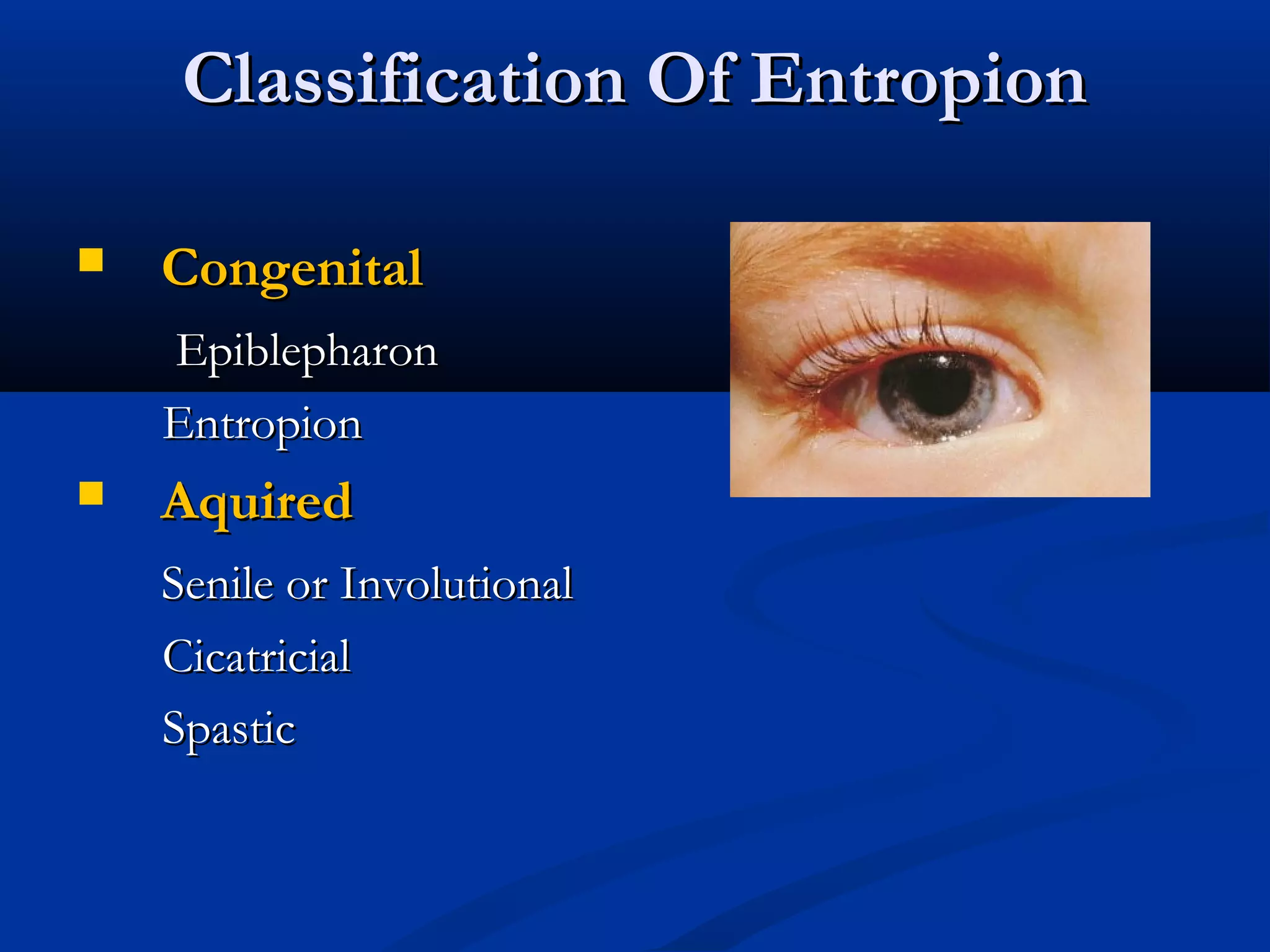
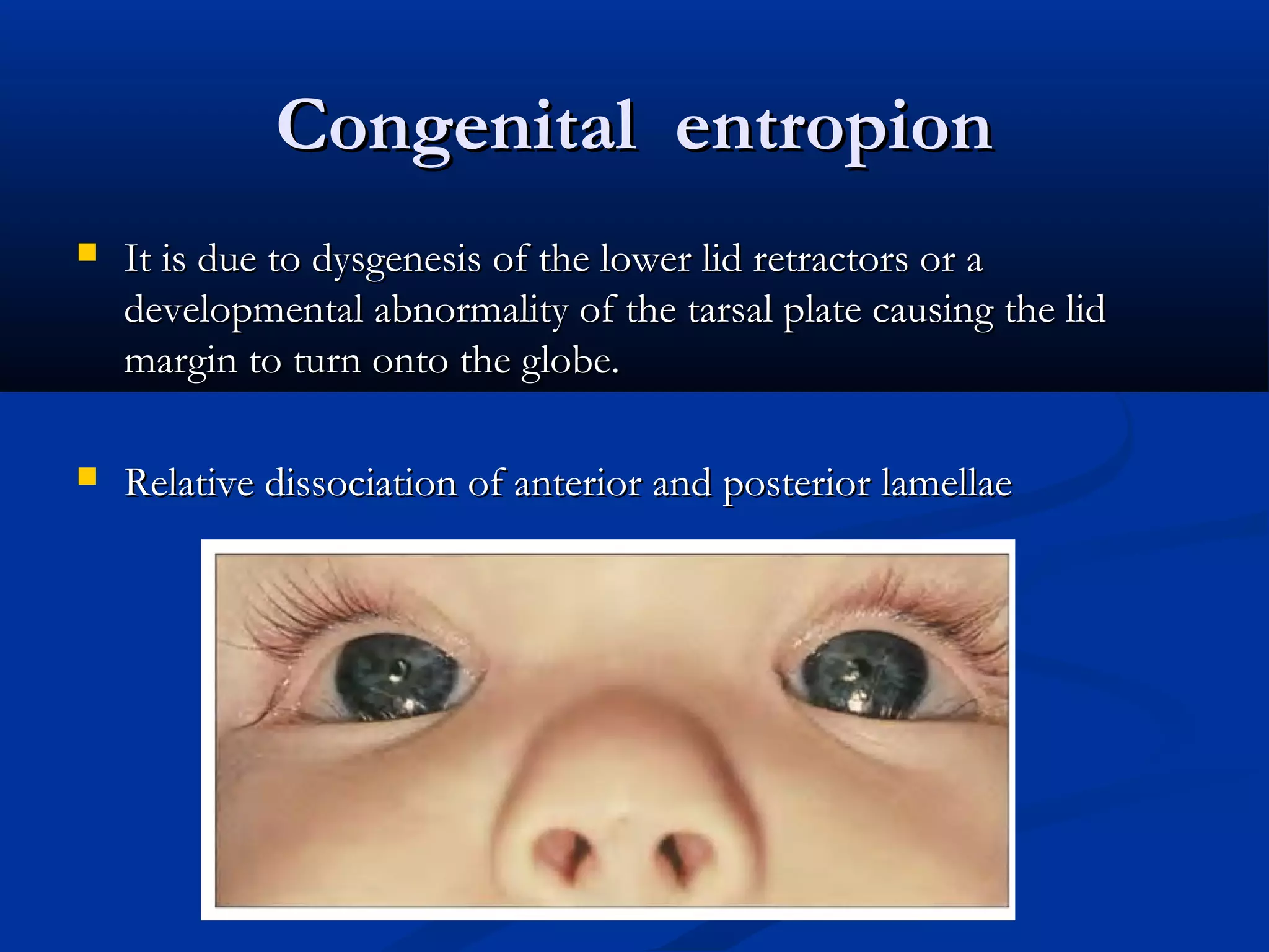
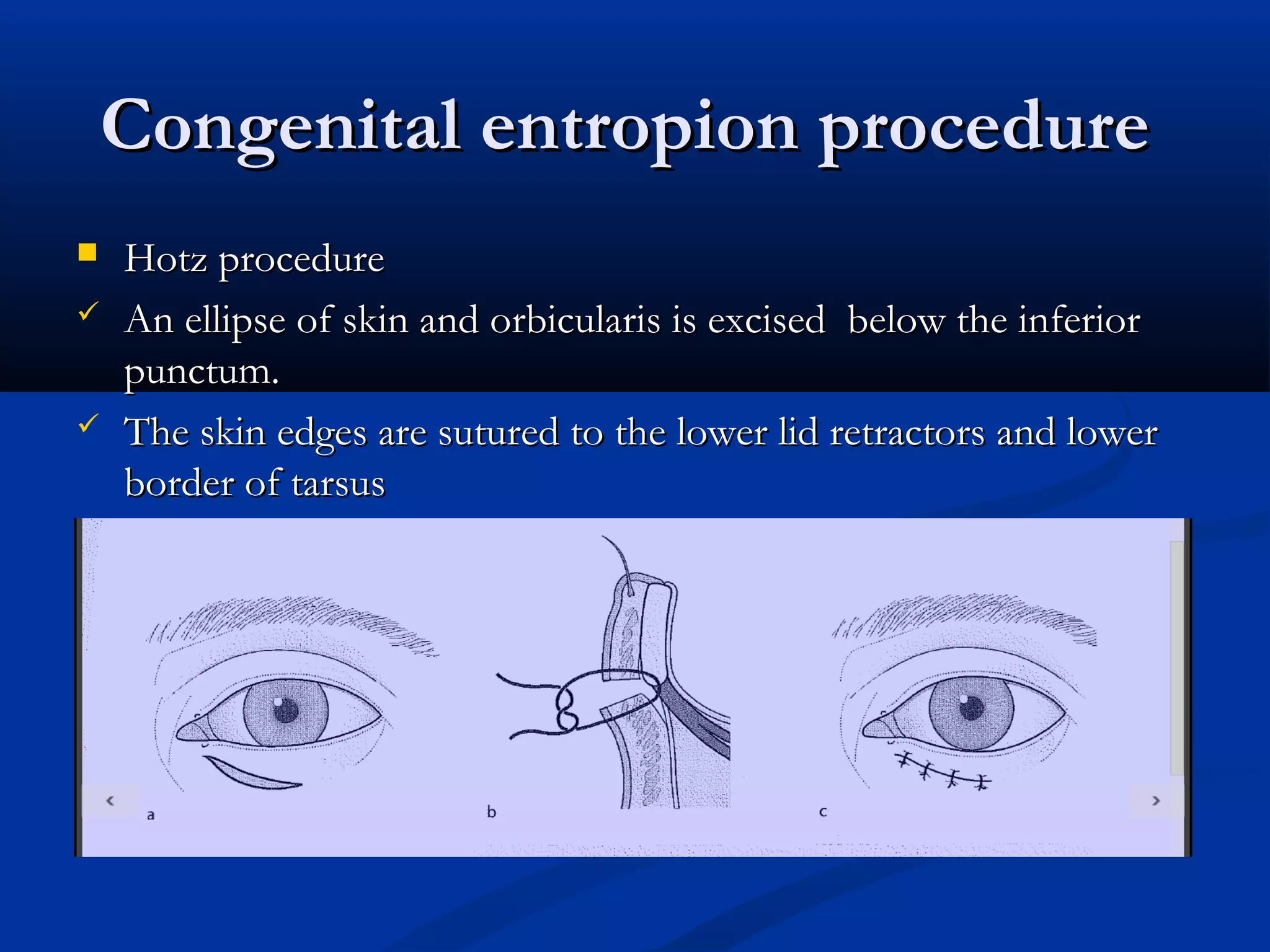
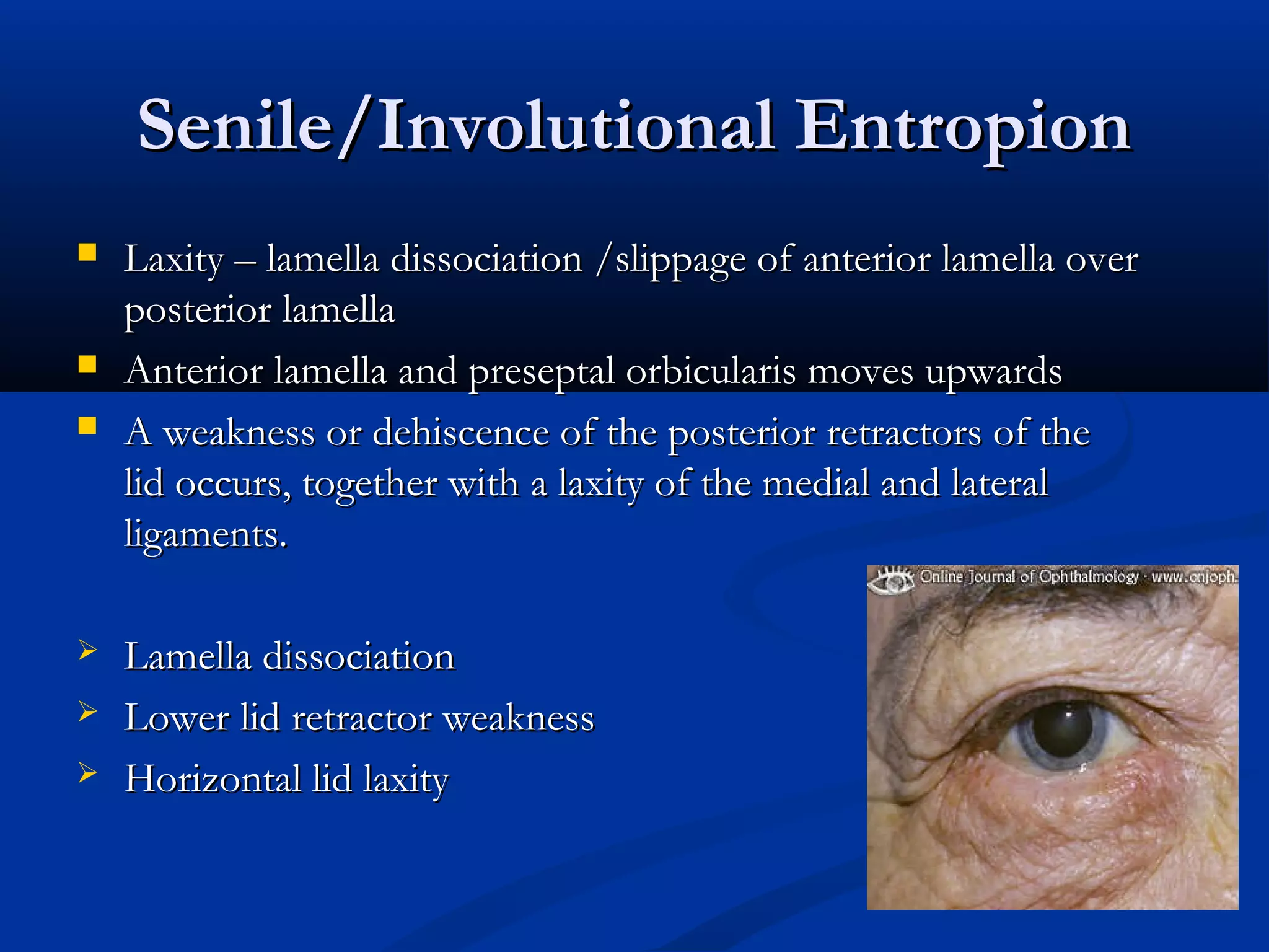
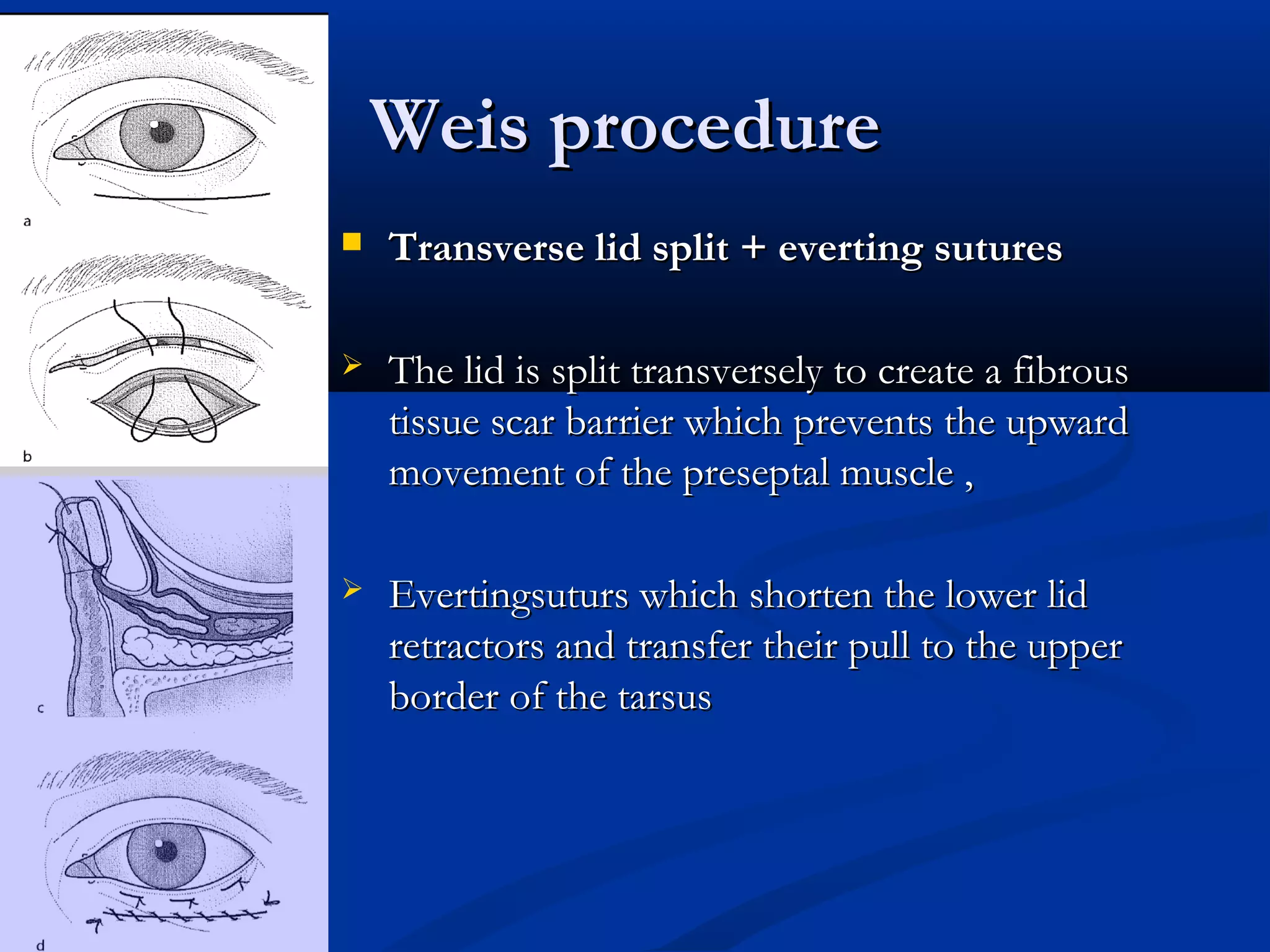
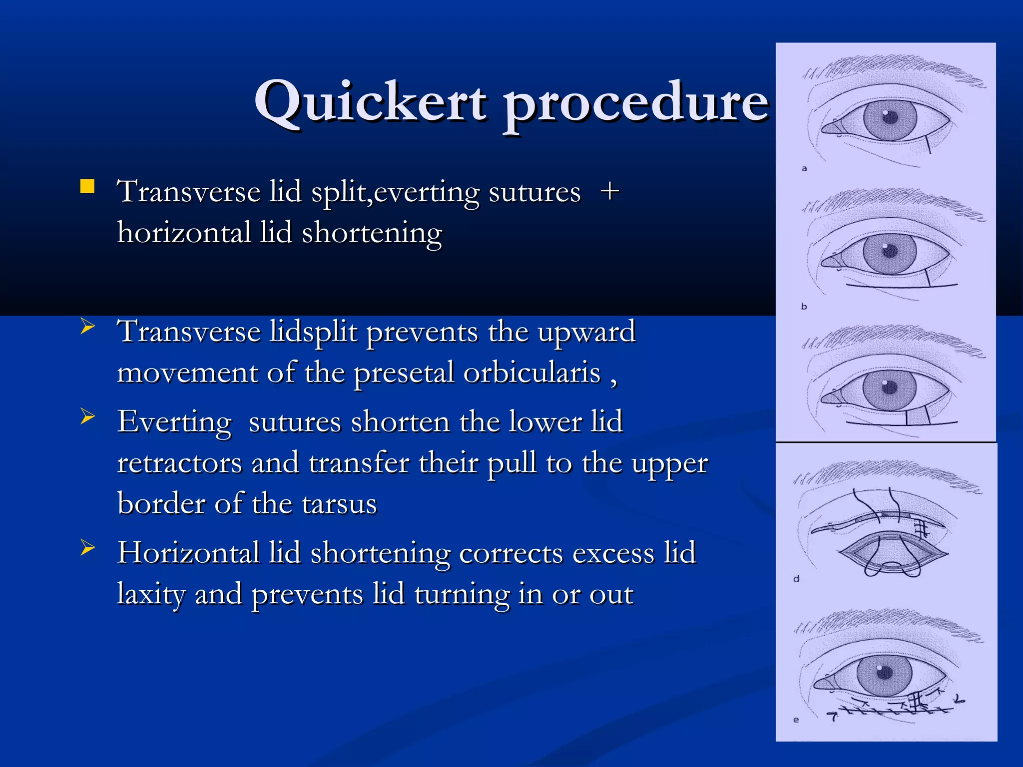
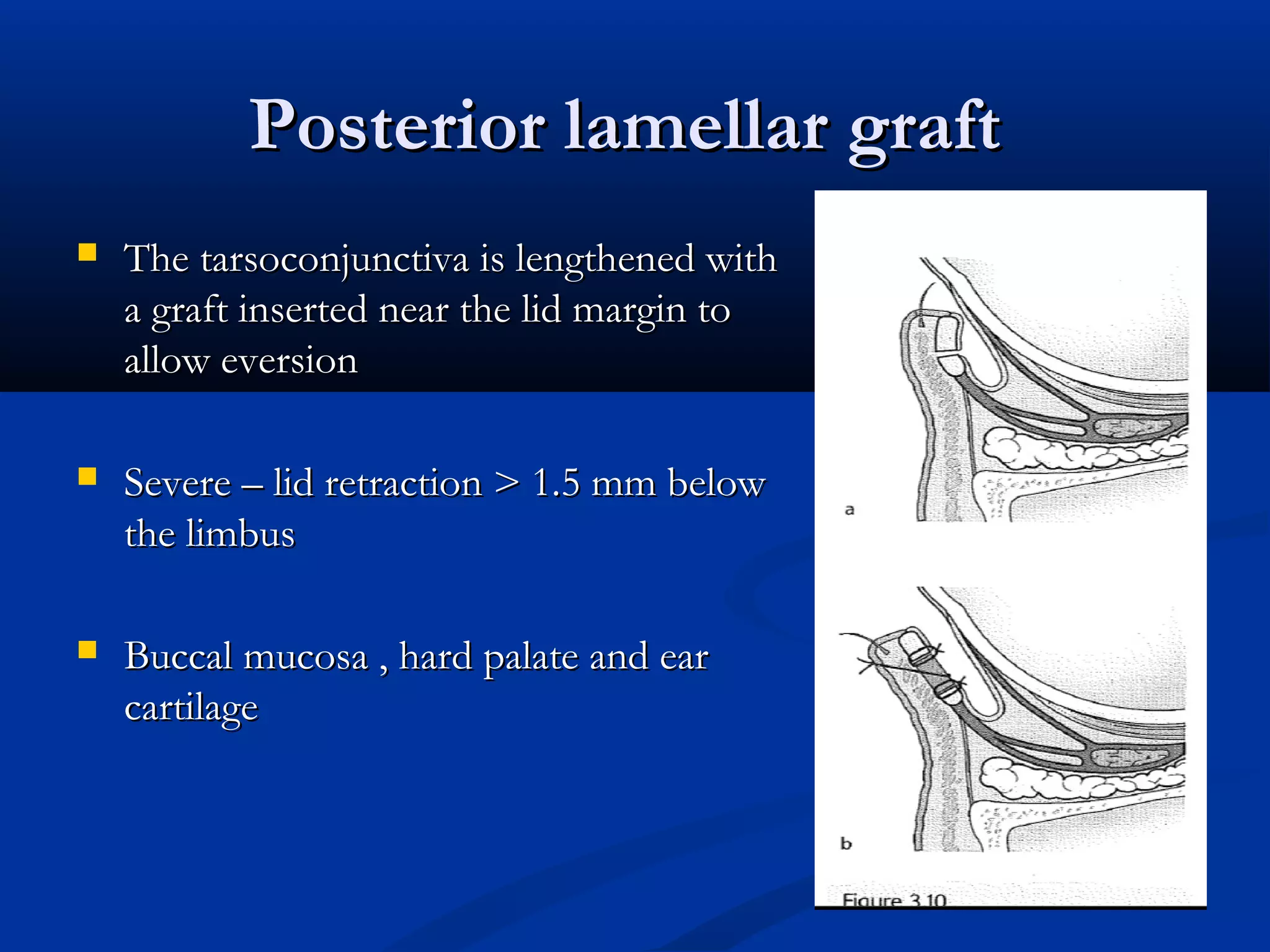
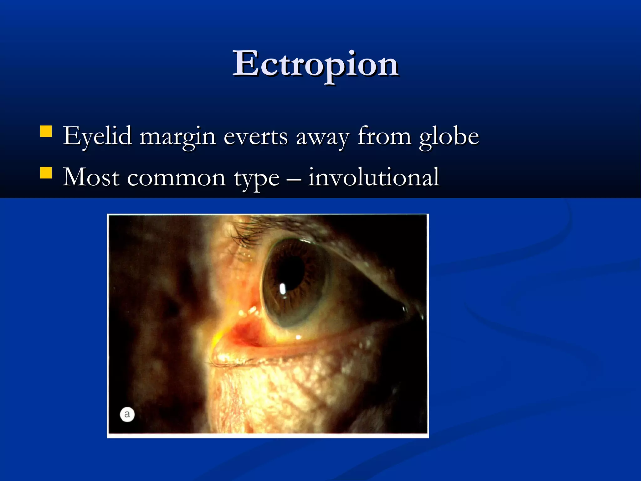
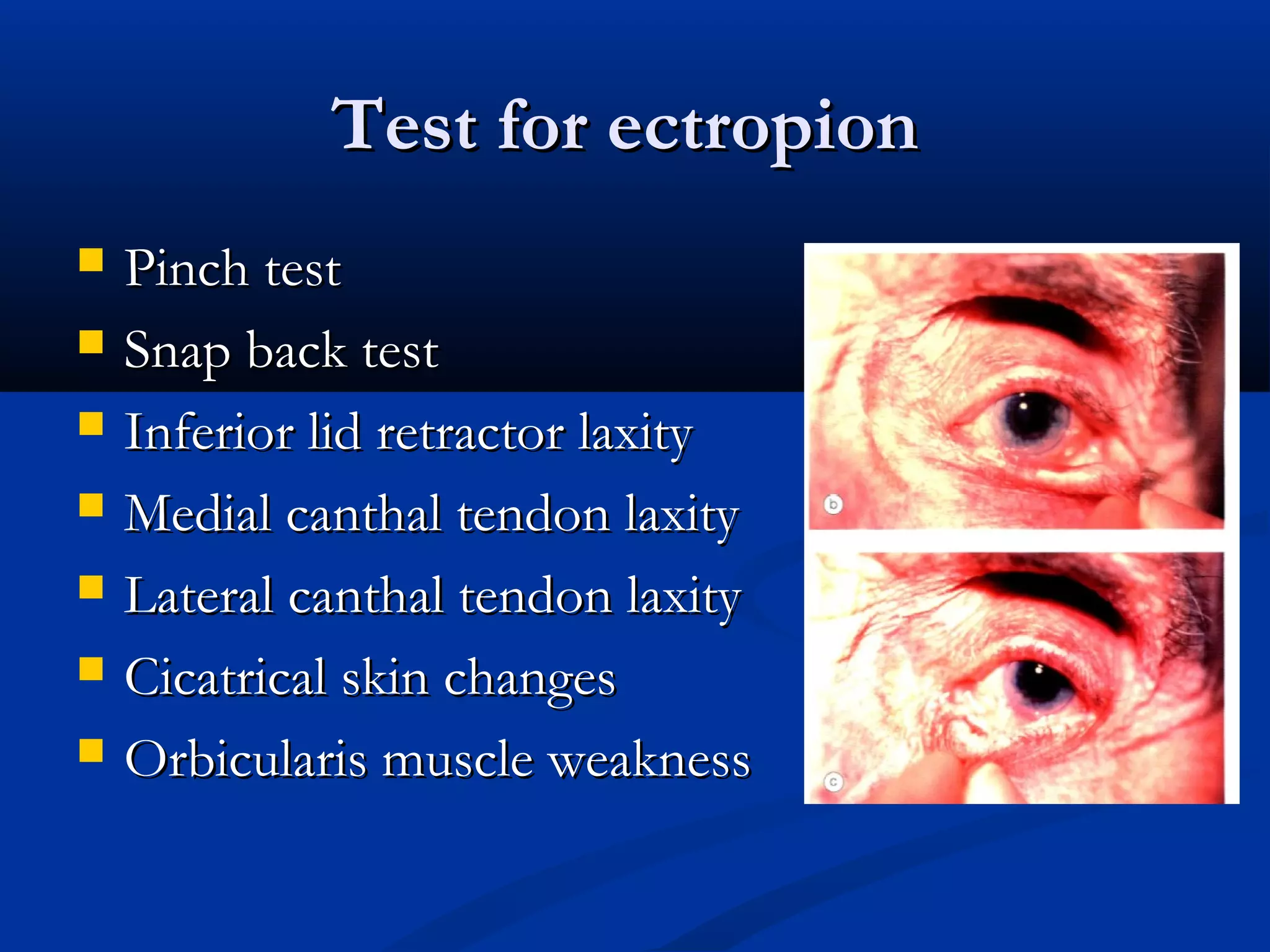
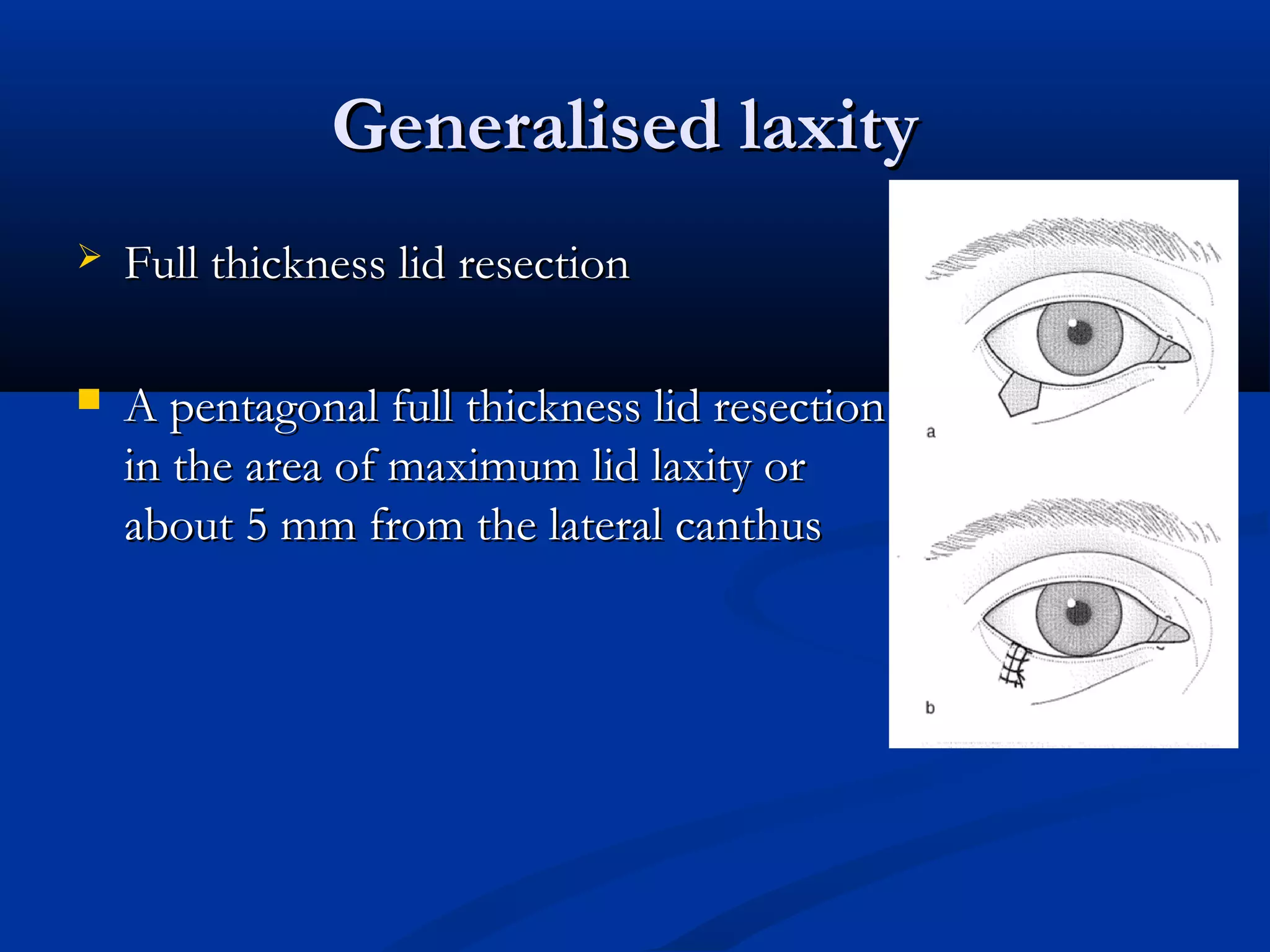
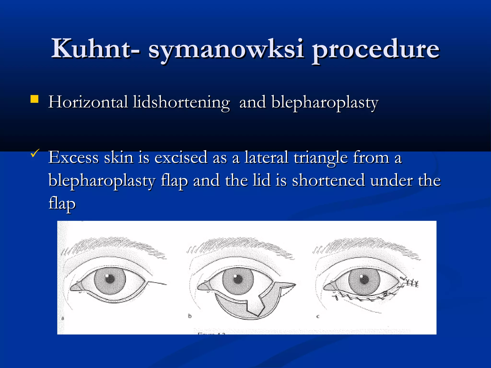
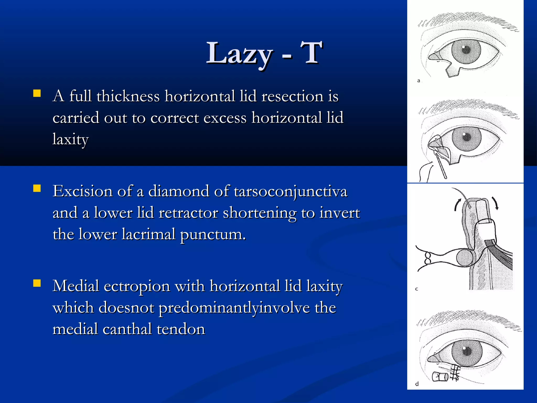
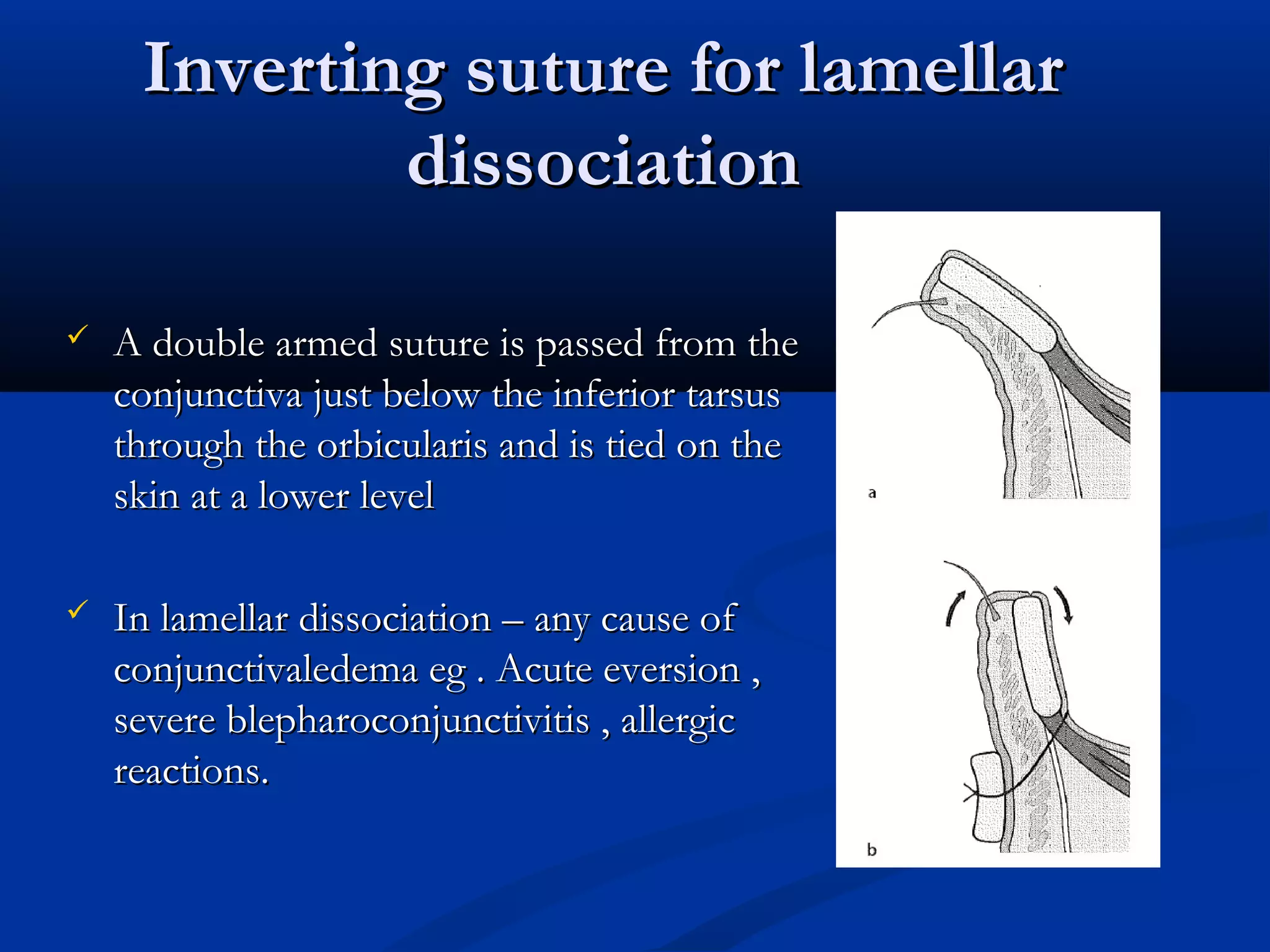
ENTROPIONENTROPION
Presenter : Dr ChethanaPresenter : Dr Chethana
Moderator : Dr HarikrishnanModerator : Dr Harikrishnan
EntropionEntropion
 It is in-turning of the eyelid margin.It is in-turning of the eyelid margin.
c/f
forEign body sEnsation
irritation
Lacrimation
photophobia
GradesGrades
 Grade 1- only posterior lid border is inrolledGrade 1- only posterior lid border is inrolled
 Grade 2- inturning upto intermarginal stripGrade 2- inturning upto intermarginal strip
 Grade 3- the whole lid margin including theGrade 3- the whole lid margin including the
anterior border is inturnedanterior border is inturned
ExaminationExamination
 Lid laxityLid laxity
o Pinch test > 8mm – LaxityPinch test > 8mm – Laxity
o Snap backSnap back
Grades features
Normal Lid returns immediately on release
1 Approx 2-3 sec
2 4-5 sec
3 > 5 sec but returns to normal position
4 Lid continues to hang down
•
Grades Features
Mild Upto nasal limbus
Moderate Upto pupil
Severe Beyond temporal limbus
 Medial canthal tendon laxityMedial canthal tendon laxity
 Lateral canthal tendon laxityLateral canthal tendon laxity
 Acute angle of lateral canthus- normalAcute angle of lateral canthus- normal
 Bell’s phenomenonBell’s phenomenon
 Orbicularis muscle toneOrbicularis muscle tone
 Digital eversion of eyelidDigital eversion of eyelid
 Slitlamp examinationSlitlamp examination
 Flurosceine stainingFlurosceine staining
 Schrimer testSchrimer test
 SyringingSyringing
Classification Of EntropionClassification Of Entropion
 CongenitalCongenital
EpiblepharonEpiblepharon
EntropionEntropion
 AquiredAquired
Senile or InvolutionalSenile or Involutional
CicatricialCicatricial
SpasticSpastic
Congenital entropionCongenital entropion
 It is due to dysgenesis of the lower lid retractors or aIt is due to dysgenesis of the lower lid retractors or a
developmental abnormality of the tarsal plate causing the liddevelopmental abnormality of the tarsal plate causing the lid
margin to turn onto the globe.margin to turn onto the globe.
 Relative dissociation of anterior and posterior lamellaeRelative dissociation of anterior and posterior lamellae
Suture correction of epiblepharonSuture correction of epiblepharon
 Sutures are passed below theSutures are passed below the
tarsal plate and tied on thetarsal plate and tied on the
apex of the epiblepharon foldapex of the epiblepharon fold
of skin to hold the 2 lamellaeof skin to hold the 2 lamellae
togethertogether
Congenital entropion procedureCongenital entropion procedure
 Hotz procedureHotz procedure
 An ellipse of skin and orbicularis is excised below the inferiorAn ellipse of skin and orbicularis is excised below the inferior
punctum.punctum.
 The skin edges are sutured to the lower lid retractors and lowerThe skin edges are sutured to the lower lid retractors and lower
border of tarsusborder of tarsus
Senile/Involutional EntropionSenile/Involutional Entropion
 Laxity – lamella dissociation /slippage of anterior lamella overLaxity – lamella dissociation /slippage of anterior lamella over
posterior lamellaposterior lamella
 Anterior lamella and preseptal orbicularis moves upwardsAnterior lamella and preseptal orbicularis moves upwards
 A weakness or dehiscence of the posterior retractors of theA weakness or dehiscence of the posterior retractors of the
lid occurs, together with a laxity of the medial and laterallid occurs, together with a laxity of the medial and lateral
ligaments.ligaments.
 Lamella dissociationLamella dissociation
 Lower lid retractor weaknessLower lid retractor weakness
 Horizontal lid laxityHorizontal lid laxity
Everting suturs to transfer the pull to
lid margins
SutursSuturs
 Transverse –Transverse –
 from just below tarsus to emerge 2 to 3mmfrom just below tarsus to emerge 2 to 3mm
below lash linebelow lash line
 To prevent upward movement of aTo prevent upward movement of a
preseptal musclepreseptal muscle
 Everting –Everting –
 from inferior fornix to emerge just belowfrom inferior fornix to emerge just below
thelash linethelash line
 To tighten the lower lid retractors and evertTo tighten the lower lid retractors and evert
the lid marginthe lid margin
Weis procedureWeis procedure
 Transverse lid split + everting suturesTransverse lid split + everting sutures
 The lid is split transversely to create a fibrousThe lid is split transversely to create a fibrous
tissue scar barrier which prevents the upwardtissue scar barrier which prevents the upward
movement of the preseptal muscle ,movement of the preseptal muscle ,
 Evertingsuturs which shorten the lower lidEvertingsuturs which shorten the lower lid
retractors and transfer their pull to the upperretractors and transfer their pull to the upper
border of the tarsusborder of the tarsus
Quickert procedureQuickert procedure
 Transverse lid split,everting sutures +Transverse lid split,everting sutures +
horizontal lid shorteninghorizontal lid shortening
 Transverse lidsplit prevents the upwardTransverse lidsplit prevents the upward
movement of the presetal orbicularis ,movement of the presetal orbicularis ,
 Everting sutures shorten the lower lidEverting sutures shorten the lower lid
retractors and transfer their pull to the upperretractors and transfer their pull to the upper
border of the tarsusborder of the tarsus
 Horizontal lid shortening corrects excess lidHorizontal lid shortening corrects excess lid
laxity and prevents lid turning in or outlaxity and prevents lid turning in or out
Jones procedureJones procedure
 Plication of lower lid retractorsPlication of lower lid retractors
 The lower lid retractors are exposed via a skinThe lower lid retractors are exposed via a skin
incision, shortened and the sutures used toincision, shortened and the sutures used to
create a barrier to the upward movement of thecreate a barrier to the upward movement of the
preseptal musclepreseptal muscle
 For recurrent entropionFor recurrent entropion
 Adequate correction – end pointAdequate correction – end point
 Avoid epiphoraAvoid epiphora
Cicatricial entropionCicatricial entropion
 It is caused by cicatricial contraction of the palpebralIt is caused by cicatricial contraction of the palpebral
conjunctiva, resulting in a relative shortening of theconjunctiva, resulting in a relative shortening of the
tarsoconjunctival lamina of the lid and an inversion of the lidtarsoconjunctival lamina of the lid and an inversion of the lid
margin.margin.
 CAUSES:CAUSES: Trachoma, trauma, chemical burns, Stevens-Trachoma, trauma, chemical burns, Stevens-
Johnsons Synd.Johnsons Synd.
Tarsal fractureTarsal fracture
 The tarsus is fractured horizontally andThe tarsus is fractured horizontally and
hinged into eversion with evertinghinged into eversion with everting
suturessutures
 In mild- moderate cases in whichIn mild- moderate cases in which
cicatrisation has not caused the lidcicatrisation has not caused the lid
margin to be retracted >1.5 mm belowmargin to be retracted >1.5 mm below
limbuslimbus
Posterior lamellar graftPosterior lamellar graft
 The tarsoconjunctiva is lengthened withThe tarsoconjunctiva is lengthened with
a graft inserted near the lid margin toa graft inserted near the lid margin to
allow eversionallow eversion
 Severe – lid retraction > 1.5 mm belowSevere – lid retraction > 1.5 mm below
the limbusthe limbus
 Buccal mucosa , hard palate and earBuccal mucosa , hard palate and ear
cartilagecartilage
Grey –line split and retractorGrey –line split and retractor
repositioningrepositioning
 The lid margin is slit at the grey line. The lower lid retractors areThe lid margin is slit at the grey line. The lower lid retractors are
attached to the anterior lamella below the lashes to forcibly evertattached to the anterior lamella below the lashes to forcibly evert
the split lid marginthe split lid margin
 Severe cicatrix with trachomaSevere cicatrix with trachoma
Spastic EntropionSpastic Entropion
 It occurs in response to ocular irritation such as inflammationsIt occurs in response to ocular irritation such as inflammations
or trauma, and is due to spasm of the orbicularis in the presenceor trauma, and is due to spasm of the orbicularis in the presence
of degeneration of the palpebral connective tissue separating theof degeneration of the palpebral connective tissue separating the
orbicularis muscle fibers.orbicularis muscle fibers.
 CAUSES:CAUSES: Chronic irritative corneal conditions, after tight ocularChronic irritative corneal conditions, after tight ocular
bandaging.bandaging.
 Treating the cause , lubricants , eye patching , botulinum toxinTreating the cause , lubricants , eye patching , botulinum toxin
Upper lid entropionUpper lid entropion
 TrachomaTrachoma
 SjsSjs
 Ocular cicatricial pemphigoidOcular cicatricial pemphigoid
 Chronic blepharoconjunctivitisChronic blepharoconjunctivitis
 Chemical burnsChemical burns
 IatrogenicIatrogenic
 Chronic anophthalmic inflammationChronic anophthalmic inflammation
 Severe eyebrow ptosisSevere eyebrow ptosis
Kemp and collinKemp and collin
Degree Clinical features Entropion
procedure
Mild Apparent migration of MG
Conjunctivalisation of lid margin
Lash globe contact on up gaze
Anterior lamellar
reposition
Moderate Mild +
Thickening of tarsal plate
Lid retraction
Tarsal wedge resection
Severe Gross lid distorsion
Metaplastic lashes
Presence of keratin plaques
Trabut procedure
(rotation of terminal
tarsus )
EctropionEctropion
EctropionEctropion
 Eyelid margin everts away from globeEyelid margin everts away from globe
 Most common type – involutionalMost common type – involutional
HistoryHistory
Test for ectropionTest for ectropion
 Pinch testPinch test
 Snap back testSnap back test
 Inferior lid retractor laxityInferior lid retractor laxity
 Medial canthal tendon laxityMedial canthal tendon laxity
 Lateral canthal tendon laxityLateral canthal tendon laxity
 Cicatrical skin changesCicatrical skin changes
 Orbicularis muscle weaknessOrbicularis muscle weakness
Position of punctaPosition of puncta
Grade Features
Mild Puncta not opposed to the globe on looking up
Moderate Puncta not opposed to the globe in primary gaze
Severe Palpabral conjunctiva and fornix are exposed
GradesGrades
 Grade 1 – punctal eversionGrade 1 – punctal eversion
 Grade 2 – ectropion of sharp posterior lidGrade 2 – ectropion of sharp posterior lid
marginmargin
 Grade 3 – palpabral conjunctival exposureGrade 3 – palpabral conjunctival exposure
 Grade 4 – exposure of fornixGrade 4 – exposure of fornix
ClassificationClassification
Congenital
• aCute eversion
• Congenital
eCtropion
aquired
• involutional
• MeChaniCal
• CiCatriCial
• paralytiC
Acute eversionAcute eversion
 Complete eversionComplete eversion
 Inclusion conjunctivitisInclusion conjunctivitis
 Anterior lamellarAnterior lamellar
inflammation or shorteninginflammation or shortening
 Lubrication and short termLubrication and short term
patcchingpatcching
Congenital ectropionCongenital ectropion
 Associated withAssociated with
blepharophimosis ,Downsblepharophimosis ,Downs
syndrome ,icthyosissyndrome ,icthyosis
 Vertical insufficiency ofVertical insufficiency of
anterior lamellaeanterior lamellae
 Horizontal thickening ofHorizontal thickening of
lateral canthal tendonlateral canthal tendon
 Vertical lengthening ofVertical lengthening of
anterior lamella by fullanterior lamella by full
thickmess skin graftthickmess skin graft
Horizontal lid shorteningHorizontal lid shortening
Generalised laxityGeneralised laxity
 Full thickness lid resectionFull thickness lid resection
 A pentagonal full thickness lid resectionA pentagonal full thickness lid resection
in the area of maximum lid laxity orin the area of maximum lid laxity or
about 5 mm from the lateral canthusabout 5 mm from the lateral canthus
Kuhnt- symanowksi procedureKuhnt- symanowksi procedure
 Horizontal lidshortening and blepharoplastyHorizontal lidshortening and blepharoplasty
 Excess skin is excised as a lateral triangle from aExcess skin is excised as a lateral triangle from a
blepharoplasty flap and the lid is shortened under theblepharoplasty flap and the lid is shortened under the
flapflap
Lateral tarsal stripLateral tarsal strip
 The lid is shortened and a new lateralThe lid is shortened and a new lateral
canthal tendon is created out of thecanthal tendon is created out of the
lateral tarsal platelateral tarsal plate
Medial canthal sutureMedial canthal suture
 Anterior limb stabilising sutureAnterior limb stabilising suture ––
 Anterior limb of medial canthal tendonAnterior limb of medial canthal tendon
is shortened by suturing the medial endis shortened by suturing the medial end
of the lower tarsal plate to the mainof the lower tarsal plate to the main
part of the medial canthal tendonpart of the medial canthal tendon
Posterior limb suturePosterior limb suture
 Suturing the medial end of lower tarsal plate to theSuturing the medial end of lower tarsal plate to the
periosteum over the posterior lacrimal crestperiosteum over the posterior lacrimal crest
 Open techniqueOpen technique
 Closed techniqueClosed technique
Lower lid retractor shortening /Lower lid retractor shortening /
reattachmentreattachment
 Excision of diamond of tarsoconjunctivaExcision of diamond of tarsoconjunctiva
 Lazy TLazy T
 Tarsal ectropion repair / tarsal eversionTarsal ectropion repair / tarsal eversion
Excision of diamond ofExcision of diamond of
tarsoconjunctivatarsoconjunctiva
 Lower lid retractor plicationLower lid retractor plication
 Lower punctum is inverted by vertically shortening the posteriorLower punctum is inverted by vertically shortening the posterior
lamella of the lid and tightening the lower lid retractorslamella of the lid and tightening the lower lid retractors
Lazy - TLazy - T
 A full thickness horizontal lid resection isA full thickness horizontal lid resection is
carried out to correct excess horizontal lidcarried out to correct excess horizontal lid
laxitylaxity
 Excision of a diamond of tarsoconjunctivaExcision of a diamond of tarsoconjunctiva
and a lower lid retractor shortening to invertand a lower lid retractor shortening to invert
the lower lacrimal punctum.the lower lacrimal punctum.
 Medial ectropion with horizontal lid laxityMedial ectropion with horizontal lid laxity
which doesnot predominantlyinvolve thewhich doesnot predominantlyinvolve the
medial canthal tendonmedial canthal tendon
Inverting suture for lamellarInverting suture for lamellar
dissociationdissociation
 A double armed suture is passed from theA double armed suture is passed from the
conjunctiva just below the inferior tarsusconjunctiva just below the inferior tarsus
through the orbicularis and is tied on thethrough the orbicularis and is tied on the
skin at a lower levelskin at a lower level
 In lamellar dissociation – any cause ofIn lamellar dissociation – any cause of
conjunctivaledema eg . Acute eversion ,conjunctivaledema eg . Acute eversion ,
severe blepharoconjunctivitis , allergicsevere blepharoconjunctivitis , allergic
reactions.reactions.
Z plasty for cicatricial ectropionZ plasty for cicatricial ectropion
 2 flaps of skin are transposed2 flaps of skin are transposed
• Increases the length of skin in the line of scar contraction at theIncreases the length of skin in the line of scar contraction at the
expense of shortening the skin at right angles to itexpense of shortening the skin at right angles to it
• Alters the line of scarAlters the line of scar
Mechanical ectropionMechanical ectropion
 Cysts or tumoursCysts or tumours
 Excise the causeExcise the cause
 Prevent cicatricial ectropion - vertical excisionPrevent cicatricial ectropion - vertical excision
 Treat associated horizontal lidlaxityTreat associated horizontal lidlaxity
THANK YOUTHANK YOU
Ectropion and entropion

Ectropion and entropion

  • 1.
    ENTROPIONENTROPION Presenter : DrChethanaPresenter : Dr Chethana Moderator : Dr HarikrishnanModerator : Dr Harikrishnan
  • 2.
    EntropionEntropion  It isin-turning of the eyelid margin.It is in-turning of the eyelid margin. c/f forEign body sEnsation irritation Lacrimation photophobia
  • 3.
    GradesGrades  Grade 1-only posterior lid border is inrolledGrade 1- only posterior lid border is inrolled  Grade 2- inturning upto intermarginal stripGrade 2- inturning upto intermarginal strip  Grade 3- the whole lid margin including theGrade 3- the whole lid margin including the anterior border is inturnedanterior border is inturned
  • 4.
    ExaminationExamination  Lid laxityLidlaxity o Pinch test > 8mm – LaxityPinch test > 8mm – Laxity o Snap backSnap back Grades features Normal Lid returns immediately on release 1 Approx 2-3 sec 2 4-5 sec 3 > 5 sec but returns to normal position 4 Lid continues to hang down
  • 5.
    • Grades Features Mild Uptonasal limbus Moderate Upto pupil Severe Beyond temporal limbus  Medial canthal tendon laxityMedial canthal tendon laxity  Lateral canthal tendon laxityLateral canthal tendon laxity  Acute angle of lateral canthus- normalAcute angle of lateral canthus- normal
  • 6.
     Bell’s phenomenonBell’sphenomenon  Orbicularis muscle toneOrbicularis muscle tone  Digital eversion of eyelidDigital eversion of eyelid  Slitlamp examinationSlitlamp examination  Flurosceine stainingFlurosceine staining  Schrimer testSchrimer test  SyringingSyringing
  • 7.
    Classification Of EntropionClassificationOf Entropion  CongenitalCongenital EpiblepharonEpiblepharon EntropionEntropion  AquiredAquired Senile or InvolutionalSenile or Involutional CicatricialCicatricial SpasticSpastic
  • 8.
    Congenital entropionCongenital entropion It is due to dysgenesis of the lower lid retractors or aIt is due to dysgenesis of the lower lid retractors or a developmental abnormality of the tarsal plate causing the liddevelopmental abnormality of the tarsal plate causing the lid margin to turn onto the globe.margin to turn onto the globe.  Relative dissociation of anterior and posterior lamellaeRelative dissociation of anterior and posterior lamellae
  • 9.
    Suture correction ofepiblepharonSuture correction of epiblepharon  Sutures are passed below theSutures are passed below the tarsal plate and tied on thetarsal plate and tied on the apex of the epiblepharon foldapex of the epiblepharon fold of skin to hold the 2 lamellaeof skin to hold the 2 lamellae togethertogether
  • 10.
    Congenital entropion procedureCongenitalentropion procedure  Hotz procedureHotz procedure  An ellipse of skin and orbicularis is excised below the inferiorAn ellipse of skin and orbicularis is excised below the inferior punctum.punctum.  The skin edges are sutured to the lower lid retractors and lowerThe skin edges are sutured to the lower lid retractors and lower border of tarsusborder of tarsus
  • 11.
    Senile/Involutional EntropionSenile/Involutional Entropion Laxity – lamella dissociation /slippage of anterior lamella overLaxity – lamella dissociation /slippage of anterior lamella over posterior lamellaposterior lamella  Anterior lamella and preseptal orbicularis moves upwardsAnterior lamella and preseptal orbicularis moves upwards  A weakness or dehiscence of the posterior retractors of theA weakness or dehiscence of the posterior retractors of the lid occurs, together with a laxity of the medial and laterallid occurs, together with a laxity of the medial and lateral ligaments.ligaments.  Lamella dissociationLamella dissociation  Lower lid retractor weaknessLower lid retractor weakness  Horizontal lid laxityHorizontal lid laxity
  • 12.
    Everting suturs totransfer the pull to lid margins
  • 13.
    SutursSuturs  Transverse –Transverse–  from just below tarsus to emerge 2 to 3mmfrom just below tarsus to emerge 2 to 3mm below lash linebelow lash line  To prevent upward movement of aTo prevent upward movement of a preseptal musclepreseptal muscle  Everting –Everting –  from inferior fornix to emerge just belowfrom inferior fornix to emerge just below thelash linethelash line  To tighten the lower lid retractors and evertTo tighten the lower lid retractors and evert the lid marginthe lid margin
  • 14.
    Weis procedureWeis procedure Transverse lid split + everting suturesTransverse lid split + everting sutures  The lid is split transversely to create a fibrousThe lid is split transversely to create a fibrous tissue scar barrier which prevents the upwardtissue scar barrier which prevents the upward movement of the preseptal muscle ,movement of the preseptal muscle ,  Evertingsuturs which shorten the lower lidEvertingsuturs which shorten the lower lid retractors and transfer their pull to the upperretractors and transfer their pull to the upper border of the tarsusborder of the tarsus
  • 15.
    Quickert procedureQuickert procedure Transverse lid split,everting sutures +Transverse lid split,everting sutures + horizontal lid shorteninghorizontal lid shortening  Transverse lidsplit prevents the upwardTransverse lidsplit prevents the upward movement of the presetal orbicularis ,movement of the presetal orbicularis ,  Everting sutures shorten the lower lidEverting sutures shorten the lower lid retractors and transfer their pull to the upperretractors and transfer their pull to the upper border of the tarsusborder of the tarsus  Horizontal lid shortening corrects excess lidHorizontal lid shortening corrects excess lid laxity and prevents lid turning in or outlaxity and prevents lid turning in or out
  • 16.
    Jones procedureJones procedure Plication of lower lid retractorsPlication of lower lid retractors  The lower lid retractors are exposed via a skinThe lower lid retractors are exposed via a skin incision, shortened and the sutures used toincision, shortened and the sutures used to create a barrier to the upward movement of thecreate a barrier to the upward movement of the preseptal musclepreseptal muscle  For recurrent entropionFor recurrent entropion  Adequate correction – end pointAdequate correction – end point  Avoid epiphoraAvoid epiphora
  • 17.
    Cicatricial entropionCicatricial entropion It is caused by cicatricial contraction of the palpebralIt is caused by cicatricial contraction of the palpebral conjunctiva, resulting in a relative shortening of theconjunctiva, resulting in a relative shortening of the tarsoconjunctival lamina of the lid and an inversion of the lidtarsoconjunctival lamina of the lid and an inversion of the lid margin.margin.  CAUSES:CAUSES: Trachoma, trauma, chemical burns, Stevens-Trachoma, trauma, chemical burns, Stevens- Johnsons Synd.Johnsons Synd.
  • 18.
    Tarsal fractureTarsal fracture The tarsus is fractured horizontally andThe tarsus is fractured horizontally and hinged into eversion with evertinghinged into eversion with everting suturessutures  In mild- moderate cases in whichIn mild- moderate cases in which cicatrisation has not caused the lidcicatrisation has not caused the lid margin to be retracted >1.5 mm belowmargin to be retracted >1.5 mm below limbuslimbus
  • 19.
    Posterior lamellar graftPosteriorlamellar graft  The tarsoconjunctiva is lengthened withThe tarsoconjunctiva is lengthened with a graft inserted near the lid margin toa graft inserted near the lid margin to allow eversionallow eversion  Severe – lid retraction > 1.5 mm belowSevere – lid retraction > 1.5 mm below the limbusthe limbus  Buccal mucosa , hard palate and earBuccal mucosa , hard palate and ear cartilagecartilage
  • 20.
    Grey –line splitand retractorGrey –line split and retractor repositioningrepositioning  The lid margin is slit at the grey line. The lower lid retractors areThe lid margin is slit at the grey line. The lower lid retractors are attached to the anterior lamella below the lashes to forcibly evertattached to the anterior lamella below the lashes to forcibly evert the split lid marginthe split lid margin  Severe cicatrix with trachomaSevere cicatrix with trachoma
  • 21.
    Spastic EntropionSpastic Entropion It occurs in response to ocular irritation such as inflammationsIt occurs in response to ocular irritation such as inflammations or trauma, and is due to spasm of the orbicularis in the presenceor trauma, and is due to spasm of the orbicularis in the presence of degeneration of the palpebral connective tissue separating theof degeneration of the palpebral connective tissue separating the orbicularis muscle fibers.orbicularis muscle fibers.  CAUSES:CAUSES: Chronic irritative corneal conditions, after tight ocularChronic irritative corneal conditions, after tight ocular bandaging.bandaging.  Treating the cause , lubricants , eye patching , botulinum toxinTreating the cause , lubricants , eye patching , botulinum toxin
  • 22.
    Upper lid entropionUpperlid entropion  TrachomaTrachoma  SjsSjs  Ocular cicatricial pemphigoidOcular cicatricial pemphigoid  Chronic blepharoconjunctivitisChronic blepharoconjunctivitis  Chemical burnsChemical burns  IatrogenicIatrogenic  Chronic anophthalmic inflammationChronic anophthalmic inflammation  Severe eyebrow ptosisSevere eyebrow ptosis
  • 23.
    Kemp and collinKempand collin Degree Clinical features Entropion procedure Mild Apparent migration of MG Conjunctivalisation of lid margin Lash globe contact on up gaze Anterior lamellar reposition Moderate Mild + Thickening of tarsal plate Lid retraction Tarsal wedge resection Severe Gross lid distorsion Metaplastic lashes Presence of keratin plaques Trabut procedure (rotation of terminal tarsus )
  • 25.
  • 26.
    EctropionEctropion  Eyelid margineverts away from globeEyelid margin everts away from globe  Most common type – involutionalMost common type – involutional
  • 27.
  • 28.
    Test for ectropionTestfor ectropion  Pinch testPinch test  Snap back testSnap back test  Inferior lid retractor laxityInferior lid retractor laxity  Medial canthal tendon laxityMedial canthal tendon laxity  Lateral canthal tendon laxityLateral canthal tendon laxity  Cicatrical skin changesCicatrical skin changes  Orbicularis muscle weaknessOrbicularis muscle weakness
  • 29.
    Position of punctaPositionof puncta Grade Features Mild Puncta not opposed to the globe on looking up Moderate Puncta not opposed to the globe in primary gaze Severe Palpabral conjunctiva and fornix are exposed
  • 30.
    GradesGrades  Grade 1– punctal eversionGrade 1 – punctal eversion  Grade 2 – ectropion of sharp posterior lidGrade 2 – ectropion of sharp posterior lid marginmargin  Grade 3 – palpabral conjunctival exposureGrade 3 – palpabral conjunctival exposure  Grade 4 – exposure of fornixGrade 4 – exposure of fornix
  • 31.
    ClassificationClassification Congenital • aCute eversion •Congenital eCtropion aquired • involutional • MeChaniCal • CiCatriCial • paralytiC
  • 32.
    Acute eversionAcute eversion Complete eversionComplete eversion  Inclusion conjunctivitisInclusion conjunctivitis  Anterior lamellarAnterior lamellar inflammation or shorteninginflammation or shortening  Lubrication and short termLubrication and short term patcchingpatcching Congenital ectropionCongenital ectropion  Associated withAssociated with blepharophimosis ,Downsblepharophimosis ,Downs syndrome ,icthyosissyndrome ,icthyosis  Vertical insufficiency ofVertical insufficiency of anterior lamellaeanterior lamellae  Horizontal thickening ofHorizontal thickening of lateral canthal tendonlateral canthal tendon  Vertical lengthening ofVertical lengthening of anterior lamella by fullanterior lamella by full thickmess skin graftthickmess skin graft
  • 34.
  • 35.
    Generalised laxityGeneralised laxity Full thickness lid resectionFull thickness lid resection  A pentagonal full thickness lid resectionA pentagonal full thickness lid resection in the area of maximum lid laxity orin the area of maximum lid laxity or about 5 mm from the lateral canthusabout 5 mm from the lateral canthus
  • 36.
    Kuhnt- symanowksi procedureKuhnt-symanowksi procedure  Horizontal lidshortening and blepharoplastyHorizontal lidshortening and blepharoplasty  Excess skin is excised as a lateral triangle from aExcess skin is excised as a lateral triangle from a blepharoplasty flap and the lid is shortened under theblepharoplasty flap and the lid is shortened under the flapflap
  • 37.
    Lateral tarsal stripLateraltarsal strip  The lid is shortened and a new lateralThe lid is shortened and a new lateral canthal tendon is created out of thecanthal tendon is created out of the lateral tarsal platelateral tarsal plate
  • 38.
    Medial canthal sutureMedialcanthal suture  Anterior limb stabilising sutureAnterior limb stabilising suture ––  Anterior limb of medial canthal tendonAnterior limb of medial canthal tendon is shortened by suturing the medial endis shortened by suturing the medial end of the lower tarsal plate to the mainof the lower tarsal plate to the main part of the medial canthal tendonpart of the medial canthal tendon
  • 39.
    Posterior limb suturePosteriorlimb suture  Suturing the medial end of lower tarsal plate to theSuturing the medial end of lower tarsal plate to the periosteum over the posterior lacrimal crestperiosteum over the posterior lacrimal crest  Open techniqueOpen technique  Closed techniqueClosed technique
  • 40.
    Lower lid retractorshortening /Lower lid retractor shortening / reattachmentreattachment  Excision of diamond of tarsoconjunctivaExcision of diamond of tarsoconjunctiva  Lazy TLazy T  Tarsal ectropion repair / tarsal eversionTarsal ectropion repair / tarsal eversion
  • 41.
    Excision of diamondofExcision of diamond of tarsoconjunctivatarsoconjunctiva  Lower lid retractor plicationLower lid retractor plication  Lower punctum is inverted by vertically shortening the posteriorLower punctum is inverted by vertically shortening the posterior lamella of the lid and tightening the lower lid retractorslamella of the lid and tightening the lower lid retractors
  • 42.
    Lazy - TLazy- T  A full thickness horizontal lid resection isA full thickness horizontal lid resection is carried out to correct excess horizontal lidcarried out to correct excess horizontal lid laxitylaxity  Excision of a diamond of tarsoconjunctivaExcision of a diamond of tarsoconjunctiva and a lower lid retractor shortening to invertand a lower lid retractor shortening to invert the lower lacrimal punctum.the lower lacrimal punctum.  Medial ectropion with horizontal lid laxityMedial ectropion with horizontal lid laxity which doesnot predominantlyinvolve thewhich doesnot predominantlyinvolve the medial canthal tendonmedial canthal tendon
  • 43.
    Inverting suture forlamellarInverting suture for lamellar dissociationdissociation  A double armed suture is passed from theA double armed suture is passed from the conjunctiva just below the inferior tarsusconjunctiva just below the inferior tarsus through the orbicularis and is tied on thethrough the orbicularis and is tied on the skin at a lower levelskin at a lower level  In lamellar dissociation – any cause ofIn lamellar dissociation – any cause of conjunctivaledema eg . Acute eversion ,conjunctivaledema eg . Acute eversion , severe blepharoconjunctivitis , allergicsevere blepharoconjunctivitis , allergic reactions.reactions.
  • 44.
    Z plasty forcicatricial ectropionZ plasty for cicatricial ectropion  2 flaps of skin are transposed2 flaps of skin are transposed • Increases the length of skin in the line of scar contraction at theIncreases the length of skin in the line of scar contraction at the expense of shortening the skin at right angles to itexpense of shortening the skin at right angles to it • Alters the line of scarAlters the line of scar
  • 47.
    Mechanical ectropionMechanical ectropion Cysts or tumoursCysts or tumours  Excise the causeExcise the cause  Prevent cicatricial ectropion - vertical excisionPrevent cicatricial ectropion - vertical excision  Treat associated horizontal lidlaxityTreat associated horizontal lidlaxity
  • 48.