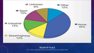
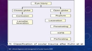
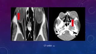
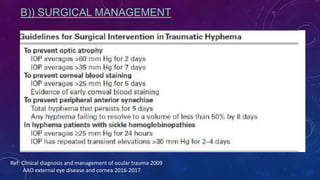
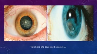
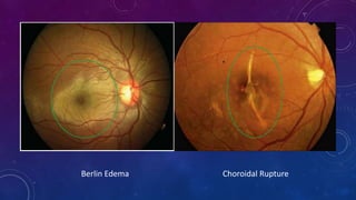
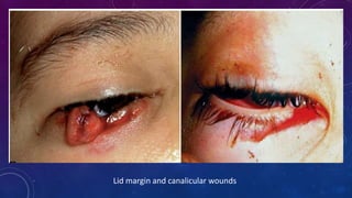
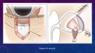
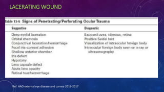
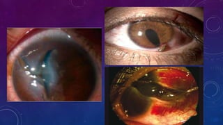
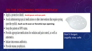
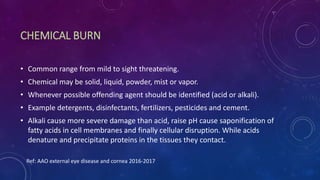
OCULAR TRAUMA
DUHOK EYE HOSPITAL
DR. NIWAR AMEEN
TOPICS
• Epidemiology
• Classification
• Clinical evaluation
• Investigation
• Closed globe injury
• Open globe injury
• Chemical burn
• References
BACKGROUND
• An injury to the eye or its surrounding tissues is the most common cause for attendance to the
emergency department.
• The extent of trauma may range from simple superficial injuries to devastating penetrating injuries of
the eyelids, lacrimal system, and globe.
• The surgical management of such injuries is directed primarily at the restoration of normal ocular
anatomy; the ultimate goal is to prevent secondary complications and maximize the patient’s visual
prognosis.
• Eye injuries are a major and under recognized cause of disabling ocular morbidity that especially
affect the young.
• Globally, more than 500.000 blinding injuries occur every year.
• In general, males are more frequently reported to have eye injuries than females.
• Results varied across studies regarding the age-specific frequency of eye injuries with some reporting
a higher incidence in older children and others in younger children.
Ref: clinical diagnosis and management of ocular trauma 2009
INJURY BY PLACE
REF: CLINICAL DIAGNOSIS AND MANAGEMENT OF OCULAR TRAUMA 2009
CLASSIFICATION
• The classification system categorizes ocular injuries at the time of initial examination. It is designed to promote the
use of standard terminology and assessment.
• Terms frequently used are :
• 1. Eyewall—For clinical and practical purposes, the term eyewall must be restricted to the rigid structures of the sclera
and cornea.
• 2. Closed-globe injury—The eyewall does not have a full-thickness wound.
• 3. Open-globe injury—The eyewall has a full-thickness wound, cornea and/or sclera. Choroid and retina may be intact,
prolapsed or damaged.
• I . Rupture—Full-thickness wound of the eyewall, caused by a blunt object; the impact results in momentary increase
of the intraocular pressure. The eyewall gives way at its weakest point (at the impact site or elsewhere; example: an
old cataract wound dehisces even though the impact occurred elsewhere); the actual wound is produced by an inside-
out mechanism.
• II . Laceration—Full-thickness wound of the eyewall, usually caused by a sharp object; the wound occurs at the impact
site by an outside-in mechanism.
• A . Penetrating injury—Single laceration of the eyewall, usually caused by a sharp object. No exit wound has occurred.
• B . Intraocular foreign body injury (IOFB)—Retained foreign object(s) causing entrance laceration(s).
• C. Perforating injury—Two full-thickness lacerations (entrance and exit) of the eyewall, usually caused by a sharp
object.
Ref: clinical diagnosis and management of ocular trauma 2009
CLINICAL EVALUATION
History
•History of presenting event (time, location, object, associations, other details)
• Medical history ( D.M , HrT, asthma, sickle thalassemia and others)
• History of allergy to any known medication
• Previous Ocular surgery and VA
General examination
 Structural examination Inspect the orbits and eyelids
 Functional examination (VA, RAPD, IOP, Ocular motility)
 Slit lamp examination (conj, cornea, AC, Lens, Fundus) mandatory almost for every trauma
with children be gentle and patience … sedation may be needed
Ref: clinical diagnosis and management of ocular trauma 2009
INVESTIGATIONS
1.B - scan ultrasonography
2.Plain orbital x-ray
3.CT-scan
Vitreous Hemorrhage and RD Net
Orbital plain x-ray Net
CT-orbit Net
CLOSED GLOBE INJURY
Contusion
- Corneal abrasion
- Conj Abrasion or
Laceration
Superficial FB
- Corneal FB
- Conj FB
Lamellar laceration
- Corneal
- Scleral
Corneal FB AAO
Conjunctival FB Net
Corneal Abrasion Net
-Distinguish from ulcer
-Rx with topical Abs with cycloplegia alone
-Patching not always necessary
-Therapeutic CL (close follow up)
- topical NSAIDs for 1st 24-48 hrs … pain relieve
Ref: AAO External eye disease and Cornea 2016-2017
Conjunctival Laceration Net
Lamellar Corneal wound Net
OPEN GLOBE INJURY
Rupture blunt trauma Laceration
-Perforation
-Penetration
-IOFB
HYPHEMA
• Hemorrhage into AC, from vessels of iris or anterior CB.
• Caused by blunt trauma most commonly.
• Most common in children 34 are boys
• Spontaneous hyphema caused by neovascul, ocular neoplasm and vascular
Anomalies.
• Grading ;
1. G1 … blood occupying less than 13 AC
2. G2 … blood occupying 13 – 12 AC
3. G3 … blood filling more than 12 AC
4. G4 … total filling AC ( eight ball)
Complications ;
1- corneal blood staining Amblyopia
2- peripheral anterior synechiae
3- posterior synechiae
4- optic atrophy … due to IOP ‘’ transiently acute or chronic ‘’
- 50 mmHg for 5 days
- 35 mmHg or more for 7 days
- with sickle patient’s 35-39 mmHg for 2-4 days
Prognostic factors is usually good even for total hyphema unless ;
1- associated damage to other ocular structures
2- secondary hemorrhage happened or not?
3- occurrence of complications
• A)) MEDICAL MANAGEMENT
1. Protective shield over the injured eye
2. Restriction of physical activity
3. Elevation of the head of the bed
4. Daily observation
5. Treatment on outpatient basis unless
• Medications:
1. Topical corticosteroids
2. Topical cycloplegic (controversy)
3. Topical IOP lowering agents
4. Oral antifibrinolytics
B)) SURGICAL MANAGEMENT
Ref: Clinical diagnosis and management of ocular trauma 2009
AAO external eye disease and cornea 2016-2017
Traumatic Mydriasis Net
Traumatic and dislocated cataract Net
Berlin Edema Choroidal Rupture
Scleral Rupture
The most common sites for scleral rupture are
1. Limbus … site of previous surgery
2. Parallel to and under insertion of rectus muscle
• Important diagnostic signs
1. Marked decrease in ocular duction
2. Very boggy conj chemosis with hemorrhage (ecchymosis)
3. Deepened AC
4. Severe Vitreous hemorrhage
 IOP usually reduced but may be normal or high
 Peritomy is required to explore site of rupture for repair
Ref: AAO external eye disease and cornea 2016-2017
Rupture Globe Net
LID MARGIN AND CANALICULAS
• Careful examination so as not to miss associated occult globe injury
• Repair is better to be done as soon as possible … still can be delayed up to 48 hrs
• Repair can be done under L.A, or in case of child G.A is required
• one tarsal vicryl suture involve 90% of tarsus
• Three non-absorbable sutures through meibomian orifices, gray line and lash line left long
• Stitches removal after two weeks
• This is for direct suturing without tissue lose … otherwise wound closure is individualized accordingly
• Canalicular injuries should not be missed and repaired under G.A with bicanalicular or monocanalicular
tube or polyprolene can be placed in for 3-6 months.
Ref: Clinical diagnosis and management of ocular trauma 2009
Lid margin and canalicular wounds
Repair of wound
LACERATING WOUND
Ref: AAO external eye disease and cornea 2016-2017
Management
• Laceration must be repaired as soon as possible although 36 hrs delay no disadvantages
documented, early repair minimize following possible complications
1. Pain control
2. Prolapse of intraocular structures
3. Suprachoroidal hemorrhage
4. Microbial contamination of the wound
5. Proliferation of projected microbes in the eye
6. Migration of epithelium in the wound
7. Intraocular inflammation
8. Lens opacity
Ref: AAO external eye disease and cornea 2016-2017
DO THE FOLLOWING PREOPERATIVELY
Such as B-scan or forceful eye opening
Don’t forget:
Legally stay safe
Avoid gauze and eye pads
During surgery
• Irrigate the wound , remove debris
• Reposit nonnecrotic uveal tissue
• Excise prolapsed vitreous and necrotic
uveal tissue
• Initiate suturing with landmarks 1st
(limbus and angles of the wound)
• Repair cornea and sclera then lid margins
• Suture cornea with 10-0 nylon
• Sclera with 9-0 nylon or 8-0 silk
• 80-90% corneal thickness with long and narrow
limbal stitches and wide and short toward visual axis
Ref: AAO external eye disease and cornea 2016-2017
CHEMICAL BURN
• Common range from mild to sight threatening.
• Chemical may be solid, liquid, powder, mist or vapor.
• Whenever possible offending agent should be identified (acid or alkali).
• Example detergents, disinfectants, fertilizers, pesticides and cement.
• Alkali cause more severe damage than acid, raise pH cause saponification of
fatty acids in cell membranes and finally cellular disruption. While acids
denature and precipitate proteins in the tissues they contact.
Ref: AAO external eye disease and cornea 2016-2017
Ref: clinical diagnosis and management of ocular trauma 2009
Acid burn
Alkali burn
Alkali burn
MANAGEMENT
First
• Instill topical anaesthetic
• Immediate and copious irrigation with BSS or N.S
• Evert eyelids with speculum or retractor
• Remove particulate chemicals cotton tipped applicator or forceps
• Overtreat to normalize the pH
Second
• Intensive nonpreserved lubricants
• Necrotic corneal epithelium should be debrided to minimize release of inflammatory mediators
• Topical Corticosteroid to inhibit PMN, intensely for 1st 10-14 days, then tapering
• Oral tetracycline and topical citric acid 10% inhibit PMN-induced collagenolysis
• Cycloplegia in case of AC reaction
• Oral Vitamin C 1-2 g per day … promote wound healing and prevent stromal ulceration
Third
• Autologus conj or limbal transplant from the fellow eye as soon as 2 wks done.
• Amniotic membrane transplantation
Ref: AAO external eye disease and cornea 2016-2017
REFERENCES
• Clinical diagnosis and management of ocular trauma 2009
• AAO external eye disease and corneal 2016 – 2017
THANK YOU

Ocular trauma

  • 1.
    OCULAR TRAUMA DUHOK EYEHOSPITAL DR. NIWAR AMEEN
  • 2.
    TOPICS • Epidemiology • Classification •Clinical evaluation • Investigation • Closed globe injury • Open globe injury • Chemical burn • References
  • 3.
    BACKGROUND • An injuryto the eye or its surrounding tissues is the most common cause for attendance to the emergency department. • The extent of trauma may range from simple superficial injuries to devastating penetrating injuries of the eyelids, lacrimal system, and globe. • The surgical management of such injuries is directed primarily at the restoration of normal ocular anatomy; the ultimate goal is to prevent secondary complications and maximize the patient’s visual prognosis. • Eye injuries are a major and under recognized cause of disabling ocular morbidity that especially affect the young. • Globally, more than 500.000 blinding injuries occur every year. • In general, males are more frequently reported to have eye injuries than females. • Results varied across studies regarding the age-specific frequency of eye injuries with some reporting a higher incidence in older children and others in younger children. Ref: clinical diagnosis and management of ocular trauma 2009
  • 4.
    INJURY BY PLACE REF:CLINICAL DIAGNOSIS AND MANAGEMENT OF OCULAR TRAUMA 2009
  • 5.
    CLASSIFICATION • The classificationsystem categorizes ocular injuries at the time of initial examination. It is designed to promote the use of standard terminology and assessment. • Terms frequently used are : • 1. Eyewall—For clinical and practical purposes, the term eyewall must be restricted to the rigid structures of the sclera and cornea. • 2. Closed-globe injury—The eyewall does not have a full-thickness wound. • 3. Open-globe injury—The eyewall has a full-thickness wound, cornea and/or sclera. Choroid and retina may be intact, prolapsed or damaged. • I . Rupture—Full-thickness wound of the eyewall, caused by a blunt object; the impact results in momentary increase of the intraocular pressure. The eyewall gives way at its weakest point (at the impact site or elsewhere; example: an old cataract wound dehisces even though the impact occurred elsewhere); the actual wound is produced by an inside- out mechanism. • II . Laceration—Full-thickness wound of the eyewall, usually caused by a sharp object; the wound occurs at the impact site by an outside-in mechanism. • A . Penetrating injury—Single laceration of the eyewall, usually caused by a sharp object. No exit wound has occurred. • B . Intraocular foreign body injury (IOFB)—Retained foreign object(s) causing entrance laceration(s). • C. Perforating injury—Two full-thickness lacerations (entrance and exit) of the eyewall, usually caused by a sharp object. Ref: clinical diagnosis and management of ocular trauma 2009
  • 6.
    Ref: clinical diagnosisand management of ocular trauma 2009
  • 7.
    CLINICAL EVALUATION History •History ofpresenting event (time, location, object, associations, other details) • Medical history ( D.M , HrT, asthma, sickle thalassemia and others) • History of allergy to any known medication • Previous Ocular surgery and VA General examination  Structural examination Inspect the orbits and eyelids  Functional examination (VA, RAPD, IOP, Ocular motility)  Slit lamp examination (conj, cornea, AC, Lens, Fundus) mandatory almost for every trauma with children be gentle and patience … sedation may be needed Ref: clinical diagnosis and management of ocular trauma 2009
  • 8.
    INVESTIGATIONS 1.B - scanultrasonography 2.Plain orbital x-ray 3.CT-scan
  • 9.
  • 10.
  • 11.
  • 12.
    CLOSED GLOBE INJURY Contusion -Corneal abrasion - Conj Abrasion or Laceration Superficial FB - Corneal FB - Conj FB Lamellar laceration - Corneal - Scleral
  • 13.
  • 14.
  • 15.
    Corneal Abrasion Net -Distinguishfrom ulcer -Rx with topical Abs with cycloplegia alone -Patching not always necessary -Therapeutic CL (close follow up) - topical NSAIDs for 1st 24-48 hrs … pain relieve Ref: AAO External eye disease and Cornea 2016-2017
  • 16.
  • 17.
  • 18.
    OPEN GLOBE INJURY Ruptureblunt trauma Laceration -Perforation -Penetration -IOFB
  • 19.
    HYPHEMA • Hemorrhage intoAC, from vessels of iris or anterior CB. • Caused by blunt trauma most commonly. • Most common in children 34 are boys • Spontaneous hyphema caused by neovascul, ocular neoplasm and vascular Anomalies. • Grading ; 1. G1 … blood occupying less than 13 AC 2. G2 … blood occupying 13 – 12 AC 3. G3 … blood filling more than 12 AC 4. G4 … total filling AC ( eight ball)
  • 21.
    Complications ; 1- cornealblood staining Amblyopia 2- peripheral anterior synechiae 3- posterior synechiae 4- optic atrophy … due to IOP ‘’ transiently acute or chronic ‘’ - 50 mmHg for 5 days - 35 mmHg or more for 7 days - with sickle patient’s 35-39 mmHg for 2-4 days Prognostic factors is usually good even for total hyphema unless ; 1- associated damage to other ocular structures 2- secondary hemorrhage happened or not? 3- occurrence of complications
  • 22.
    • A)) MEDICALMANAGEMENT 1. Protective shield over the injured eye 2. Restriction of physical activity 3. Elevation of the head of the bed 4. Daily observation 5. Treatment on outpatient basis unless • Medications: 1. Topical corticosteroids 2. Topical cycloplegic (controversy) 3. Topical IOP lowering agents 4. Oral antifibrinolytics
  • 23.
    B)) SURGICAL MANAGEMENT Ref:Clinical diagnosis and management of ocular trauma 2009 AAO external eye disease and cornea 2016-2017
  • 24.
  • 25.
  • 26.
  • 27.
    Scleral Rupture The mostcommon sites for scleral rupture are 1. Limbus … site of previous surgery 2. Parallel to and under insertion of rectus muscle • Important diagnostic signs 1. Marked decrease in ocular duction 2. Very boggy conj chemosis with hemorrhage (ecchymosis) 3. Deepened AC 4. Severe Vitreous hemorrhage  IOP usually reduced but may be normal or high  Peritomy is required to explore site of rupture for repair Ref: AAO external eye disease and cornea 2016-2017
  • 28.
  • 29.
    LID MARGIN ANDCANALICULAS • Careful examination so as not to miss associated occult globe injury • Repair is better to be done as soon as possible … still can be delayed up to 48 hrs • Repair can be done under L.A, or in case of child G.A is required • one tarsal vicryl suture involve 90% of tarsus • Three non-absorbable sutures through meibomian orifices, gray line and lash line left long • Stitches removal after two weeks • This is for direct suturing without tissue lose … otherwise wound closure is individualized accordingly • Canalicular injuries should not be missed and repaired under G.A with bicanalicular or monocanalicular tube or polyprolene can be placed in for 3-6 months. Ref: Clinical diagnosis and management of ocular trauma 2009
  • 30.
    Lid margin andcanalicular wounds
  • 31.
  • 32.
    LACERATING WOUND Ref: AAOexternal eye disease and cornea 2016-2017
  • 35.
    Management • Laceration mustbe repaired as soon as possible although 36 hrs delay no disadvantages documented, early repair minimize following possible complications 1. Pain control 2. Prolapse of intraocular structures 3. Suprachoroidal hemorrhage 4. Microbial contamination of the wound 5. Proliferation of projected microbes in the eye 6. Migration of epithelium in the wound 7. Intraocular inflammation 8. Lens opacity Ref: AAO external eye disease and cornea 2016-2017
  • 36.
    DO THE FOLLOWINGPREOPERATIVELY Such as B-scan or forceful eye opening Don’t forget: Legally stay safe Avoid gauze and eye pads
  • 37.
    During surgery • Irrigatethe wound , remove debris • Reposit nonnecrotic uveal tissue • Excise prolapsed vitreous and necrotic uveal tissue • Initiate suturing with landmarks 1st (limbus and angles of the wound) • Repair cornea and sclera then lid margins • Suture cornea with 10-0 nylon • Sclera with 9-0 nylon or 8-0 silk • 80-90% corneal thickness with long and narrow limbal stitches and wide and short toward visual axis Ref: AAO external eye disease and cornea 2016-2017
  • 38.
    CHEMICAL BURN • Commonrange from mild to sight threatening. • Chemical may be solid, liquid, powder, mist or vapor. • Whenever possible offending agent should be identified (acid or alkali). • Example detergents, disinfectants, fertilizers, pesticides and cement. • Alkali cause more severe damage than acid, raise pH cause saponification of fatty acids in cell membranes and finally cellular disruption. While acids denature and precipitate proteins in the tissues they contact. Ref: AAO external eye disease and cornea 2016-2017
  • 39.
    Ref: clinical diagnosisand management of ocular trauma 2009
  • 40.
  • 41.
  • 42.
  • 43.
    MANAGEMENT First • Instill topicalanaesthetic • Immediate and copious irrigation with BSS or N.S • Evert eyelids with speculum or retractor • Remove particulate chemicals cotton tipped applicator or forceps • Overtreat to normalize the pH
  • 44.
    Second • Intensive nonpreservedlubricants • Necrotic corneal epithelium should be debrided to minimize release of inflammatory mediators • Topical Corticosteroid to inhibit PMN, intensely for 1st 10-14 days, then tapering • Oral tetracycline and topical citric acid 10% inhibit PMN-induced collagenolysis • Cycloplegia in case of AC reaction • Oral Vitamin C 1-2 g per day … promote wound healing and prevent stromal ulceration Third • Autologus conj or limbal transplant from the fellow eye as soon as 2 wks done. • Amniotic membrane transplantation Ref: AAO external eye disease and cornea 2016-2017
  • 45.
    REFERENCES • Clinical diagnosisand management of ocular trauma 2009 • AAO external eye disease and corneal 2016 – 2017
  • 46.