
Recommended
More Related Content
What's hot
What's hot (20)
Similar to Ocular trauma
Similar to Ocular trauma (20)
More from Supun Dhanasekara
Recently uploaded
Recently uploaded (20)
Ocular trauma
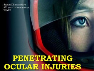
- 1. PENETRATING OCULAR INJURIES Supun Dhanasekara 5TH year 2nd semmaster TSMU
- 2. OCULAR TRAUMA • The eye is protected from direct injury by lids, eyelashes and the projecting margins of the orbit. • It can be injured in a variety of ways; by chemicals, heat, radiation and mechanical trauma.
- 3. OCULAR INJURIES Mechanical Non mechanical • Extra ocular foriegn bodies · Chemical burns • Blunt trauma Acid burns • Perforating injuries Alkali burns • Intra ocular foriegn bodies · Thermal injuries • Penetrating Injuries · Electrical injuries • Sympathetic ophthalmitis Raditional injuries UV radiations
- 5. • Eyewall: It consists of the Sclera and the Cornea • Closed globe injury; No full-thickness wound of eye wall,but there is intr-ocular damage. • Open globe injury: It refers to the full thickness injury of the eye wall and the intra-ocular structures. • Contusion: It is a result of direct energy delivary to the eye by a blunt object.injury may be at the site of impact or at a distant site. • Lamellar laceration: Partial-thickness wound of the eyewall. • Laceration Full-thickness wound of the eyewall, caused by a sharp object. • Penetrating injury: is an injury where a foriegn object has been embedded in the eye.It is usually a full thickness wound & it has a site of Entrance. Perforating injury has both an Entrance and exit wounds. Both wounds caused by the same agent.
- 6. PENETRATING INJURY • Trauma: Usually by a sharp and pointed instruments like needles,sticks,pencils,knives arows,pens,glass and any object with sharp edges. • the most common causes of penetrating ocular injuries are due to trauma caused by wood, metal and stone .Most of the injuries occurred during chopping or cutting wood, hammering metals or nails and carving stone. • These are associated with professions such as farming, garage work and carpentry in adults. • Children, on the other hand, mostly sustain accidental injuries by rubber bands, needles, pencils, sticks while playing with others.
- 7. EFFECTS OF PENETRATING OCULAR INJURIES • Mechanical effects:such as Laceration of the conjunctiva,corneal lacerations,Vitreous haemorrage,rupture of globe,retinal tears and detachments,scarring which leads to cataract and glaucoma. And Intra ocular foriegn bodies. • Introduction of infection:the entrance of the wound may serve as a route of entry for pyogenic bacteria,which may lead to the fromation of abscess of cornea,purulent iridocyclitis or Endophthalmitis • Sympathetic opthalmitis:it is a complication of penetrating injury. • Visual impairment and Enucleation
- 8. MAIN SYMPTOMS • Redness of eye, • Haemorrages • Congestion • Lacrimation • Photophobia Inability to Open Eye • Raised Eyelids • Itchy/Watery Eyes • Blurring or Loss of Vision • Change in Pupil Shape • Blood or Fluid Leakage from the Eye • Foreign Object Penetrating Eye
- 9. DON’TS AND DO’S • DO NOT flush the eye with any liquids other than saline or warm water or even better just do not touch the eye • DO NOT remove the object out of the eye • DO NOT put any pressure on the eye • Do NOT rub your eye. • Flush the eye with copious amounts of saline or warm water until symptoms resolve unless severe, penetrating or bleeding injury. • Reassure the person and advise against rubbing or moving their eye as this can cause further damage • If the injury is severe, place a moist pad and loosely bandage the eye. • Transport the patient to the nearest Hospital as fast as possible • In the case of small penetrating objects, use a cup to cover the object and keep the person calm and lying down until help arrives.
- 10. COMMON DIAGNOSTIC PROCEDURES FOR OCULAR INJURIES• External examination of the eye • Measurement of intra ocular pressure using tonometer • Ophthalmoscopy: • Direct ophthalmoscopy: allows the examiner to view the back of the eyeball. • Indirect ophthalmoscopy: You will either lie or sit in a semi-reclined position.. • Slit-lamp examination :The slit-lamp examination looks at structures that are at the front of the eye • Visual acuity test • Ultrasound :Ultrasound involves the use of high-frequency sound waves to create images of organs and systems within the eye • Electroretinogram (a record of the electrical currents in the retina produced by visual stimuli)
- 11. COMMON DIAGNOSTIC PROCEDURES FOR OCULAR INJURIES High-resolution ultrasound image of the anterior segment obtained with arc-scan geometry. Visualized structures include the cornea (C), sclera (S) , iris (I), anterior lens surface (L) and ciliary body (CB). Direct opthalmoscopy Slit lamp examintaion Indirect opthalmoscopy
- 13. • A corneal laceration is a partial- or full-thickness injury to the cornea,caused by flying metal fragments, sharp objects, fingernails, air-bag deployment, fireworks, explosions, blunt force trauma, pellets • The main symptoms are intense pain initially which may diminish slightly due to corneal desensitization. • Patients are photophobic and lacrimate profusely. • There is a significant attendant uveitis and the anterior chamber is shallow or even flat in a full thickness laceration. • Intraocular pressure generally ranges from 2 to 6 mmHg. • Bubbles within the anterior chamber are a key finding. • There is significantly reduced visual acuity
- 14. CONJUNCTIVAL LACERATION Trauma to the ocular surface often involves the conjunctiva. Mechanisms of injury to the conjunctiva include thermal and chemical burns and blunt or penetrating trauma.
- 15. Clinical features: • May be isolated or part of more severe intraocular injuries. • Symptoms: ocular irritation, pain and foreign body sensation. • Signs include chemosis, subconjunctival hemorrhage and torn conjunctiva. • eye examination under topical or general anesthesia includes dilated fundus examination to rule out intraocular foreign body. • CT scan to rule out intraocular foreign body. Ultrasonography. • Management: • Observation. • Prophylactic topical antibiotics for small lacerations. • Surgical repair(suturing) may be required for large lacerations >2mm
- 16. GLOBE RUPTURE Globe rupture occurs when the integrity of the outer membranes of the eye is disrupted by blunt or penetrating trauma. Notice the dark arc in the bottom of the photo representing the ciliary body visible through the scleral breach. Subconjunctival hemorrhage of this severity should raise suspicion of occult globe rupture
- 17. • Globe rupture may occur when a blunt object impacts the orbit, causing anterior-posterior compression of the globe and raising intraocular pressure to a point that the sclera tears. • Ruptures from blunt/penetrating trauma usually occur at the sites where the sclera is thinnest, at the insertions of the extraocular muscles, at the limbus, and around the optic nerve. • Sharp objects or those traveling at high velocity may penetrate the globe directly. • Small foreign bodies may penetrate the eye and remain within the globe. • The possibility of globe rupture should be considered and ruled out during the evaluation of all blunt and penetrating orbital traumas as well as in all cases involving high-speed projectiles with potential for ocular penetration.
- 18. • Globe rupture in adults may occur after blunt/penetrating injuries during motor vehicle accidents, sports activity, assault, or other trauma. • Globe penetration or perforation may occur with gunshot and stab wounds, workplace accidents, and other accidents involving sharps or projectiles. • .
- 19. Symptoms • Pain • Pain may be difficult to assess in patients with obtundation or distracting injuries. • Pain may not be severe initially in sharp injuries, with or without intraocular foreign body. • Vision - Usually greatly decreased • Diplopia • If present, diplopia is usually due to dysfunction of extraocular muscles with associated orbital floor blowout fractures. • Diplopia may be due to traumatic cranial nerve palsy from associated head injury. • Monocular diplopia may be due to associated lens dislocation or subluxation.
- 20. RETINAL DETACHMENT • Retinal detachment is a disorder of the eye in which the retina peels away from its underlying layer of support tissue. without rapid treatment the entire retina may detach, leading to vision loss and blindness • Rhegmatogenous retinal detachment –Detachment occurs due to a hole, tear, or break in the retina that allows fluid to pass from the vitreous space into the sub retinal space between the sensory retina and the retinal pigment epithelium. • Exudative, or secondary retinal detachment –Due to inflammation, injury or vascular abnormalities that results in fluid accumulating underneath the retina without the presence of a hole, tear, or break. • Tractional retinal detachment – A tractional retinal detachment occurs when fibrovascular tissue, caused by an injury, inflammation or neovascularization, pulls the sensory retina from the retinal pigment epithelium.
- 21. Symptoms of Retinal Detachment • The sudden appearance of many floaters — tiny specks that seem to drift through your field of vision • Flashes of light in one or both eyes • Blurred vision • Gradually reduced side (peripheral) vision • A curtain-like shadow over your visual field
- 22. TREATMENT • Surgery is the only effective therapy for a retinal tear, hole or detachment • If a tear or a hole is treated before detachment develops or if a retinal detachment is treated before the central part of the retina (macula) detaches, you'll probably retain much of your vision. • Laser surgery. The laser makes burns around the retinal tear, and the scarring that results usually "welds" the retina to the underlying tissue. This procedure requires no surgical incision, and it causes less irritation to your eye than does cryopexy. • Freezing (cryopexy). After a local anesthetic numbs your eye, a freezing probe to the outer surface of the eye directly over the retinal defect. This freezes the area around the hole, leaving a delicate scar that helps secure the retina to the eye wall • Injecting air or gas into your eye.(pneumatic retinopexy) injects a bubble of air or gas into the the vitreous cavity. bubble pushes the area of the
- 25. ORBITAL FRACTURES Types : • •Blow-out orbital floor fracture • •Blow-out medial wall fracture • •Roof fracture • •Lateral wall fracture
- 26. • Blow-out orbital floor fracture • Cause: • Sudden increase in orbital pressure by an impacting object greater in diameter than the orbital aperture (>5 cm) • e.g.- Fist, tennis ball etc.
- 27. Mechanism of an orbital floor blow-out fracture
- 28. Signs of orbital floor blow-out fracture • •Periorbital ecchymosis, oedema and emphysema may also present • •Infraorbital nerve anaesthesia • •Ophthalmoplegia tipically in up and down-gaze (double diplopia) • •Enophthalmos – if severe
- 29. INVESTIGATIONS Coronal CT scan Right blow-out fracture with ‘tear-drop’ sign Restriction of right upgaze and downgaze • Secondary overaction of left eye Hess test
- 31. • Release of entrapped tissue • • Repair of bony defect TREATMENT Orbital floor reconstructed using rib graft harvested from right sixth rib and titanium mesh. Note that the right enophthalmos was a secondary deformity with severe volume loss requiring augmentation with rib graft in addition to the mesh
- 32. FOREIGN BODIES(FB) • The seriousness of the injury depends upon the retention of the intraocular frreign body • Common foreign bodies maybe chips of iron or steel,particles of glass,stone,lead pellets,wood chips,plastic • The symptoms of a foreign body may range from irritation to intense, excruciating pain. This is dependent on the location, material, and type of injury. • Mild to extreme irritation • Scratching • Burning • Intense pain • Redness • Tearing • Light sensitivity
- 33. SUPERFICIAL FOREIGN BODY Subtarsal foreign body Corneal foreign body with surrounding cellular infiltration
- 34. Management • a. Careful slit-lamp examination for exact position & depth • b. Removal under slit-lamp with 26-gause needle • c. Magnetic removal for a deeply embedded metallic foreign body • c. Residual ‘rust ring’ may remove with rotating sterile burr • d. Antibiotic oint. with cycloplegic and/or NSAIDs
- 35. INTRAOCULAR FOREIGN BODY • The location and damage caused by an IOFB will depends on several factors including the size, shape, and composition
- 36. Management: • a. Accurate history- helpful for nature of FB • b. Examination • - Entry exit point • - Gonioscopy & fundoscopy • - Documentation for damaged structure • c. CT scan • d. MRI contraindicated for metalic FB
- 37. CHEMICAL INJURY • •Majority of injuries are accidental • •Few due to assault • •2/3 rd of accidental burns occur at work place • •Alkali burns are more common. • •Alkali burns more severe than acid
- 38. GRADING OF SEVERITY OF CHEMICAL INJURIES Grade I (excellent prognosis) •Clear cornea •Limbal ischaemia - nil Grade II (good prognosis) •Cornea hazy but visible iris details •Limbal ischaemia <1/3 Grade III (guarded prognosis) •Hazy cornea with no iris details •Limbal ischaemia 1/3 to 1/2 Grade IV (very poor prognosis) •Opaque cornea •Limbal ischaemia >1/2
- 39. • Grade 1 ocular surface burn. Large corneal burn following accidental exposure to ammonia. There is no limbal or conjunctival involvement. Fluorescein stained diffuse view of the cornea.
- 40. (A) Grade 3 ocular surface burn. involvement with 30% conjunctival involvement following ocular surface burn with a domestic cleansing (alkali) injury. (B) (C) the surviving limbal epithelium demonstrates circumferential migration of tongue-shaped projections affording limbal epithelial cover to denuded limbus. (D) The entire limbus has healed with limbal epithelium and the corneal surface too is almost completely healed with corneal epithelium.
- 41. • Grade 3 (5/35%) ocular surface burn following an accident involving an industrial alkaline chemical (A) Diffuse view with patient looking straight illustrating the extent of limbal involvement (B) Diffuse view with patient looking up and out and (C) looking up and in to show the extent of conjunctival involvement.
- 42. • Grade 5 (9.5/60%) ocular surface burn following alkali injury. limbus and 60% of the conjunctiva were involved
- 43. • Grade 6 (12/100%) ocular surface burn with a “fish pond cleaning liquid” following an assault. The entire limbus and the entire conjunctiva were involved
- 44. • Grade 6 (12/100%) ocular surface burn following injury with cement powder. The entire limbus and conjunctiva were involved. This picture was taken 7 months after the injury.
- 45. MEDICAL TREATMENT OF CHEMICAL INJURIES • 1.Copious irrigation (15-30 min) – to restore normal pH • 2.Topical steroids (first 7-10 days) – to reduce inflamation • 3.Topical and systemic ascorbic acid – to enhance collagen production • 4.Topical citric acid – to inhibit neutrophil activity • 5.Topical and systemic tetracycline – to inhibit collagenase and neutrophil activity • 6.Cycloplegia – to improve comfort
- 46. • References: • www.mayoclinic.com • www. emedicine.medscape.com • www.wikipedia.com • www.ecureme.com • www.medicinenet.com