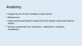
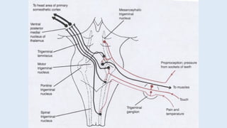
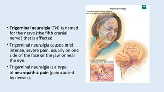
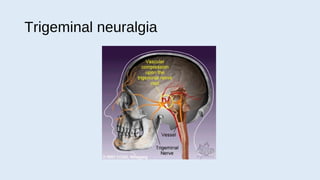
The trigeminal nerve is the largest and most complex cranial nerve, with both sensory and motor functions. Trigeminal neuralgia causes severe, stabbing, intermittent facial pain and is caused by compression or irritation of the trigeminal nerve. Symptoms include episodes of unilateral pain in the face, mouth, or eye that are triggered by mundane activities like eating or talking. Diagnosis is clinical without a confirmatory test. Treatment involves drug therapy with anticonvulsants or surgical procedures like microvascular decompression to relieve nerve compression.
The presentation explain white lesions in oral cavity and the classification the demonstrate the etiology, histopathology, diagnosis and treatment for each one.
Class about 5th cranial nerve, Introduction, Nucleus, Details of Nucleus, Functional component & Clinical anatomy it is useful for Medical ,Dental UG & PG Students including Bsc & Msc Nursing Students
Trigeminal nerve (V):
Responsible for sensation in the face and motor functions such as chewing. The trigeminal nerve has both sensory and Medial Motor roots that emerges from the pons and enlarge forming trigeminal ganglia.
Trigeminal nerve (V):
Responsible for sensation in the face and motor functions such as chewing. The trigeminal nerve has both sensory and Medial Motor roots that emerges from the pons and enlarge forming trigeminal ganglia.
TEST BANK for Operations Management, 14th Edition by William J. Stevenson, Ve...kevinkariuki227
TEST BANK for Operations Management, 14th Edition by William J. Stevenson, Verified Chapters 1 - 19, Complete Newest Version.pdf
Couples presenting to the infertility clinic- Do they really have infertility...Sujoy Dasgupta
Dr Sujoy Dasgupta presented the study on "Couples presenting to the infertility clinic- Do they really have infertility? – The unexplored stories of non-consummation" in the 13th Congress of the Asia Pacific Initiative on Reproduction (ASPIRE 2024) at Manila on 24 May, 2024.
Explore natural remedies for syphilis treatment in Singapore. Discover alternative therapies, herbal remedies, and lifestyle changes that may complement conventional treatments. Learn about holistic approaches to managing syphilis symptoms and supporting overall health.
- Video recording of this lecture in English language: https://youtu.be/lK81BzxMqdo
- Video recording of this lecture in Arabic language: https://youtu.be/Ve4P0COk9OI
- Link to download the book free: https://nephrotube.blogspot.com/p/nephrotube-nephrology-books.html
- Link to NephroTube website: www.NephroTube.com
- Link to NephroTube social media accounts: https://nephrotube.blogspot.com/p/join-nephrotube-on-social-media.html
The prostate is an exocrine gland of the male mammalian reproductive system
It is a walnut-sized gland that forms part of the male reproductive system and is located in front of the rectum and just below the urinary bladder
Function is to store and secrete a clear, slightly alkaline fluid that constitutes 10-30% of the volume of the seminal fluid that along with the spermatozoa, constitutes semen
A healthy human prostate measures (4cm-vertical, by 3cm-horizontal, 2cm ant-post ).
It surrounds the urethra just below the urinary bladder. It has anterior, median, posterior and two lateral lobes
It’s work is regulated by androgens which are responsible for male sex characteristics
Generalised disease of the prostate due to hormonal derangement which leads to non malignant enlargement of the gland (increase in the number of epithelial cells and stromal tissue)to cause compression of the urethra leading to symptoms (LUTS
Flu Vaccine Alert in Bangalore Karnatakaaddon Scans
As flu season approaches, health officials in Bangalore, Karnataka, are urging residents to get their flu vaccinations. The seasonal flu, while common, can lead to severe health complications, particularly for vulnerable populations such as young children, the elderly, and those with underlying health conditions.
Dr. Vidisha Kumari, a leading epidemiologist in Bangalore, emphasizes the importance of getting vaccinated. "The flu vaccine is our best defense against the influenza virus. It not only protects individuals but also helps prevent the spread of the virus in our communities," he says.
This year, the flu season is expected to coincide with a potential increase in other respiratory illnesses. The Karnataka Health Department has launched an awareness campaign highlighting the significance of flu vaccinations. They have set up multiple vaccination centers across Bangalore, making it convenient for residents to receive their shots.
To encourage widespread vaccination, the government is also collaborating with local schools, workplaces, and community centers to facilitate vaccination drives. Special attention is being given to ensuring that the vaccine is accessible to all, including marginalized communities who may have limited access to healthcare.
Residents are reminded that the flu vaccine is safe and effective. Common side effects are mild and may include soreness at the injection site, mild fever, or muscle aches. These side effects are generally short-lived and far less severe than the flu itself.
Healthcare providers are also stressing the importance of continuing COVID-19 precautions. Wearing masks, practicing good hand hygiene, and maintaining social distancing are still crucial, especially in crowded places.
Protect yourself and your loved ones by getting vaccinated. Together, we can help keep Bangalore healthy and safe this flu season. For more information on vaccination centers and schedules, residents can visit the Karnataka Health Department’s official website or follow their social media pages.
Stay informed, stay safe, and get your flu shot today!
Title: Sense of Smell
Presenter: Dr. Faiza, Assistant Professor of Physiology
Qualifications:
MBBS (Best Graduate, AIMC Lahore)
FCPS Physiology
ICMT, CHPE, DHPE (STMU)
MPH (GC University, Faisalabad)
MBA (Virtual University of Pakistan)
Learning Objectives:
Describe the primary categories of smells and the concept of odor blindness.
Explain the structure and location of the olfactory membrane and mucosa, including the types and roles of cells involved in olfaction.
Describe the pathway and mechanisms of olfactory signal transmission from the olfactory receptors to the brain.
Illustrate the biochemical cascade triggered by odorant binding to olfactory receptors, including the role of G-proteins and second messengers in generating an action potential.
Identify different types of olfactory disorders such as anosmia, hyposmia, hyperosmia, and dysosmia, including their potential causes.
Key Topics:
Olfactory Genes:
3% of the human genome accounts for olfactory genes.
400 genes for odorant receptors.
Olfactory Membrane:
Located in the superior part of the nasal cavity.
Medially: Folds downward along the superior septum.
Laterally: Folds over the superior turbinate and upper surface of the middle turbinate.
Total surface area: 5-10 square centimeters.
Olfactory Mucosa:
Olfactory Cells: Bipolar nerve cells derived from the CNS (100 million), with 4-25 olfactory cilia per cell.
Sustentacular Cells: Produce mucus and maintain ionic and molecular environment.
Basal Cells: Replace worn-out olfactory cells with an average lifespan of 1-2 months.
Bowman’s Gland: Secretes mucus.
Stimulation of Olfactory Cells:
Odorant dissolves in mucus and attaches to receptors on olfactory cilia.
Involves a cascade effect through G-proteins and second messengers, leading to depolarization and action potential generation in the olfactory nerve.
Quality of a Good Odorant:
Small (3-20 Carbon atoms), volatile, water-soluble, and lipid-soluble.
Facilitated by odorant-binding proteins in mucus.
Membrane Potential and Action Potential:
Resting membrane potential: -55mV.
Action potential frequency in the olfactory nerve increases with odorant strength.
Adaptation Towards the Sense of Smell:
Rapid adaptation within the first second, with further slow adaptation.
Psychological adaptation greater than receptor adaptation, involving feedback inhibition from the central nervous system.
Primary Sensations of Smell:
Camphoraceous, Musky, Floral, Pepperminty, Ethereal, Pungent, Putrid.
Odor Detection Threshold:
Examples: Hydrogen sulfide (0.0005 ppm), Methyl-mercaptan (0.002 ppm).
Some toxic substances are odorless at lethal concentrations.
Characteristics of Smell:
Odor blindness for single substances due to lack of appropriate receptor protein.
Behavioral and emotional influences of smell.
Transmission of Olfactory Signals:
From olfactory cells to glomeruli in the olfactory bulb, involving lateral inhibition.
Primitive, less old, and new olfactory systems with different path
Pulmonary Thromboembolism - etilogy, types, medical- Surgical and nursing man...VarunMahajani
Disruption of blood supply to lung alveoli due to blockage of one or more pulmonary blood vessels is called as Pulmonary thromboembolism. In this presentation we will discuss its causes, types and its management in depth.
These lecture slides, by Dr Sidra Arshad, offer a quick overview of physiological basis of a normal electrocardiogram.
Learning objectives:
1. Define an electrocardiogram (ECG) and electrocardiography
2. Describe how dipoles generated by the heart produce the waveforms of the ECG
3. Describe the components of a normal electrocardiogram of a typical bipolar leads (limb II)
4. Differentiate between intervals and segments
5. Enlist some common indications for obtaining an ECG
Study Resources:
1. Chapter 11, Guyton and Hall Textbook of Medical Physiology, 14th edition
2. Chapter 9, Human Physiology - From Cells to Systems, Lauralee Sherwood, 9th edition
3. Chapter 29, Ganong’s Review of Medical Physiology, 26th edition
4. Electrocardiogram, StatPearls - https://www.ncbi.nlm.nih.gov/books/NBK549803/
5. ECG in Medical Practice by ABM Abdullah, 4th edition
6. ECG Basics, http://www.nataliescasebook.com/tag/e-c-g-basics
Anti ulcer drugs and their Advance pharmacology ||
Anti-ulcer drugs are medications used to prevent and treat ulcers in the stomach and upper part of the small intestine (duodenal ulcers). These ulcers are often caused by an imbalance between stomach acid and the mucosal lining, which protects the stomach lining.
||Scope: Overview of various classes of anti-ulcer drugs, their mechanisms of action, indications, side effects, and clinical considerations.
3. Anatomy:
• Largest & one of most complex cranial nerves
• Mixed nerve
• Large sensory part (portio major) & much smaller motor part (portio
minor)
• Sensory component has 3 divisions : ophthalmic, maxillary,
mandibular.
4. Function:
• The sensory function of the trigeminal nerve:
- provide the tactile, proprioceptive, and nociceptive afferent of the
face and mouth.
• The motor function:
- activates the muscles of mastication, the tensor tympani, tensor veli
palatini, mylohyoid, and anterior belly of the digastric.
5. Peripheral Anatomy:
• The three major branches converge on
the trigeminal ganglion (also called the
semilunar ganglion or gasserian
ganglion), located within Meckel's cave,
and contains the cell bodies of incoming
sensory nerve fibers.
• The trigeminal ganglion is analogous to
the dorsal root ganglia of the spinal
cord, which contain the cell bodies of
incoming sensory fibers from the rest of
the body.
6. • From the trigeminal ganglion, a single
large sensory root enters the brainstem
at the level of the pons. Immediately
adjacent to the sensory root, a smaller
motor root emerges from the pons at
the same level.
• Motor fibers pass through the
trigeminal ganglion on their way to
peripheral muscles, but their cell bodies
are located in the nucleus of the fifth
nerve, deep within the pons.
7. Sensory Branches of the Vth nerve:
Ophthalmic div
• Skull foramen : superior orbital
fissure
• Terminal br: : frontal , lacrimal,
nasociliary, meningeal
• Cutaneous innervation : bridge &
side of nose, upper eyelid,
forehead, scalp back to vertex,
eyeball, lacrimal gland, nasal
septum, lat wall of nasal cavity,
ethmoid sinus, tentorium
cerebelli
8. Sensory Branches of the Vth nerve:
Maxillary div
• Skull foramen : foramen
rotundum
• Terminal br : infraorbital,
zygomatic, sup.alveolar,
pterygopalatine, meningeal
• Cutaneous innervation : cheek,
lat.forehead, side of nose, upper
lip, upper teeth & gums, palate,
nasopharynx, post.nasal cavity,
meninges of ant & middle
cranial fossae
10. Motor branches of Vth nerve:
• Distributed in the mandibular nerve.
• These fibers originate in the motor nucleus of the fifth nerve, which is
located near the main trigeminal nucleus in the pons.
• The motor branches of the trigeminal nerve control the movement of
eight muscles, including the four muscles of mastication.
-Masseter
-Temporalis
-Medial pterygoids
-Lateral pterygoids
11. • Others:
-tensor veli palatine
-mylohyoid
-anterior belly of digastric
-tensor tympani
• With the exception of tensor tympani, all of these muscles are involved in
biting, chewing and swallowing.
• All have 'bilateral' cortical representation.
• A unilateral central lesion (e.g., a stroke), no matter how large, is unlikely to
produce any observable deficit.
• Injury to the peripheral nerve can cause paralysis of muscles on one side of
the jaw. The jaw deviates to the paralyzed side when it opens.
• This direction of the mandible is due to the action of normal pterygoids on
the opposite side.
12. Trigeminal nucleus
• The trigeminal nucleus extends throughout the entire brainstem, from the
midbrain to the medulla, and continues into the cervical cord, where it merges
with the dorsal horn cells of the spinal cord.
• The nucleus is divided anatomically into three parts, visible in microscopic
sections of the brainstem.
• They are the spinal trigeminal nucleus, the main trigeminal nucleus, and the
mesencephalic trigeminal nucleus.
• The three parts of the trigeminal nucleus receive different types of sensory
information.
-The spinal trigeminal nucleus receives pain/temperature fibers.
-The main trigeminal nucleus receives touch/position fibers.
-The mesencephalic nucleus receives proprioceptor and mechanoreceptor fibers
from the jaws and teeth
13.
14. Clinical examination
sensory functions
• Pain, touch, heat, cold – tested on face & mucous membranes
• Each of the 3 divisions of Vth.N is tested individually and compared
with the opposite side.
15. Clinical examination
motor functions
• Bulk & power of masseters & pterygoids – palpating as pt clinches the
jaw
• Ask pt – to protrude & retract the jaw
• Pt bite on tongue depressors with molar teeth
• U/L Trigeminal motor weakness –
deviation of jaw towards the weak side on opening
pt will be unable to move the jaw contralaterally.
Lesion inv brainstem, gasserian ganglion, motor root
16. Clinical examination
motor functions
• B/L Weakness of muscles of
mastication with inability to close
the mouth ( dangling jaw ) –
motor neuron ds, neuromuscular
transmission disorder, myopathy
18. • Trigeminal neuralgia (TN) is named
for the nerve (the fifth cranial
nerve) that is affected.
• Trigeminal neuralgia causes brief,
intense, severe pain, usually on one
side of the face or the jaw or near
the eye.
• Trigeminal neuralgia is a type
of neuropathic pain (pain caused
by nerves).
19. Trigeminal Neuralgia
• Initiating pathologic events include:
• nerve compression by tortuous arteries of the posterior fossa blood vessels
• demyelinating plaques
• herpes virus infection
• infection of teeth and jaw
• a brainstem infarct
21. Clinical manifestations
• Abrupt onset with excruciating pain!!
• Pain described as burning, knifelike, or lightinglike shock in the lips,
upper or lower gums, cheek, forehead, or side of the nose.
• Patient may twitch, grimace, frequent blinking and tearing of eye (tic)
may occur.
22. Clinical manifestations
• Attacks may be brief (2 or 3 minutes)
• Unilateral
• Episodes may be initiated by triggering mechanism of light cutaneous
stimulation as a specific point (trigger zone) along nerve branches.
23. Precipitating stimuli
• Chewing, brushing teeth, hot or cold blast of air on the face, washing
the face, yawning, or talking.
• Patient may eat improperly, neglect hygiene practices, wear cloth
over face, withdraw from interaction with others.
24. Diagnostic studies
• Need to rule out other neurological causes of facial and cephalic pain.
• CT scan will rule out brain lesions, vascular malformations. LP and
MRI will r/o MS.
• There is no specific diagnostic test for TN.
25. Drug Therapy
• Antiseizure meds may prevent and acute attack or promote
remission-mechanism unknown.
• Carbamazeprine (Tegretol)…most common
• Phenytoin (Dilantin)
• Valproate (Depakene)
26. Drug Therapy
• Carbamazeprine has side effects:
• Bone marrow suppression leading to blood abnormalities (CBC counts
needed)
• Pain relief not permanent
28. Surgery
• Percutaneous radiofrequency rhizotomy (electrocoagulation)- placing
a needle into the trigeminal nerve to destroy the area by
radiofrequency currents.
• May lose corneal reflex
• Easily performed; minimal risk
• Pain relieved, but face is numb
29. Microvascular decompression
• Most common surgical procedure
• Blood vessels that are compressing the nerve are displaced and
repositioned. This relieves pain without residual sensory loss.
• Long-term success rate
• Safe without residual sequelae
• Recurrence occurs in 30% of patients within 6 years
Sympathetic and parasympathetic fibers join the three divisions and are distributed to the pupil, to the nasal mucosa causing mucus secretion, to the lacrimal, submaxillary, and sublingual glands, and to the arterioles of the face.
Motor root: Passes forward in posterior fossa Pierces the duramater beneath attachment of tentorium to tip of petrous part of temporal bone. Enters the meckel’s cave leaves skull via Foramen Ovale. It joins the mandibular div of Vth N to form mandibular nerve – supplies masticatory muscles .