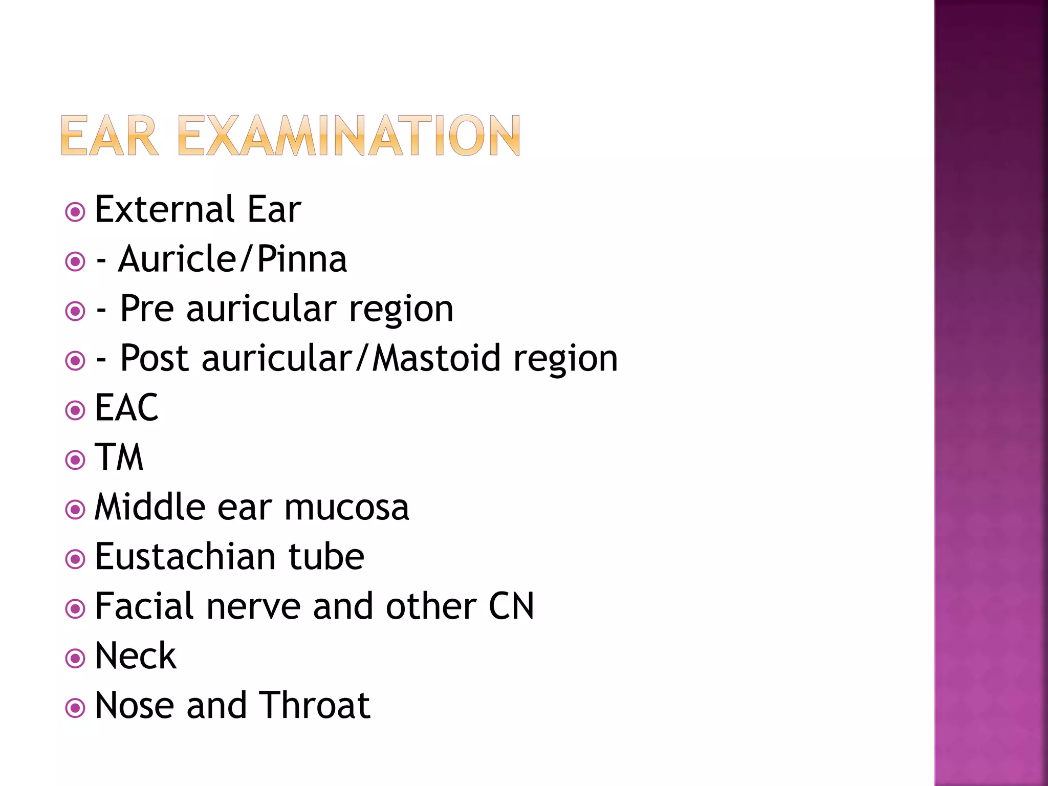
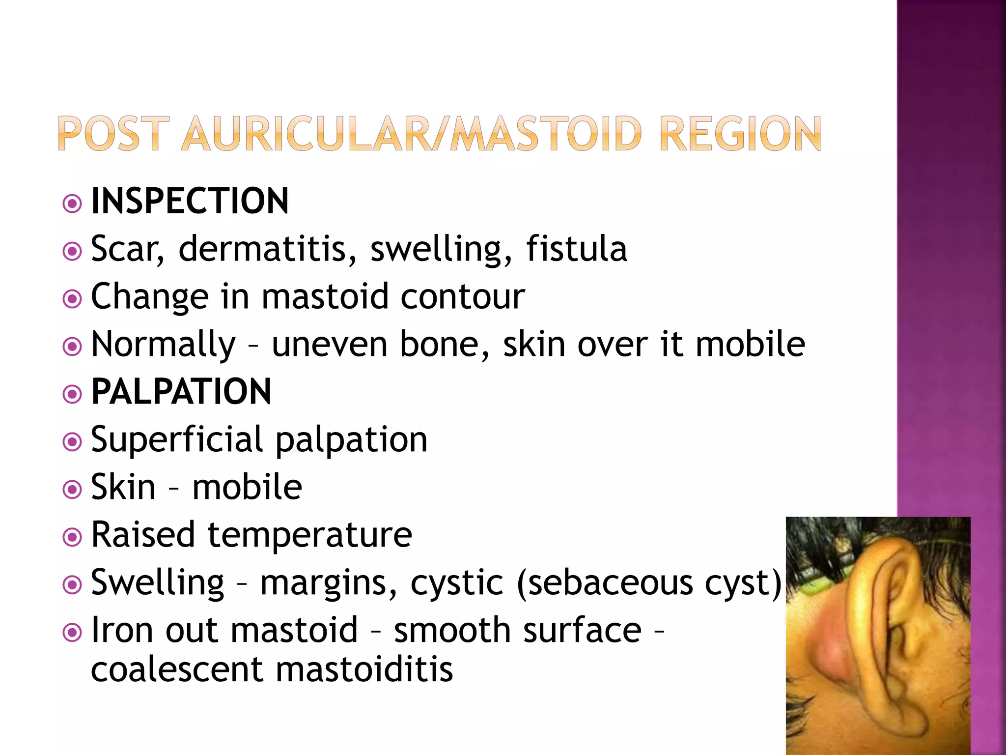
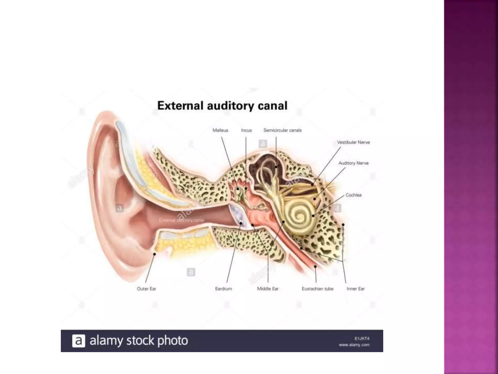
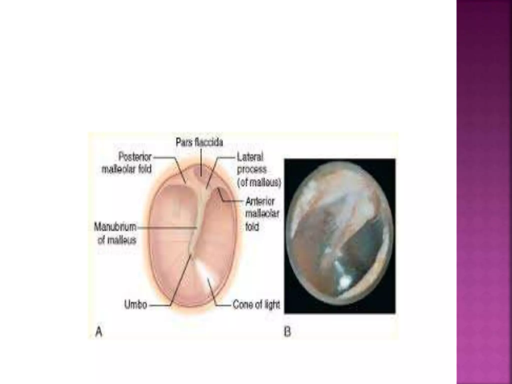
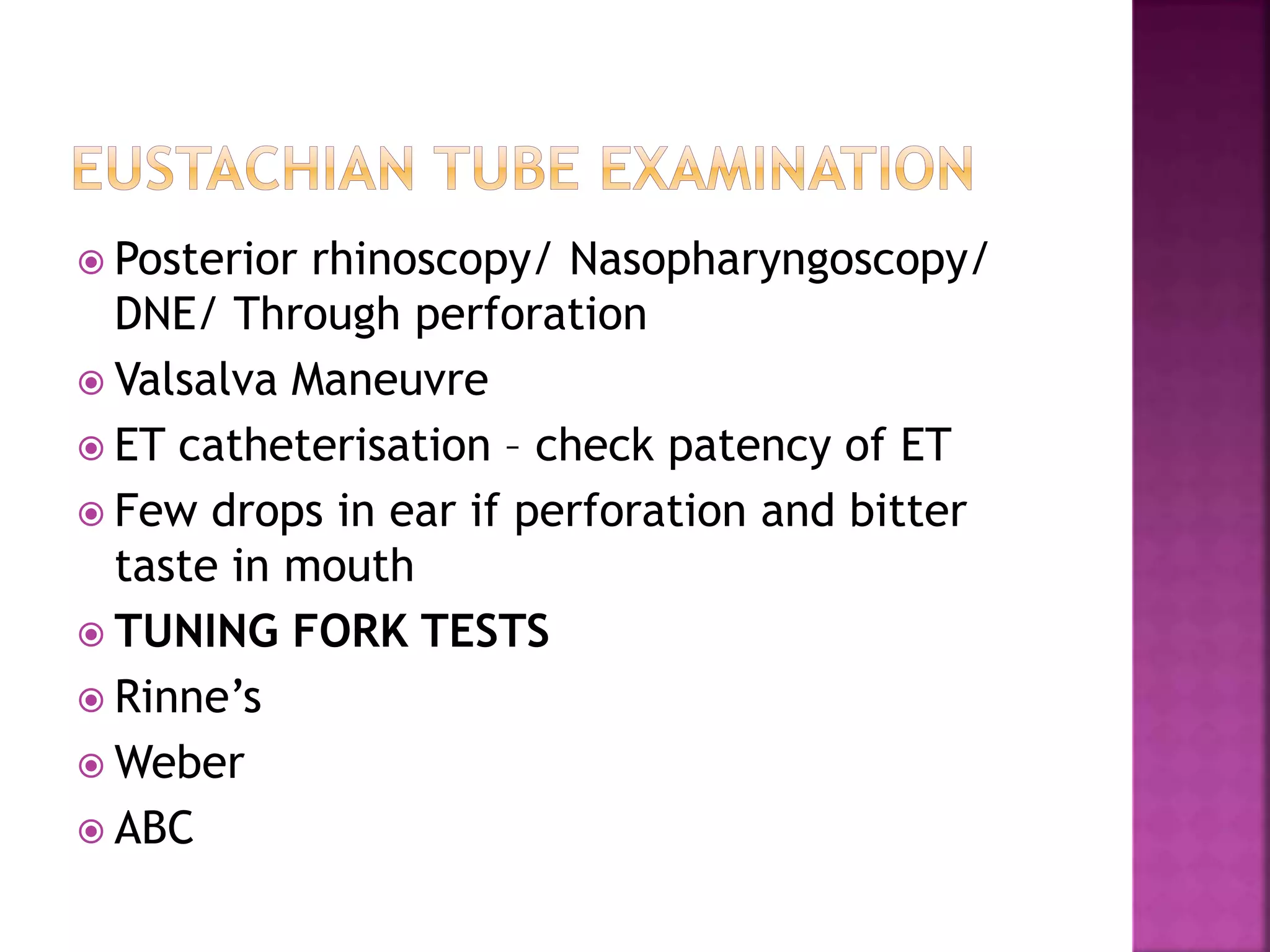
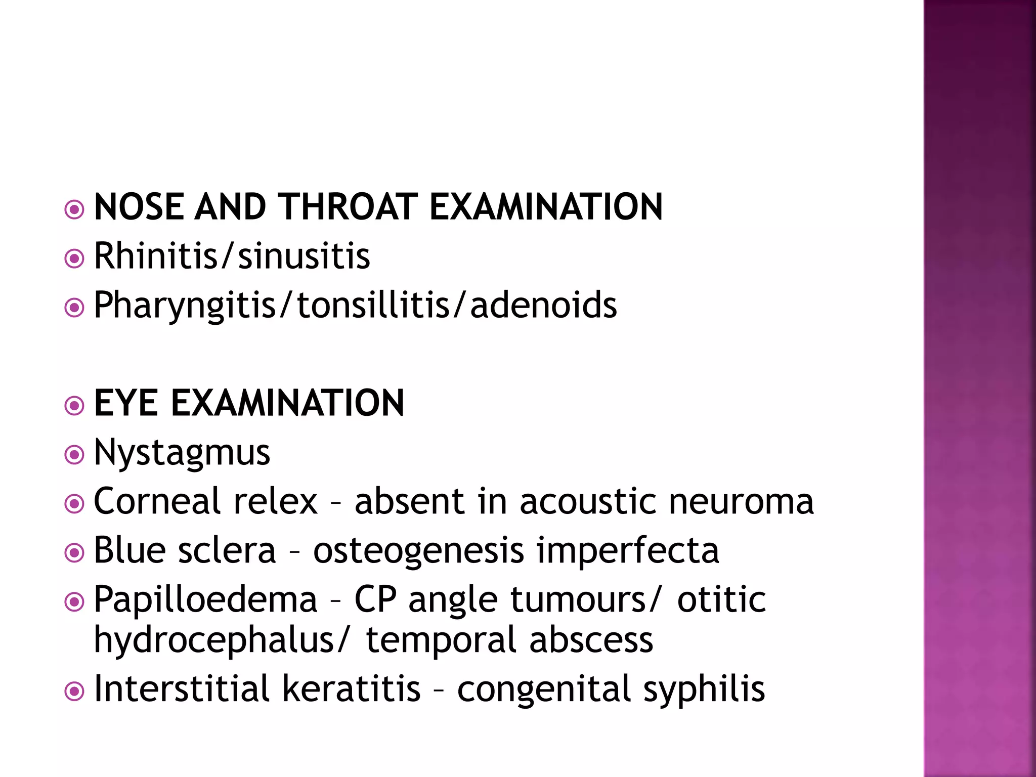
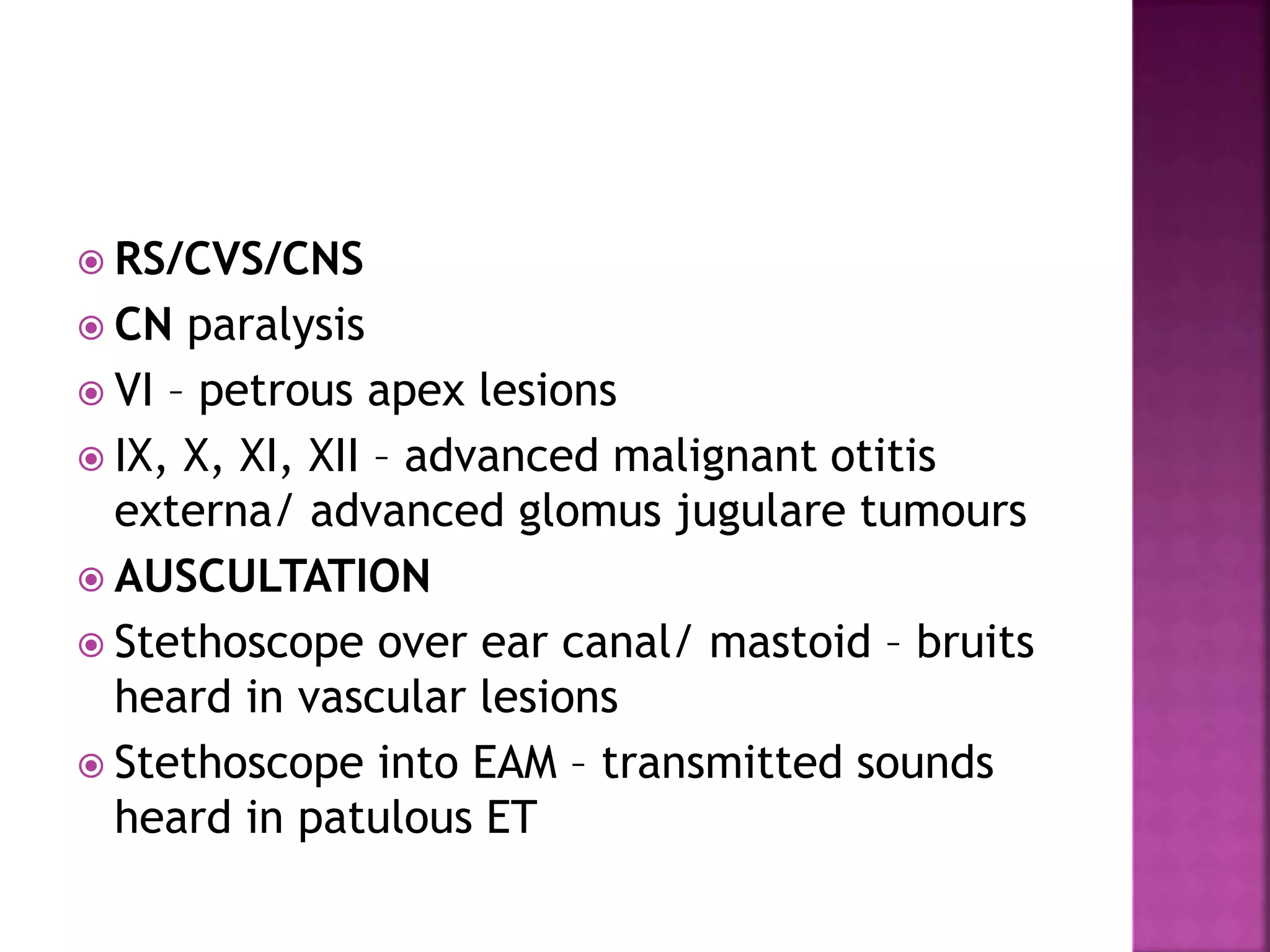
 Mental state
 Consciousness
 Built and nutrition
 Attitude and Gait
 Facies
 Pallor – palmar creases, mm lips cheeks, conjuctiva,
nail beds
 Cyanosis – tongue (central), nail bed , tip of nose,
palmar skin
 Jaundice – sclera, nail bed , ear lobule, tip of nose
 Skin eruptions – macules ( change in skin colour),
papules , vesicles, pustules (solid projections)
 Neck nodes
 Pulse – pulse rate, rhythm (regular, irregular)
 Respiratory rate – fast, slow
 Temperature – continous ( fluctuates less
than 1 deg), remittent ( fluctuates more than
2 deg), intermittent
 Blood Pressure
 Examine the affected region
 Inspection
 Palpation
 Movements
 Measurements’
 LYMPH NODES
 General Examination- Cranial Nerves,
Respiratory
 CVS for surgery purpose
 External Ear
 - Auricle/Pinna
 - Pre auricular region
 - Post auricular/Mastoid region
 EAC
 TM
 Middle ear mucosa
 Eustachian tube
 Facial nerve and other CN
 Neck
 Nose and Throat
 INSPECTION
 Size – anotia (absence), microtia (small)
 Shape – cauliflower ear, bat ear (auricle
protrudes anteriorly)
 Colour- red (perichondritis)
 Position- displacement of auricle forwards,
laterally or inferiorly – mastoid abscess
 Swelling
 Scar
 Ulcer
 PALPATION
 Superficial palpation
 Using fingers (digital palpation) of cartilage and
soft tissue
 Soft tissue – mobility of skin (lost in malignancy),
thickening, swelling
 Raised temperature, tenderness
 Cartilage – defect or loss
 Deep palpation
 Tragal tenderness- inflammation
 Painful movement of pinna – acute otitis externa
 Sinus – pre auricular sinus
 Fistula
 Scar
 Swelling – cystic, lymphadenitis
 INSPECTION
 Scar, dermatitis, swelling, fistula
 Change in mastoid contour
 Normally – uneven bone, skin over it mobile
 PALPATION
 Superficial palpation
 Skin – mobile
 Raised temperature
 Swelling – margins, cystic (sebaceous cyst)
 Iron out mastoid – smooth surface –
coalescent mastoiditis
 Deep palpation
 Tenderness – mastoiditis
 1. Cymba concha – bony landmark for mastoid
antrum
 2. Midpoint of posterior border of mastoid
 3. Tip of mastoid
 NECK EXAMINATION
 Bezold’s abscess – pus track along SCM inferiorly
 Citelli’s abscess – pus track along digastric into
submandibular triangle
 Luc’s abscess – pus around zygoma
 IJV as hard cord on palpation along SCM – IJV
thrombosis
 Direct examination
 Without speculum
 Size of meatus – atresia/wide/narrow
 Wide – post op, syphilis, otosclerosis
 Narrow – congenital atresia, scar due to trauma,
burns tumour - osteoma
 Content of meatus – wax, discharge, FB, Polyp
 Digital examination –
 Adults – pinna pulled upwards, backwards and
laterally, tragus pulled forwards
 Children – pinna pulled downwards and laterally
 Look for furuncle, swelling
 Fungal infection (otomycosis) – black –
aspergillus niger, yellow – candida albicans
 Polyp – probe test – if probe all around – arise
from middle ear
 Impacted wax – whole EAC occluded
 Tumours
 With speculum examination – deep meatus to
straighten the canal
 Largest speculum which can enter
 Black coated
 Introduce in slow rotatory fashion upto cartilage
only
 Cough – vagal irritation
 Sagging of posterior superior EAC –
cholesteatoma, mastoid abscess, mastoiditis
 Absence of sensation in post sup EAC –
Hitselberger’s sign – mass lesion in CP angle
 Normal – pearly white colour, obliquely set,
anterior and posterior malleolar fold (longer)
 Handle of malleus – whitish bony landmark
 Umbo
 Cone of light/light relex – triangular in shape
anterio inferiorly – unreliable landmark
 Short or lateral process of malleus – small
yellowish prominence – at 12 o clock position -
always present – last landmark to be destroyed
in disease
 Quadrants – 4 – imaginary line through tip of
umbo, 2 nd vertically along handle of malleus –
ant sup/inf, post sup/inf
 Colour of TM – normal pearly white/greyish
white, red in ASOM, acute myringitis
 Congestion with yellowish tint – ASOM
 Diffuse congestion/ localised at handle of
malleus – ASOM
 Dark grey colour/dull appearance – tubal
occlusion
 Dull white/thickened cotton like drum – senile
sclerosis, scarring
 Chalky white appearance – tympanosclerosis
after otitis media
 Dull lusterless/bulging – secretory otitis media
 Blue drum – transudative otitis media
 Dark blue drum - haemotympanum
 TM normally inclined downwards and medially
 Protrude outwards – bulging drum – acute otitis
media (pus),Haemotympanum (blood),OME with
good ET function (air) – increase length of handle
of malleus, less prominent short process and
malleolar folds, absence of cone of light
 Pulled inwards – retracted TM- OME with poor ET
function, ET obstruction, atelectasis – shortening
of handle of malleus, more prominent short
process, anterior and post malleolar folds,
distorted cone of light
 Retraction – attic region (retraction pocket) if
deepens – cholesteatoma sac
 Siegle’s pneumatic speculum/ Valsalva
maneuvre
 Normal – change in shape of triangular light
reflex/ handle of malleus movement
 Decreased/absent – ET dysfunction, adhesive
otitis media (fixed), ankylosis of ossicular
chain, SOM
 Hypermobility – Patulous ET, atrophy of TM
 Perforation – ovoid/ kidney shaped/ round,
small, medium, large, sub total, total,
central/marginal, pars tensa/attic, dry/wet,
single/multiple (TB, measles, wegner’s
granulomatosis), margins – regular/irregular,
edge of perforation – thick (CSOM)/ thin
(ASOM)
 Scars
 Bulla – grey/red/bluish pearls like structures
attached to surface of TM
 Chalky white patch - tympanosclerosis
 Only through perforation/ if TM thinned out/
semitransparent
 Middle ear mucosa – oedema/ polyp
 Granulation tissue
 Ossicles, ET, round window, oval window
 FB
 Fistula
 Posterior rhinoscopy/ Nasopharyngoscopy/
DNE/ Through perforation
 Valsalva Maneuvre
 ET catheterisation – check patency of ET
 Few drops in ear if perforation and bitter
taste in mouth
 TUNING FORK TESTS
 Rinne’s
 Weber
 ABC
 FISTULA TEST
 Apply intermittent pressure on tragus/
siegle’s pneumatic speculum – ask patient to
look straight – check for vertigo/ nystagmus
towards opposite side
 FACIAL NERVE EXAMINATION
 Paralysis in ASOM, CSOM, Malignant otitis
externa, herpes zoster, tumours and trauma
 Wrinking of forehead/ closure of eyes/ loss
of naso labial folds/ deviation of angle of
mouth/ cant whistle or blow
 NOSE AND THROAT EXAMINATION
 Rhinitis/sinusitis
 Pharyngitis/tonsillitis/adenoids
 EYE EXAMINATION
 Nystagmus
 Corneal relex – absent in acoustic neuroma
 Blue sclera – osteogenesis imperfecta
 Papilloedema – CP angle tumours/ otitic
hydrocephalus/ temporal abscess
 Interstitial keratitis – congenital syphilis
 RS/CVS/CNS
 CN paralysis
 VI – petrous apex lesions
 IX, X, XI, XII – advanced malignant otitis
externa/ advanced glomus jugulare tumours
 AUSCULTATION
 Stethoscope over ear canal/ mastoid – bruits
heard in vascular lesions
 Stethoscope into EAM – transmitted sounds
heard in patulous ET

Examination of ear

  • 2.
     Mental state Consciousness  Built and nutrition  Attitude and Gait  Facies  Pallor – palmar creases, mm lips cheeks, conjuctiva, nail beds  Cyanosis – tongue (central), nail bed , tip of nose, palmar skin  Jaundice – sclera, nail bed , ear lobule, tip of nose  Skin eruptions – macules ( change in skin colour), papules , vesicles, pustules (solid projections)  Neck nodes
  • 3.
     Pulse –pulse rate, rhythm (regular, irregular)  Respiratory rate – fast, slow  Temperature – continous ( fluctuates less than 1 deg), remittent ( fluctuates more than 2 deg), intermittent  Blood Pressure
  • 4.
     Examine theaffected region  Inspection  Palpation  Movements  Measurements’  LYMPH NODES  General Examination- Cranial Nerves, Respiratory  CVS for surgery purpose
  • 5.
     External Ear - Auricle/Pinna  - Pre auricular region  - Post auricular/Mastoid region  EAC  TM  Middle ear mucosa  Eustachian tube  Facial nerve and other CN  Neck  Nose and Throat
  • 6.
     INSPECTION  Size– anotia (absence), microtia (small)  Shape – cauliflower ear, bat ear (auricle protrudes anteriorly)  Colour- red (perichondritis)  Position- displacement of auricle forwards, laterally or inferiorly – mastoid abscess  Swelling  Scar  Ulcer
  • 8.
     PALPATION  Superficialpalpation  Using fingers (digital palpation) of cartilage and soft tissue  Soft tissue – mobility of skin (lost in malignancy), thickening, swelling  Raised temperature, tenderness  Cartilage – defect or loss  Deep palpation  Tragal tenderness- inflammation  Painful movement of pinna – acute otitis externa
  • 9.
     Sinus –pre auricular sinus  Fistula  Scar  Swelling – cystic, lymphadenitis
  • 10.
     INSPECTION  Scar,dermatitis, swelling, fistula  Change in mastoid contour  Normally – uneven bone, skin over it mobile  PALPATION  Superficial palpation  Skin – mobile  Raised temperature  Swelling – margins, cystic (sebaceous cyst)  Iron out mastoid – smooth surface – coalescent mastoiditis
  • 11.
     Deep palpation Tenderness – mastoiditis  1. Cymba concha – bony landmark for mastoid antrum  2. Midpoint of posterior border of mastoid  3. Tip of mastoid  NECK EXAMINATION  Bezold’s abscess – pus track along SCM inferiorly  Citelli’s abscess – pus track along digastric into submandibular triangle  Luc’s abscess – pus around zygoma  IJV as hard cord on palpation along SCM – IJV thrombosis
  • 12.
     Direct examination Without speculum  Size of meatus – atresia/wide/narrow  Wide – post op, syphilis, otosclerosis  Narrow – congenital atresia, scar due to trauma, burns tumour - osteoma  Content of meatus – wax, discharge, FB, Polyp  Digital examination –  Adults – pinna pulled upwards, backwards and laterally, tragus pulled forwards  Children – pinna pulled downwards and laterally
  • 14.
     Look forfuruncle, swelling  Fungal infection (otomycosis) – black – aspergillus niger, yellow – candida albicans  Polyp – probe test – if probe all around – arise from middle ear  Impacted wax – whole EAC occluded  Tumours  With speculum examination – deep meatus to straighten the canal  Largest speculum which can enter  Black coated  Introduce in slow rotatory fashion upto cartilage only
  • 15.
     Cough –vagal irritation  Sagging of posterior superior EAC – cholesteatoma, mastoid abscess, mastoiditis  Absence of sensation in post sup EAC – Hitselberger’s sign – mass lesion in CP angle
  • 16.
     Normal –pearly white colour, obliquely set, anterior and posterior malleolar fold (longer)  Handle of malleus – whitish bony landmark  Umbo  Cone of light/light relex – triangular in shape anterio inferiorly – unreliable landmark  Short or lateral process of malleus – small yellowish prominence – at 12 o clock position - always present – last landmark to be destroyed in disease  Quadrants – 4 – imaginary line through tip of umbo, 2 nd vertically along handle of malleus – ant sup/inf, post sup/inf
  • 18.
     Colour ofTM – normal pearly white/greyish white, red in ASOM, acute myringitis  Congestion with yellowish tint – ASOM  Diffuse congestion/ localised at handle of malleus – ASOM  Dark grey colour/dull appearance – tubal occlusion  Dull white/thickened cotton like drum – senile sclerosis, scarring  Chalky white appearance – tympanosclerosis after otitis media  Dull lusterless/bulging – secretory otitis media  Blue drum – transudative otitis media  Dark blue drum - haemotympanum
  • 19.
     TM normallyinclined downwards and medially  Protrude outwards – bulging drum – acute otitis media (pus),Haemotympanum (blood),OME with good ET function (air) – increase length of handle of malleus, less prominent short process and malleolar folds, absence of cone of light  Pulled inwards – retracted TM- OME with poor ET function, ET obstruction, atelectasis – shortening of handle of malleus, more prominent short process, anterior and post malleolar folds, distorted cone of light  Retraction – attic region (retraction pocket) if deepens – cholesteatoma sac
  • 21.
     Siegle’s pneumaticspeculum/ Valsalva maneuvre  Normal – change in shape of triangular light reflex/ handle of malleus movement  Decreased/absent – ET dysfunction, adhesive otitis media (fixed), ankylosis of ossicular chain, SOM  Hypermobility – Patulous ET, atrophy of TM
  • 23.
     Perforation –ovoid/ kidney shaped/ round, small, medium, large, sub total, total, central/marginal, pars tensa/attic, dry/wet, single/multiple (TB, measles, wegner’s granulomatosis), margins – regular/irregular, edge of perforation – thick (CSOM)/ thin (ASOM)  Scars  Bulla – grey/red/bluish pearls like structures attached to surface of TM  Chalky white patch - tympanosclerosis
  • 25.
     Only throughperforation/ if TM thinned out/ semitransparent  Middle ear mucosa – oedema/ polyp  Granulation tissue  Ossicles, ET, round window, oval window  FB  Fistula
  • 26.
     Posterior rhinoscopy/Nasopharyngoscopy/ DNE/ Through perforation  Valsalva Maneuvre  ET catheterisation – check patency of ET  Few drops in ear if perforation and bitter taste in mouth  TUNING FORK TESTS  Rinne’s  Weber  ABC
  • 27.
     FISTULA TEST Apply intermittent pressure on tragus/ siegle’s pneumatic speculum – ask patient to look straight – check for vertigo/ nystagmus towards opposite side  FACIAL NERVE EXAMINATION  Paralysis in ASOM, CSOM, Malignant otitis externa, herpes zoster, tumours and trauma  Wrinking of forehead/ closure of eyes/ loss of naso labial folds/ deviation of angle of mouth/ cant whistle or blow
  • 28.
     NOSE ANDTHROAT EXAMINATION  Rhinitis/sinusitis  Pharyngitis/tonsillitis/adenoids  EYE EXAMINATION  Nystagmus  Corneal relex – absent in acoustic neuroma  Blue sclera – osteogenesis imperfecta  Papilloedema – CP angle tumours/ otitic hydrocephalus/ temporal abscess  Interstitial keratitis – congenital syphilis
  • 29.
     RS/CVS/CNS  CNparalysis  VI – petrous apex lesions  IX, X, XI, XII – advanced malignant otitis externa/ advanced glomus jugulare tumours  AUSCULTATION  Stethoscope over ear canal/ mastoid – bruits heard in vascular lesions  Stethoscope into EAM – transmitted sounds heard in patulous ET