

Dr.Mohd Najmussadiq Khan
 Patient profile :- name, sex, age, occupation,
residence, address
 Complaint: - main complaints pain, lacrimation
decrease, visual acuity
 Present history: - each symptom in details.
 Past history: -medical and surgical history e.g.
DM, HT
 Family history:- of same illness, ocular
diseases(e.g. squint, ptosis, congenital
cataract, corneal dystrophy, refractive errors,
glaucoma) & D.M ,HT , allergy
 Drug history. e. g. steroids, chloroquine,
ethambutol
 Central retinal artery occlusion
 Central retinal vein occlusion
 Retinal detachment
 Vitreous haemorrhage
 Spontaneous lens dislocation
 Central serous retinopahty
 Optic neuritis
 Acute attack of primary angle closure
glaucoma
 Acute attack of secondary angle closure
glaucoma
 Acute iridocyclitis
 Mechanical,chemical or thermal injury
 Senile cataract
 Chronic simple glaucoma(POAG)
 Refractive errors
 Choroiditis
 Corneal degeneration
 Age related macular degeneration
 Chronic toxic amblyopia
 Optic atrophy
 Diabetic or hypertensive retinopathy
 Chronic iridocyclitis
 Chronic glaucoma
 Entropion,trichiasis
 Interstitial keratitis
 Orbital cellulitis
 Optic nerve compression
 Temporal arteritis
 Vitamin-A deficiency
 Advanced OAG
 Retinitis pigmentosa
 Congenital high myopia
 Peripheral corneal or lenticular opacities
 Portal cirrhosis & Oguchis disease
 Congenital deficiency of cones
 Central corneal & lenticular opacities
 Central macular choroiditis
 Macular burn & coloboma of the macula
 Presbyopia
 Cycloplegia
 Internal/total ophthalmoplegia
primary hyperlacrimation
Reflux hyperlacrimation (v nerve irritation)
 Lid – stye,internal hordeolum,acute
meibomitis,trichiasis,concretions,entropion
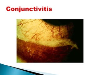
 Conjunctiva-conjuctivitis
 Cornea-abrasions,ulcers,nonulcerative keratitis
 Sclera-episcleritis,scleritis
 Uveal tissue-iritis, cyclitis,iridocyclitis
 Acute glaucoma
 Endophthalmitis,panophthalamitis
 Orbital cellulitis
Central lacrimation-emotional,hysterical
lacrimal pump failure
eversion of lower punctum
 Old age,chronic conjuctivitis,chronic blepharitis,ectropion
punctal obstruction by
 Congenital.injury,burn,foreign
body,concretion,cilia,pilocarpine,idoxuridine
canalicular obstruction by
 Foreign body,trauma,stricture,canaliculitis
lacrimal sac
 Congenital mucous membrane fold
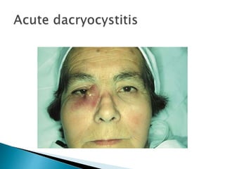
 Traumatic stricture,dacryocystitis
 Tuberculosis,syphilis
 Dacryolithiasis,tumours of sac
 Atonia of the sac
in nasolacrimal duct
 Non canalisation,strictures,inflammation,tumours,
 Surrounding bony disease
 CONJUNCTIVITIS,KERATITIS,SCLERITIS,EPISCLE
RITIS, IRIDOCYCLITIS,ACUTE GLAUCOMA
Pain & discharge
General & systemic examination if needed
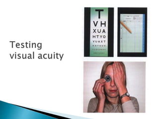
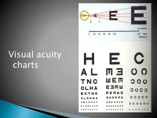
Visual acuity (distant &near)
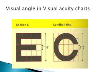
Visual acuity
charts
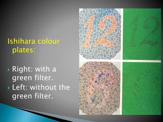
Ishihara colour
plates:
 Right: with a
green filter.
 Left: without the
green filter.
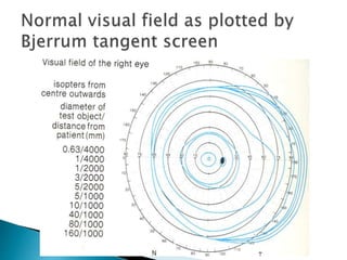
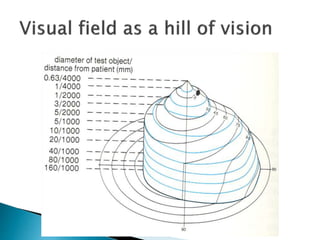
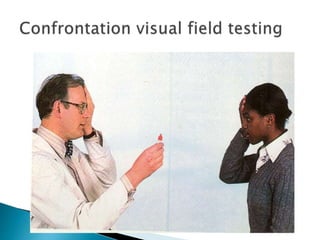
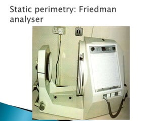
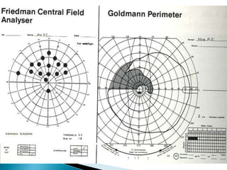
Kinetic
visual field
testing with
Goldmann
perimeter
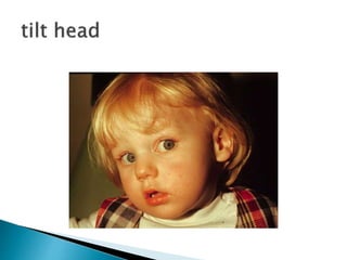
 The head is turned in the direction of action
of the paralysed muscle to avoid diplopia
 In ptosis the chin is elevated to uncover
pupillary area
 Increased wrinkling due to frontalis
contraction in ptosis
 Absent wrinkling in one half of fore head in
LMN facial falsy
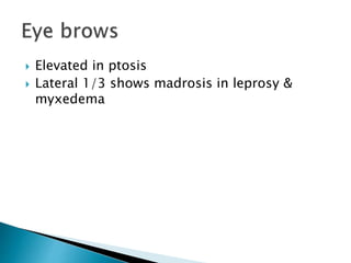
 Elevated in ptosis
 Lateral 1/3 shows madrosis in leprosy &
myxedema
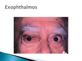
 Proptosis/exophthalmos-axial or
eccentric,reducible or non reducible ,
pulsatile or non pulsatile
 Enophthalmos
 Visual axis of eye balls(squint)
 Size of eye balls increased in
buphthalmos,myopia and decreased in
microphthalmos,pthisis bulbi,atrophic bulbi
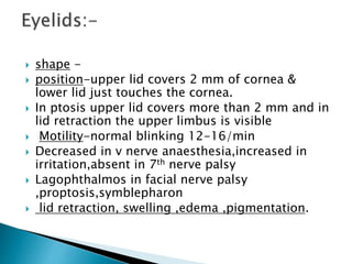
 shape -
 position-upper lid covers 2 mm of cornea &
lower lid just touches the cornea.
 In ptosis upper lid covers more than 2 mm and in
lid retraction the upper limbus is visible
 Motility-normal blinking 12-16/min
 Decreased in v nerve anaesthesia,increased in
irritation,absent in 7th nerve palsy
 Lagophthalmos in facial nerve palsy
,proptosis,symblepharon
 lid retraction, swelling ,edema ,pigmentation.
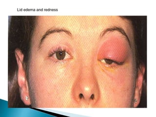
Lid edema and redness
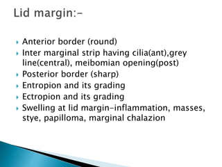
 Anterior border (round)
 Inter marginal strip having cilia(ant),grey
line(central), meibomian opening(post)
 Posterior border (sharp)
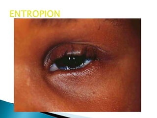
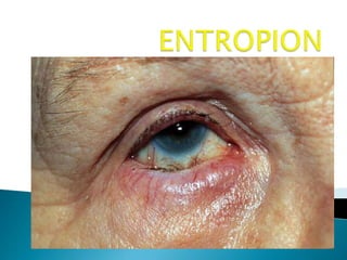
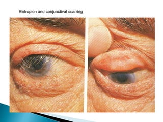
 Entropion and its grading
 Ectropion and its grading
 Swelling at lid margin-inflammation, masses,
stye, papilloma, marginal chalazion
 triciasis(misdirected)-
trachoma,stye,blepharitis,trauma
 Distichiasis (extra row)
 madrosis(loss of cilia)-chronic
blepharitis,leprosy,myxedema
 poliosis(greying of cilia)-old age,Vogt-
Koyanagi-Hardas disease
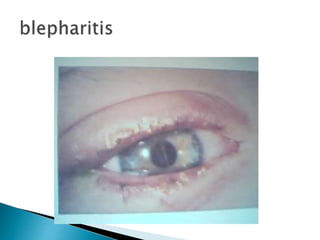
 scales at lid margin-blepharitis
 Ankyloblepharon(horizontally narrow) in
ulcerative blepharitis,burns
 Blepharophimosis(all arround narrow)
 Vertical narrow-
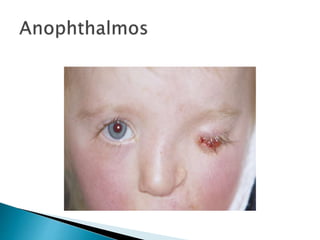
blepharospasm,ptosis,enophthalmos,anophth
almos,microphthalmos,phthisis bulbi,atrophic
bulbi
 Vertical wide-proptosis , large eye ball,upper
lid retraction,facial nerve palsy
 inspection , palpation examined by pulling up
the outer part of the upper lid while asking
the patient to look downwards and
inwards[note swelling or mass].
 Do regurgitation test with pressure on
lacrimal sac with eyes of the examiner on
puncti.
both bulbar and palpebral are examined.
 Palpebral conjunctiva of lower lid:- is examined
by pulling the lower lid down while asking the
patient to look upwards .
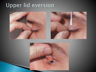
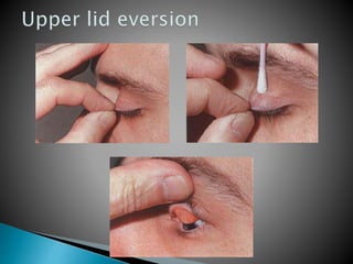
 Palpebral conjunctiva of upper lid:- is examined
by asking the patient to look downward s then
place the thumb and index finger on upper lid
pull out and down –slightly then press on the lid
which a thin stick ---- exposed surface ,exam.
For dryness ,defects , discharge, follicles ,
papillae .
 Bulbar conjunctiva :- paleness , injection ,
discharge , edema , color .
 Color -normally pink in color
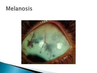
 Brownish -melanosis & argyrosis
 pale -anaemia
 bluish -cyanosis
 bright red –subconjuctival haemorrhage
 greyish -muscara
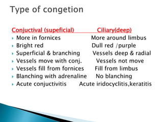
Conjuctival (supeficial) Ciliary(deep)
 More in fornices More around limbus
 Bright red Dull red /purple
 Superficial & branching Vessels deep & radial
 Vessels move with conj. Vessels not move
 Vessels fill from fornices Fill from limbus
 Blanching with adrenaline No blanching
 Acute conjuctivitis Acute iridocyclitis,keratitis
Ciliary congestion
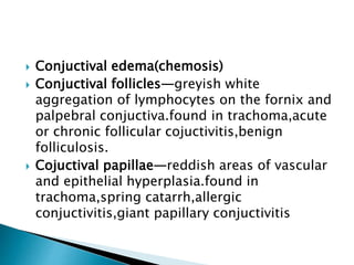
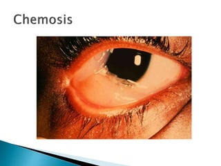
 Conjuctival edema(chemosis)
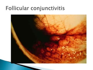
 Conjuctival follicles—greyish white
aggregation of lymphocytes on the fornix and
palpebral conjuctiva.found in trachoma,acute
or chronic follicular cojuctivitis,benign
folliculosis.
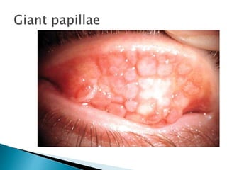
 Cojuctival papillae—reddish areas of vascular
and epithelial hyperplasia.found in
trachoma,spring catarrh,allergic
conjuctivitis,giant papillary conjuctivitis
 Concretions—yellowish white hard looking
raised areas.trachoma,cojuctival
degeneration,idiopathic
 Conjuctival foreign body
 Conjuctival scarring—
trachoma,membranous/pseudomembranous
conjuctivitis,healed traumatic wound,operated
scar
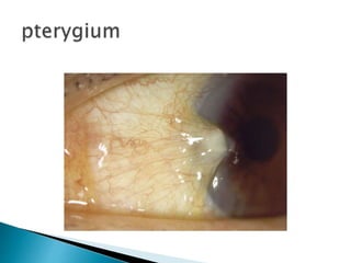
 Pinguiculla & pterygium
 Conjuctival cysts—
retention,implantation,lymphatic,cysticercosis
 Conjuctival tumours—
dermoid,papilloma,squamous cell carcinoma
Entropion and conjunctival scarring
Sub conjunctival haemorrhage
Color
 white but yellow in jaundice,
 blue in child,high myopia,scleritis,marfans
synd,pseudoxanthoma elasticum,osteitis
deformans
 pigmentation in melanosis,naevus of ota
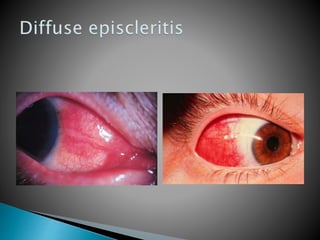
Inflammation –scleritis,episcleritis
Staphyloma-thinned out buldging areas of
sclera lined by uveal tissue.
Traumatic perforation
Size—diameter 11.7mmvertically and 12.6mm
horizontally.
 microcornea <9mm(microphthalmos,phthisis
bulbi)
 megalocornea >13mm(congenital
megalocornea,buphthalmos)
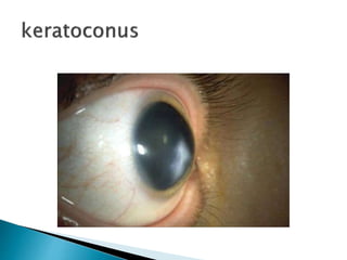
Shape—like a watch glass but abnormal in
keratoconus,keratoglobus,cornea plana
Surface—irregular surface in ulcer,abrasion
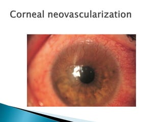
Transparency—absent in edema
,opacity,ulcer,degeneration,dystrophy,deposites,v
ascularisation
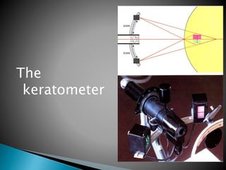
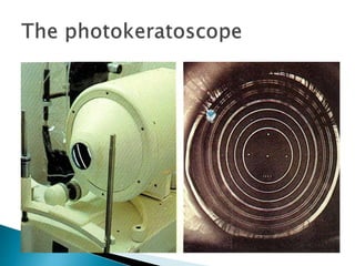
The
keratometer
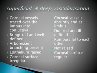
 Corneal vessels
traced over the
limbus into
conjuctiva
 Brihgt red and well
defined
 Arborescent
branching present
 Epithelium raised
 Corneal surface
irregular
 Corneal vessels
abruptly end at
limbus
 Dull red and ill
defined
 Run parallel to each
other
 Not raised
 Corneal surface
regular
Sensitivity—reduced in herpetic
keratitis,neuroparalytic keratitis,leprosy,absolute
glaucoma.It is checked by a thin cotton wool
touching cornea -----reflex lid closure .
Sheen—lost in dry eye
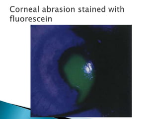
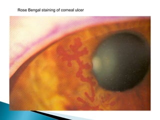
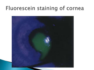
Ulcer—size,site,shape,depth,floor,edges,stain
Opacity—
number,site,size,shape,density[nebular,macular,l
eucomatous]
Back of cornea—Kps
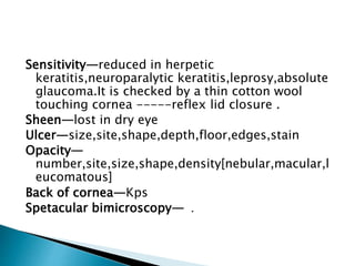
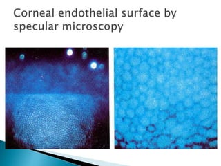
Spetacular bimicroscopy— .
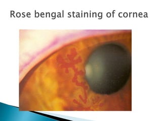
Rose Bengal staining of corneal ulcer
 Shallow A/C—PACG,extreme young and old
age,hypermetropia,iris bombe,perforating
corneal ulcer/injury,post operative,malignant
gluacoma,anterior lens
subluxation,intumescent cataract,pupllary
block
 Deep A/C—
aphakia,myopia,keratoglobus,keratoconus,bu
phthalmos,total posterior synechiae,lens
dislocation into A/C,posterior globe
perforation,partially absorbed traumatic lens
 Irregular A/C—adherent leucoma,iris bombe due to
annular synechiae,tilting of lens
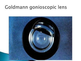
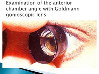
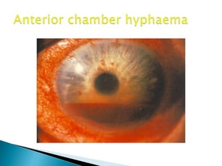
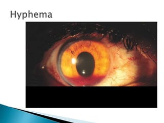
 Blood in A/C[hyphaema]—trauma,surgery,herpes
zooster,gonococcal iridocyclitis
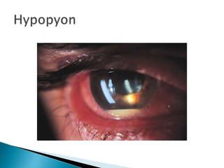
 Pus in A/C[hypopyon]—corneal
ulcer,iridocyclitis,endophthalmitis,panophthalmitis
 Aqueous flare in A/C[inflammatory cells+protein]
 Pseudohypopyon[collection of tumour cells in A/C]—
retinoblastoma,malignant melanoma
 Foreign body in A/C
 Lens particle in A/C after surgery
 Paracytic cyst in A/C—cysticercus
 Artificial lens in A/C
Color- between two eyes , appearance of anterior surface.Blue
green in caucacians,dark brown in orientals
 Congenital heterochromia iridium-different colour of two eye
 Hetrochromia iridis-different colour sectors of the same iris
 Greyish atrophic patches in healed iridocyclitis
 Dark pigmented spots in naevi
Iris pattern—normal patter due to collarette,crypts,and radial
striationsin its anterior surface.
 Muddy iris in acute iridocyclitis
 Atrophy of iris in haeled iridocyclitis
Persistent pupillary membrane(PPM)—abnormal congenital tags
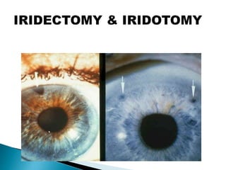
Any defect as iridectomy , iridodialysis (lost in inflammation ,
glaucoma .
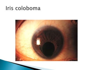
Congenital defect:- as coloboma aniridia .
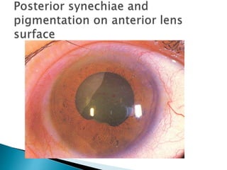
 Adhesion to cornea k/a posterior or to lens k/a anterior
synechiae.The posterior synechiae may be
annular,segmental,total
 Iris support (normally to lens) if lost k/a iridodonesis or
tremulous iris in aphakia,subluxation.
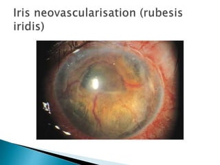
 New vessels (Rubeosis iridis)—D.M.,CRVO,chronic
iridocyclitis
 Hole in iris—congenital coloboma,surgical iridectomy,laser
iridotomy
 Nodule on iris surface—granulomatous
uveitis,melanoma,tuberculoma,gumma of iris
 Iridodialysis—separation of iris from ciliary body
 Aniridia-congenital absence of iris
 Iridraemia
 Iris cyst
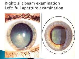
Posterior synechiae and pigmentation on anterior lens capsule