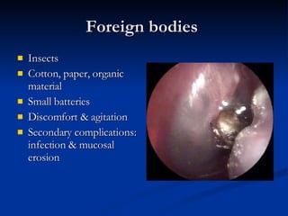
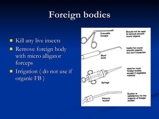
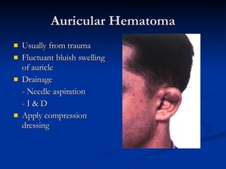
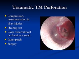
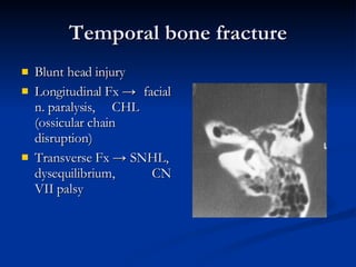
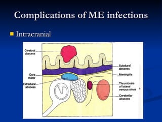
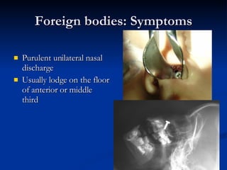
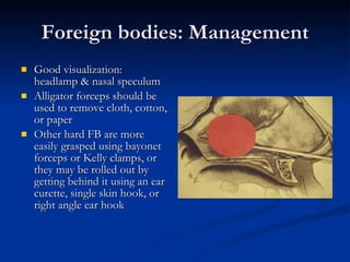
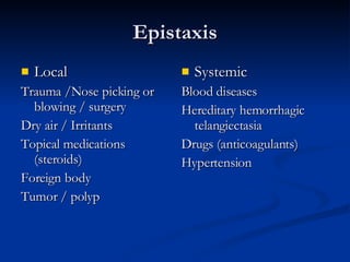
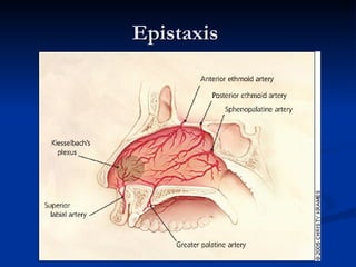
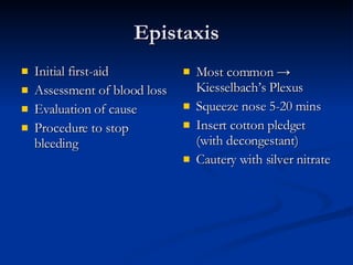
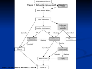
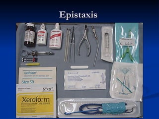
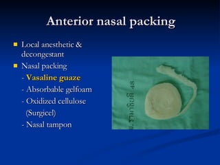
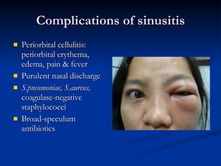
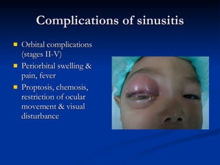
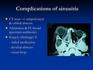
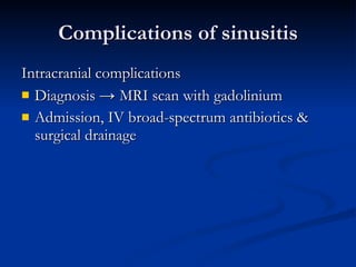
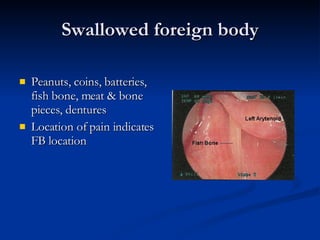
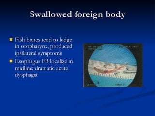
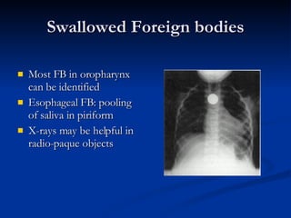
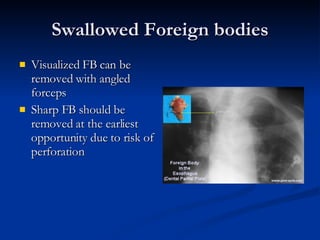
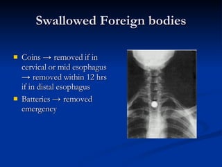
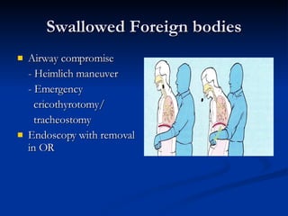
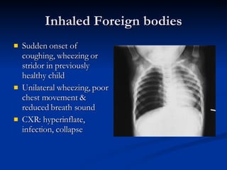
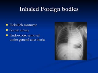
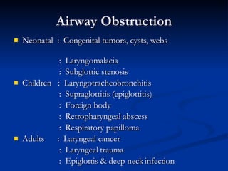
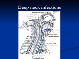
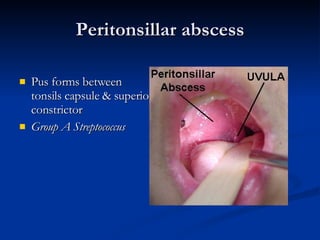
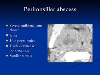
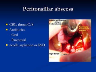
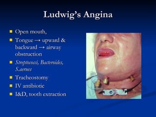
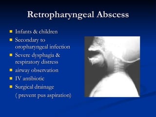
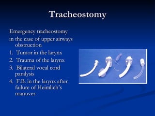
COMMON ENT EMERGENCIES Thongchai Luxameechanporn ENT department Ramathibodi hospital
Common ENT emergencies Foreign bodies Trauma Complications of ENT infections
ear
Foreign bodies  Insects Cotton, paper, organic material Small batteries Discomfort & agitation Secondary complications: infection & mucosal erosion
Foreign bodies Kill any live insects Remove foreign body with micro alligator forceps Irrigation ( do not use if organic FB )
Auricular Hematoma Usually from trauma Fluctuant bluish swelling of auricle Drainage - Needle aspiration - I & D Apply compression dressing
Traumatic TM Perforation Compression, instrumentation &  blast injuries Hearing test Close observation if perforation is small Paper patch Surgery
Temporal bone fracture Blunt head injury Longitudinal Fx  ->  facial n. paralysis,  CHL (ossicular chain disruption) Transverse Fx  ->  SNHL, dysequilibrium,  CN VII palsy
Temporal bone fracture  Battle’s sign (bluish discoloration of postauricular region), raccoon eyes, hemotympanum, hearing loss, dizziness, CSF otorrhea, CN VII palsy CT temporal bone
Acoustic trauma Sudden exposure (impact or blast) to noise SHNL, tinnitus Avoidance/ ear protection Corticosteroids, carbogen, vasodilators, diuretics, anticoagulants, plasma expanders
Otitic Barotrauma Inability to ventilate middle ear  ->  abnormal dysfunction of ET Occur in rising ambient pressure (descent in flight / scuba diving) Can produce hemotympanum
Barotrauma Repeated Valsalva maneuver Topical nasal decongestants Myringotomy & PE tube insertion may be needed
Sudden Hearing Loss SNHL ≥ 30 dB over 3 contiguous frequencies within 3 days or less Etiology : Viral & Infectious, Vascular, Trauma, Autoimmune, Neurologic
Complications of ME infections Extracranial
Acute Mastoiditis preceded by AOM young children severe pain, fever, edema over mastoid area intravenous ATB Myringotomy ± PE tube
Subperiosteal Abscess pinna pushed  down & outward  intravenous ATB I&D mastoidectomy
Complications of ME infections Intracranial
nose
Foreign bodies: Symptoms Purulent unilateral nasal discharge Usually lodge on the floor of anterior or middle third
Foreign bodies: Management Good visualization: headlamp & nasal speculum Alligator forceps should be used to remove cloth, cotton, or paper  Other hard FB are more easily grasped using bayonet forceps or Kelly clamps, or they may be rolled out by getting behind it using an ear curette, single skin hook, or right angle ear hook
Nasal Fracture Hx of fall or force directed to midface Deformity of nose Swelling, ecchymosis, epistaxis Close or open reduction
Septal hematoma/abscess Trauma, surgery Soft, fluctuant swelling of septum Needle  a spiration  or  I&D   Bilateral nasal packing for several days   Prophylactic antibiotics
Septal hematoma/abscess
Epistaxis Local Trauma /Nose picking or blowing / surgery Dry air / Irritants  Topical medications (steroids) Foreign body  Tumor / polyp Systemic Blood diseases Hereditary hemorrhagic telangiectasia Drugs (anticoagulants) Hypertension
Epistaxis
Epistaxis Initial first-aid Assessment of blood loss Evaluation of cause  Procedure to stop bleeding Most common  ->  Kiesselbach’s Plexus  Squeeze nose 5-20 mins Insert cotton pledget (with decongestant) Cautery with silver nitrate
Pope, L E R et al. Postgrad Med J 2005;81:309-314 Figure 1  Epistaxis management protocol.
Epistaxis
Anterior nasal packing Local anesthetic & decongestant  Nasal packing -  Vasaline guaze - Absorbable gelfoam - Oxidized cellulose (Surgicel)  - Nasal tampon
Anterior nasal packing
Anterior nasal packing Nasal packing -  Vasaline guaze -  Absorbable gelfoam - Oxidized cellulose (Surgicel)   - Nasal tampon
Anterior nasal packing Nasal packing -  Vasaline guaze -  Absorbable gelfoam - Oxidized cellulose (Surgicel)  - Nasal tampon
Anterior nasal packing Nasal packing -  Vasaline guaze - Absorbable gelfoam - Oxidized cellulose (Surgicel)  -  Nasal tampon
Copyright ©2005 BMJ Publishing Group Ltd. Pope, L E R et al. Postgrad Med J 2005;81:309-314 Figure 2  Correct insertion of a nasal tampon (note that the direction is along the floor of the nasal cavity).
Posterior nasal packing Topical anesthetic & decongestant Posterior nasal packing   Double balloon device Foley catheter
Posterior nasal packing Topical anesthetic & decongestant Posterior nasal packing  Double balloon device Foley catheter
Complications of sinusitis Orbital complications Intracranial complications
Classification of orbital inflammation Stage I II III IV V Inflammation Inflammatory edema (periorbital cellulitis) Orbital cellulitis Subperiosteal abscess Orbital abscess Cavernous sinus thrombosis
Complications of sinusitis Periorbital cellulitis: periorbital erythema, edema, pain & fever Purulent nasal discharge S.pneumoniae, S.aureus,  coagulase-negative staphylococci Broad-speculum antibiotics
Complications of sinusitis Orbital complications (stages II-V) Periorbital swelling & pain, fever Proptosis, chemosis, restriction of ocular movement & visual disturbance
Complications of sinusitis CT scan  ->  subperiosteal & orbital abscess Admission & IV broad- spectrum antibiotics Surgery (drainage) if - failed medication  - develop abscess - visual drop
Complications of sinusitis Intracranial complications Cavernous sinus thrombosis, meningitis, extradural abscess, intracranial abscess & subdural empyema Purulent rhinorrhea, fever, frontal/retro-orbital headache Personality change/lethargy, seizures, N/V, focal neurological deficits
Complications of sinusitis Intracranial complications Diagnosis  ->  MRI scan with gadolinium Admission, IV broad-spectrum antibiotics & surgical drainage
throat
Swallowed foreign body Peanuts, coins, batteries, fish bone, meat & bone pieces, dentures Location of pain indicates FB location
Swallowed foreign body Fish bones tend to lodge in oropharynx, produced ipsilateral symptoms Esophagus FB localize in midline: dramatic acute dysphagia
Swallowed Foreign bodies Most FB in oropharynx can be identified Esophageal FB: pooling of saliva in piriform X-rays may be helpful in radio-paque objects
Swallowed Foreign bodies Visualized FB can be removed with angled forceps Sharp FB should be removed at the earliest opportunity due to risk of perforation
Swallowed Foreign bodies Coins  ->  removed if in cervical or mid esophagus  ->  removed within 12 hrs if in distal esophagus Batteries  ->  removed emergency
Swallowed Foreign bodies Airway compromise - Heimlich maneuver - Emergency cricothyrotomy/  tracheostomy Endoscopy with removal in OR
Inhaled Foreign bodies Sudden onset of coughing, wheezing or stridor in previously healthy child Unilateral wheezing, poor chest movement & reduced breath sound CXR: hyperinflate, infection, collapse
Inhaled Foreign bodies Heimlich manuver Secure airway  Endoscopic removal under general anesthesia
Airway Obstruction Neonatal  :   Congenital tumors, cysts, webs  :  Laryngomalacia :  Subglottic stenosis Children  :  Laryngotracheobronchitis :  Supraglottitis (epiglottitis) :  Foreign body :  Retropharyngeal abscess :  Respiratory papilloma Adults  :  Laryngeal cancer  :  Laryngeal trauma :  Epiglottis & deep neck infection
Deep neck infections
Peritonsillar abscess Pus forms between tonsils capsule & superior constrictor Group A Streptococcus
Peritonsillar abscess Severe, unilateral sore throat fever Hot potato voice Uvula deviates to opposite side Swollen tonsils
Peritonsillar abscess CBC, throat C/S Antibiotics  - Oral  - Parenteral  needle aspiration or I&D
Ludwig’s Angina Rapid swelling cellulitis of sublingual & submaxillary spaces Dental infection, floor of mouth, salivary gland Fever, edema & erythema of neck under chin & floor of mouth
Ludwig’s Angina Open mouth,  Tongue  ->  upward & backward   ->   airway obstruction Streptococci, Bacteroides, S.aerues Tracheostomy IV antibiotic I&D, tooth extraction
Epiglottitis Age 3-7 yrs old H. influenzae  type B, Group A  Streptococcus  severe sore throat & fever, dysphagia, drooling  Stridor Breathing with raised chin & open mouth
Epiglottitis CBC: leukocytosis Film lateral neck  ->  thumb shaped epiglottis Avoid tongue depressor Controlled intubation Intravenous ATB
Retropharyngeal Abscess Infants & children Secondary to oropharyngeal infection Severe dysphagia & respiratory distress airway observation IV antibiotic Surgical drainage ( prevent pus aspiration)
Tracheostomy Emergency tracheostomy in the case of upper airways obstruction 1.  Tumor in the larynx 2.  Trauma of the larynx 3.  Bilateral vocal cord paralysis 4.  F.B. in the larynx after failure of Heimlich’s manuver

Common ENT emergencies

  • 1.
    COMMON ENT EMERGENCIESThongchai Luxameechanporn ENT department Ramathibodi hospital
  • 2.
    Common ENT emergenciesForeign bodies Trauma Complications of ENT infections
  • 3.
  • 4.
    Foreign bodies Insects Cotton, paper, organic material Small batteries Discomfort & agitation Secondary complications: infection & mucosal erosion
  • 5.
    Foreign bodies Killany live insects Remove foreign body with micro alligator forceps Irrigation ( do not use if organic FB )
  • 6.
    Auricular Hematoma Usuallyfrom trauma Fluctuant bluish swelling of auricle Drainage - Needle aspiration - I & D Apply compression dressing
  • 7.
    Traumatic TM PerforationCompression, instrumentation & blast injuries Hearing test Close observation if perforation is small Paper patch Surgery
  • 8.
    Temporal bone fractureBlunt head injury Longitudinal Fx -> facial n. paralysis, CHL (ossicular chain disruption) Transverse Fx -> SNHL, dysequilibrium, CN VII palsy
  • 9.
    Temporal bone fracture Battle’s sign (bluish discoloration of postauricular region), raccoon eyes, hemotympanum, hearing loss, dizziness, CSF otorrhea, CN VII palsy CT temporal bone
  • 10.
    Acoustic trauma Suddenexposure (impact or blast) to noise SHNL, tinnitus Avoidance/ ear protection Corticosteroids, carbogen, vasodilators, diuretics, anticoagulants, plasma expanders
  • 11.
    Otitic Barotrauma Inabilityto ventilate middle ear -> abnormal dysfunction of ET Occur in rising ambient pressure (descent in flight / scuba diving) Can produce hemotympanum
  • 12.
    Barotrauma Repeated Valsalvamaneuver Topical nasal decongestants Myringotomy & PE tube insertion may be needed
  • 13.
    Sudden Hearing LossSNHL ≥ 30 dB over 3 contiguous frequencies within 3 days or less Etiology : Viral & Infectious, Vascular, Trauma, Autoimmune, Neurologic
  • 14.
    Complications of MEinfections Extracranial
  • 15.
    Acute Mastoiditis precededby AOM young children severe pain, fever, edema over mastoid area intravenous ATB Myringotomy ± PE tube
  • 16.
    Subperiosteal Abscess pinnapushed down & outward intravenous ATB I&D mastoidectomy
  • 17.
    Complications of MEinfections Intracranial
  • 18.
  • 19.
    Foreign bodies: SymptomsPurulent unilateral nasal discharge Usually lodge on the floor of anterior or middle third
  • 20.
    Foreign bodies: ManagementGood visualization: headlamp & nasal speculum Alligator forceps should be used to remove cloth, cotton, or paper Other hard FB are more easily grasped using bayonet forceps or Kelly clamps, or they may be rolled out by getting behind it using an ear curette, single skin hook, or right angle ear hook
  • 21.
    Nasal Fracture Hxof fall or force directed to midface Deformity of nose Swelling, ecchymosis, epistaxis Close or open reduction
  • 22.
    Septal hematoma/abscess Trauma,surgery Soft, fluctuant swelling of septum Needle a spiration or I&D Bilateral nasal packing for several days Prophylactic antibiotics
  • 23.
  • 24.
    Epistaxis Local Trauma/Nose picking or blowing / surgery Dry air / Irritants Topical medications (steroids) Foreign body Tumor / polyp Systemic Blood diseases Hereditary hemorrhagic telangiectasia Drugs (anticoagulants) Hypertension
  • 25.
  • 26.
    Epistaxis Initial first-aidAssessment of blood loss Evaluation of cause Procedure to stop bleeding Most common -> Kiesselbach’s Plexus Squeeze nose 5-20 mins Insert cotton pledget (with decongestant) Cautery with silver nitrate
  • 27.
    Pope, L ER et al. Postgrad Med J 2005;81:309-314 Figure 1 Epistaxis management protocol.
  • 28.
  • 29.
    Anterior nasal packingLocal anesthetic & decongestant Nasal packing - Vasaline guaze - Absorbable gelfoam - Oxidized cellulose (Surgicel) - Nasal tampon
  • 30.
  • 31.
    Anterior nasal packingNasal packing - Vasaline guaze - Absorbable gelfoam - Oxidized cellulose (Surgicel) - Nasal tampon
  • 32.
    Anterior nasal packingNasal packing - Vasaline guaze - Absorbable gelfoam - Oxidized cellulose (Surgicel) - Nasal tampon
  • 33.
    Anterior nasal packingNasal packing - Vasaline guaze - Absorbable gelfoam - Oxidized cellulose (Surgicel) - Nasal tampon
  • 34.
    Copyright ©2005 BMJPublishing Group Ltd. Pope, L E R et al. Postgrad Med J 2005;81:309-314 Figure 2 Correct insertion of a nasal tampon (note that the direction is along the floor of the nasal cavity).
  • 35.
    Posterior nasal packingTopical anesthetic & decongestant Posterior nasal packing Double balloon device Foley catheter
  • 36.
    Posterior nasal packingTopical anesthetic & decongestant Posterior nasal packing Double balloon device Foley catheter
  • 37.
    Posterior nasal packingTopical anesthetic & decongestant Posterior nasal packing Double balloon device Foley catheter
  • 38.
    Complications of sinusitisOrbital complications Intracranial complications
  • 39.
    Classification of orbitalinflammation Stage I II III IV V Inflammation Inflammatory edema (periorbital cellulitis) Orbital cellulitis Subperiosteal abscess Orbital abscess Cavernous sinus thrombosis
  • 40.
    Complications of sinusitisPeriorbital cellulitis: periorbital erythema, edema, pain & fever Purulent nasal discharge S.pneumoniae, S.aureus, coagulase-negative staphylococci Broad-speculum antibiotics
  • 41.
    Complications of sinusitisOrbital complications (stages II-V) Periorbital swelling & pain, fever Proptosis, chemosis, restriction of ocular movement & visual disturbance
  • 42.
    Complications of sinusitisCT scan -> subperiosteal & orbital abscess Admission & IV broad- spectrum antibiotics Surgery (drainage) if - failed medication - develop abscess - visual drop
  • 43.
    Complications of sinusitisIntracranial complications Cavernous sinus thrombosis, meningitis, extradural abscess, intracranial abscess & subdural empyema Purulent rhinorrhea, fever, frontal/retro-orbital headache Personality change/lethargy, seizures, N/V, focal neurological deficits
  • 44.
    Complications of sinusitisIntracranial complications Diagnosis -> MRI scan with gadolinium Admission, IV broad-spectrum antibiotics & surgical drainage
  • 45.
  • 46.
    Swallowed foreign bodyPeanuts, coins, batteries, fish bone, meat & bone pieces, dentures Location of pain indicates FB location
  • 47.
    Swallowed foreign bodyFish bones tend to lodge in oropharynx, produced ipsilateral symptoms Esophagus FB localize in midline: dramatic acute dysphagia
  • 48.
    Swallowed Foreign bodiesMost FB in oropharynx can be identified Esophageal FB: pooling of saliva in piriform X-rays may be helpful in radio-paque objects
  • 49.
    Swallowed Foreign bodiesVisualized FB can be removed with angled forceps Sharp FB should be removed at the earliest opportunity due to risk of perforation
  • 50.
    Swallowed Foreign bodiesCoins -> removed if in cervical or mid esophagus -> removed within 12 hrs if in distal esophagus Batteries -> removed emergency
  • 51.
    Swallowed Foreign bodiesAirway compromise - Heimlich maneuver - Emergency cricothyrotomy/ tracheostomy Endoscopy with removal in OR
  • 52.
    Inhaled Foreign bodiesSudden onset of coughing, wheezing or stridor in previously healthy child Unilateral wheezing, poor chest movement & reduced breath sound CXR: hyperinflate, infection, collapse
  • 53.
    Inhaled Foreign bodiesHeimlich manuver Secure airway Endoscopic removal under general anesthesia
  • 54.
    Airway Obstruction Neonatal : Congenital tumors, cysts, webs : Laryngomalacia : Subglottic stenosis Children : Laryngotracheobronchitis : Supraglottitis (epiglottitis) : Foreign body : Retropharyngeal abscess : Respiratory papilloma Adults : Laryngeal cancer : Laryngeal trauma : Epiglottis & deep neck infection
  • 55.
  • 56.
    Peritonsillar abscess Pusforms between tonsils capsule & superior constrictor Group A Streptococcus
  • 57.
    Peritonsillar abscess Severe,unilateral sore throat fever Hot potato voice Uvula deviates to opposite side Swollen tonsils
  • 58.
    Peritonsillar abscess CBC,throat C/S Antibiotics - Oral - Parenteral needle aspiration or I&D
  • 59.
    Ludwig’s Angina Rapidswelling cellulitis of sublingual & submaxillary spaces Dental infection, floor of mouth, salivary gland Fever, edema & erythema of neck under chin & floor of mouth
  • 60.
    Ludwig’s Angina Openmouth, Tongue -> upward & backward -> airway obstruction Streptococci, Bacteroides, S.aerues Tracheostomy IV antibiotic I&D, tooth extraction
  • 61.
    Epiglottitis Age 3-7yrs old H. influenzae type B, Group A Streptococcus severe sore throat & fever, dysphagia, drooling Stridor Breathing with raised chin & open mouth
  • 62.
    Epiglottitis CBC: leukocytosisFilm lateral neck -> thumb shaped epiglottis Avoid tongue depressor Controlled intubation Intravenous ATB
  • 63.
    Retropharyngeal Abscess Infants& children Secondary to oropharyngeal infection Severe dysphagia & respiratory distress airway observation IV antibiotic Surgical drainage ( prevent pus aspiration)
  • 64.
    Tracheostomy Emergency tracheostomyin the case of upper airways obstruction 1. Tumor in the larynx 2. Trauma of the larynx 3. Bilateral vocal cord paralysis 4. F.B. in the larynx after failure of Heimlich’s manuver