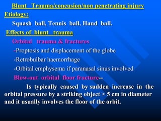
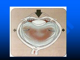
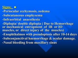
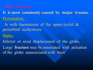
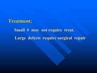
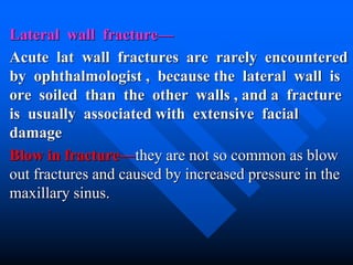
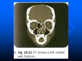
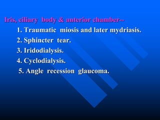
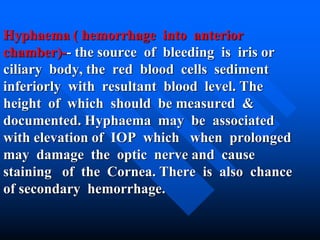
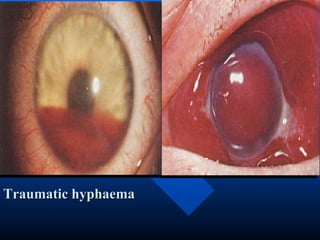
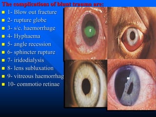
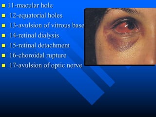
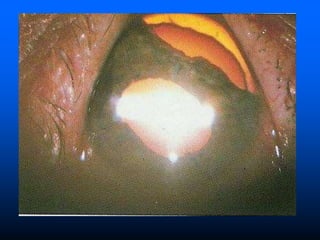
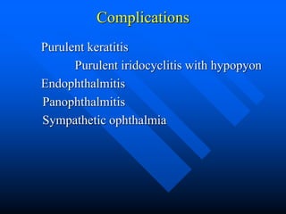
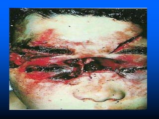
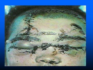
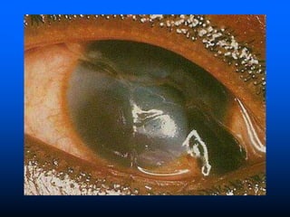
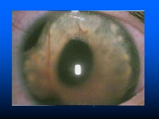
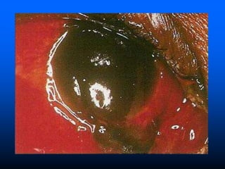
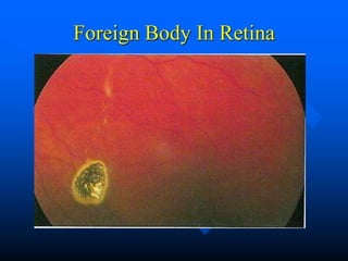
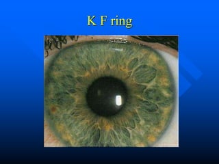
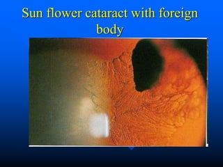
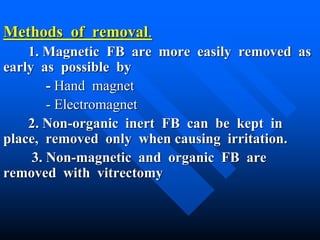
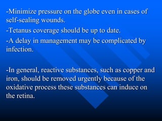
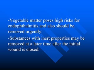
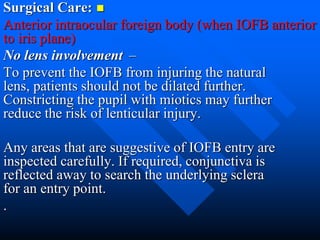
Ocular trauma
Dr. Mohd Najmussadiq Khan
M S (Ophth) DiSSO(ESASO)
Trauma
Trauma can be divided to -Extraocular foreign body.
-Blunt trauma.
- Penetrating trauma.
- Chemical injury.
Extraocular foreign body
Etiology;
Small particle of coal, dust, steel and glass.
.
Symptoms;
1. Sudden discomfort in the eye.
2. Reflex blinking.
3. Irritation if the FB is embedded
in the Cornea.
4. Lacrimation and photophobia
Signs;
1. Reflex blepharospasm.
2. FB is visible on conjunctiva, limbus ,
Cornea , Sulcus subtarsalis .
3. It may be single or multiple , superficial
or deep.
Complication;
1. Conjunctivitis.
2.Corneal ulcer.
3. Brown ring or stain on the Cornea.
4. Sharp FB may penetrate the eye wall.
Treatment;
1. Do not rub the eye.
2. Wash the eye with plenty of
water.
3. Remove the FB after LA
instillation.
4. Give antibiotic drops & oint.
Blunt Trauma/concusion/non penetrating injury
Etiology;
Squash ball, Tennis ball, Hand ball.
Effects of blunt trauma
Orbital trauma & fractures
-Proptosis and displacement of the globe
-Retrobulbar haemorrhage
-Orbital emphysema if paranasal sinus involved
Blow-out orbital floor fracture--
Is typically caused by sudden increase in the
orbital pressure by a striking object > 5 cm in diameter
and it usually involves the floor of the orbit.
Signs;
-Periocular ecchymosis, oedema
-Subcutaneous emphysema
-Infraorbital anaesthesia
-Diplopia/ double diplopia ( Due to Hemorrhage
or mechanical entrapment of IR or IO
muscles, or direct injury of the muscles)
-Enophthalmos with pseudoptosis after 10-14 days
-Subconjunctival haemorrhage & ocular damage.
-Nasal bleeding from maxillary sinus
Blow out fracture
Investigation; X-ray, CT, Hess test, Field
of binocular vision, forced duction test
(FDT).
Treatment;
1. Initial treatment is conservative with
antibiotics, and the patient should be
instructed not to blow the nose.
2. surgical repair
Subsequent treatment--
a. Small cracks unassociated with herniation
do not require treatment.
b. Fracture involving less than half the orbital
floor with no or little herniation and improving
diplopia also do not require treatment.
c. Fracture involving half or more of the
orbital floor, with entrapment orbital contents and
persistent diplopia should be repaired within 2
week.
b. Blow-out medial wall fracture—
Periorbital subcutaneous emphysema,
defective ocular motility.
Treatment—
Release of the entrapped tissue and
repair of the bony defect.
c. Roof fracture.
It is most commonly caused by major trauma.
Presentation,
Is with haematoma of the upper eyelid &
periorbital ecchymosis.
Signs,
Inferior or axial displacement of the globe,
Large fracture may be associated with pulsation
of the globe unassociated with bruit
Treatment;
Small # may not require treat.
Large defects require surgical repair
Lateral wall fracture—
Acute lat wall fractures are rarely encountered
by ophthalmologist , because the lateral wall is
ore soiled than the other walls , and a fracture
is usually associated with extensive facial
damage
Blow in fracture—they are not so common as blow
out fractures and caused by increased pressure in the
maxillary sinus.
Eyelid
Haematoma/ ecchymosis/ black eye, laceration, swelling .
Conjunctiva
Subconjunctival Haemorrhage, laceration, chemosis.
Cornea
Corneal abrasion ( treat is with topical antibiotics and
cycloplegic )
Acute Corneal oedema due to dysfunction of the corneal
endothelium
Staining of the Cornea
Rupture of the Descemet's membrane
Sclera
Rupture of the globe may occur, from very sever blunt
trauma.
Iris, ciliary body & anterior chamber--
1. Traumatic miosis and later mydriasis.
2. Sphincter tear.
3. Iridodialysis.
4. Cyclodialysis.
5. Angle recession glaucoma.
Hyphaema ( hemorrhage into anterior
chamber)-- the source of bleeding is iris or
ciliary body, the red blood cells sediment
inferiorly with resultant blood level. The
height of which should be measured &
documented. Hyphaema may be associated
with elevation of IOP which when prolonged
may damage the optic nerve and cause
staining of the Cornea. There is also chance
of secondary hemorrhage.
Traumatic hyphaema
Hyphaema Treatment
-Bed rest, Oral tranexamic acid 25 mg/kg
-Daily observation of level of hyphaema and the IOP until
it resolve spontaneously (hospital admission is indicated
only for large hyphaema.)
-When IOP is elevated we give beta blockers and or
carbonic anhydrase inhibitors
-Topical steroids are useful .
-Surgical evacuation is indicated if
1- the IOP >50 mmHg for 2 days or > 35mmHg for 7
days
2- Early corneal staining
3- Total hyphaema for more than 5 days.
Lens
1. Vossius ring.
2. Traumatic or concussion Cataract.
3. Dislocation or Subluxation.
4. Lens capsule rupture.
Vitreous
1. Vitreous haemorrhage/liquefaction.
2. Vitreous herniation in the anterior
chamber.
3. anterior or posterior vitreous detachment
.
Choroid
1. Choroidal rupture.
2. Choroidal haemorrhage.
3. Choroidal detachment.
Retina
1. Macular oedema (Berlin’ oedema or
commotio retinae)- there is milky white
cloudiness at the posterior pole with cherry red
spot in the center. It disappears after few days
or may be followed by pigmentary deposits
Macular cyst/ macular hole
Retinal haemorrhage/tears
Retinal detachment
Traumatic retinopathy
Optic Nerve.
1. Optic atrophy.
2. Avulsion of optic nerve
3. Optic nerve sheath haemorrhage
Raised IOP may be due to
-Trabecular edema
-Hyphaema
-Subluxation/dislocation of the lens
-Ghost cell glaucoma
-Angle recession glaucoma
The complications of blunt trauma are:
 1- Blow out fracture
 2- rupture globe
 3- s/c. haemorrhage
 4- Hyphaema
 5- angle recession
 6- sphincter rupture
 7- iridodialysis
 8- lens subluxation
 9- vitreous haemorrhage
 10- commotio retinae
 11-macular hole
 12-equatorial holes
 13-avulsion of vitrous base
 14-retinal dialysis
 15-retinal detachment
 16-choroidal rupture
 17-avulsion of optic nerve
Pentrating/perforating trauma
They are 3 times more common in males than
in females. The most common causes are
assault , domestic accidents and sport. They
are likely to cause severe damage to the eye.
The seriousness arises from
-The immediate effects of the trauma
-The introduction of the infections inside the eye
-Sympathetic ophthalmia
Entry Of Foreign Body Through Iris
Signs of eyeball perforation.
1. Decreased visual acuity.
2. Marked hypotony or low IOP.
3. Shallow AC.
4. Alteration in pupil size, size &
location.
5. Marked conjunctival oedema.
6. Subconjunctival haemorrhage.
7. Hole in the iris.
8. Wound track in the Cornea.
9. Hyphaema or haemophthalmos.
10. Entery wound present generally
Aim of traetment.
a. To save vision and anatomy.
b. To prevent the occurrence of sympathetic
ophthalmia.
Principle of treat.
It should be treated immediately by
1. Proper suturing and apposition of the
ocular tissue.
2. Control and treat of infection by broad
spectrum antibiotics (tetanus vaccination may
be required).
3. Close follow up.
Complications
Purulent keratitis
Purulent iridocyclitis with hypopyon
Endophthalmitis
Panophthalmitis
Sympathetic ophthalmia
Principles of primary repaire.
The technique of primary repair depends on the
severity of injury and any associated complications such as
iris incarceration, flat anterior chamber, and damage to
intraocular contents.
1. Small corneal laceration with formed AC may not
require suturing.
2. Medium-sized corneal laceration usual require suturing.
3. Corneal laceration with iris involvement. Management
depends on the duration and extent of incarceration.
- Recently incarcerated iris may be reposted.
- Large incarceration of iris should be abscised especially
if of more than few days duration or the iris appear non-
viable.
4. Corneal laceration with lenticular damage, is
reated with suturing the laceration and
removing the lens by phacoemulsification or
with a vitreous cutter.
5. Anterior Scleral laceration every attempt
should be made to suture the sclera, reposit
exposed viable uveal tissue and cut prolapsed
vitreous.
6. Posterior scleral laceration, should be
exposed and sutured.
Eyelid Laceration.
The presence of lid laceration, however
insignificant, mandate careful exploration of the
wound and examination of the globe.
Any lid defect should be repaired by direct
horizontal closure whenever possible.
1. Superficial laceration parallel the lid margin
without gaping can be sutured.
2. Lid margin laceration invariably gape and
must be carefully sutured.
3. Lid laceration with little tissue loss is
managed by performing lateral cantholysis and
suturing.
4. Laceration with extensive tissue loss may
require major reconstructive procedures.
5. Canalicular laceration, should be repaired
within 24 h.
a. The laceration is bridged by silicon
tubing.
b. The laceration is sutured.
c. The tubing is left in situ for 3-6 months.
Perforating injury with retained foreign body
(intraocular foreign body)--
The FB which are likely to penetrate the
eye and are retained include
Minute chips of iron, steel, stones, glass,
lead pellet, wood splinter.
Effects of retained FB.
1. Mechanical effects.
2. Infection.
3. Specific chemical action of the metal. it varies with
the chemical nature of the metals
some metals are inert like glass, plastic, gold, silver,
platinum.
-some cause little local reaction with encapsulation
reaction like Lead
-some cause local suppuration like Aluminum, nickel.
-some cause proliferative reaction like organic
materials like wood splinter
-some cause degenerative reaction like Iron and
Copper
Corneal foreign body
Foreign Body In Retina
History:
As with any traumatic injury, the history is
important to determine the exact circumstances
for medical-legal and possibly workman
compensation.
A high degree of suspicion is required based on the
history especially when a description of metal
striking metal is given.
Patients frequently feel something enter the eye with
no obvious external changes; hence, the incident
usually is dismissed quickly.
Patients may complain of an irritation and
sometimes
conjunctival injection.
Initial decrease in visual acuity occurs if damage is
present in the visual axis.
Determining if the IOFB is magnetic is important to
the surgical management.
Air gun pellet
Siderosis bulbi
It is a chronic irreversible degenerative change in the ocular
tissue. It is due to electrolytic dissociation of Iron
metal. Iron get deposited in----
1. Lens-The anterior lens capsule wherein oval
patches of rusty deposits are arranged radially in a
ring. The lens become opaque eventually.
2. Iris- is stained greenish and later reddish brown
(heterochromia of iris).
3. Cornea-Rusty discoloration of the cornea
4. Retina- Retinal degeneration with generalized
deposition of pigments.
5. Secondary open angle glaucoma of chronic type.
6. Blindness occur unless the FB is removed early.
Chalcosis bulbi— it is a reversible condition if
particle is removed
Copper gets deposited in the cornea, lens &
retina.
1. Kayser-Flischer ring- there is deposition
of copper in the deeper parts of periphery of
cornea in the Descemet’s membrane forming a
golden brown ring.
2. Sunflower Cataract – The deposition of
copper is in the form of petals of a flower
and is golden green in colour.
3. Retina- there are golden plaques deposited
in the posterior pole.
K F ring
Sun flower cataract with foreign
body
Diagnosis and localization of intraocular FB.
1. Slit-lamp examination and gonioscopy.
2. Ophthalmoscopic examination.
3. Radiographic examination with limbal
ring X- ray.
4. Electro-acoustic location.
5. Ultrasonography.
6. CT scan.
7. Berman / Roper hall metal locators
8. MRI scan if it is not a metallic foreign
body
Methods of removal.
1. Magnetic FB are more easily removed as
early as possible by
- Hand magnet
- Electromagnet
2. Non-organic inert FB can be kept in
place, removed only when causing irritation.
3. Non-magnetic and organic FB are
removed with vitrectomy
-Minimize pressure on the globe even in cases of
self-sealing wounds.
-Tetanus coverage should be up to date.
-A delay in management may be complicated by
infection.
-In general, reactive substances, such as copper and
iron, should be removed urgently because of the
oxidative process these substances can induce on
the retina.
-Vegetable matter poses high risks for
endophthalmitis and also should be
removed urgently.
-Substances with inert properties may be
removed at a later time after the initial
wound is closed.
Surgical Care:
Anterior intraocular foreign body (when IOFB anterior
to iris plane)
–No lens involvement
To prevent the IOFB from injuring the natural
lens, patients should not be dilated further.
Constricting the pupil with miotics may further
reduce the risk of lenticular injury.
Any areas that are suggestive of IOFB entry are
inspected carefully. If required, conjunctiva is
reflected away to search the underlying sclera
for an entry point.
.
-Any wound that is not self-sealing is closed
using the suture of choice for the specific
tissue. If the wound is in the cornea 10-0
nylon is used; if in the sclera, the use of 8-0
nylon or vicryl is used.
Reforming and maintaining the AC depth is
established prior to removing the IOFB.
Usually, injection of balanced salt solution
or viscoelastic through a paracentesis port is
recommended
Lens involvement
-If the IOFB has damaged the lens, the lens
should be removed.
-Generally, patients are young with a soft
nucleus, which enables phacoemulsification
to be performed but extracapsular lens
extraction also may be performed.
Concurrent intraocular lens insertion usually is
not performed because calculation of the
intraocular lens power may not be exact.
Sympathetic Ophthalmitis
Is a very rare , bilateral , granulomatous panuveitis , which
occurs after penetrating ocular trauma usually associated
with uveal prolepse or less frequently , following intraocular
surgery.
Presentation.
in 65% of cases is between 2 weeks – 3 months after initial
injury. 90 % of all cases occur wit hin the first yr .
Signs.
- The exciting eye show evidence of initial trauma
and is frequently very red and irritable.
- The sympathizing eye becomes photophobic and
irritable.
- Both eyes develop a chronic granulomatous
anterior uveitis.
- Optic disc swelling and multifocal choroiditis
involve the entire fundus.
Course.
rarely the uveitis runs a relatively mild and self
limmiting course.
Usually the intraocular inflammaton become chronic
and unless appropriarly treated may lead to
Cataract , glaucoma , pthisis bulbi.
Treatment
With systemic steroids .
Steroid-sparing immunosuppressive agents.
Enucleation,
-Primary enucleation should be performed
only for very severe injury and with no prospect
of retention of vision.
-Secondary enucleation may be considered
following primary injury if the eye is severely and
irreversibly damaged.
Chemical injuries and Burns
They range in severity from trivial to potentially
blinding. The majority are accidental, a few the
result of assault. Two thirds of accidental burns
occur at work. Alkali burns are twice as common
as acid.
Etiology.
- Thermal injury can be caused by hot water,
steam, hot ashes, molten metals etc.
- The most common alkali are Ammonia, sodium
hydroxide and lime.
-The most common acids are sulphuric,
sulphurous, hydrofluoric, acetic, chromic and
Hydrochloric.
-The severity of chemical burn is related to
the properties of the chemical, the area of
affected ocular surface, duration of exposure
and relate effects such as thermal effect.
-Alkalis tend to penetrate deeper than do
acids, which coagulate surface proteins,
resulting in a protective barrier.
Symptoms
1. There is red eye with marked swelling of lids
and conjunctiva.
2. Marked reflex blepharospasm.
3. Photophobia and lacrimation.
Signs.
1.Sever congestion and chemosis of conjunctiva.
2. Marks of burn over surrounding skin.
3. Cornea is dull and opaque or may get
sloughed off.
4.Anterior chamber penetration result in iris
and lens damage.
5. Hypotony and phthisis bulbi.
Alkali burn
Complication
1. Symblepharon is adhesion of the lid to
the globe, A glass rod well-coated with a
lubricant or ointment is swept around the
upper and lower fornix.
2. Corneal ulcer is usually present which
may perforate.
3. Uveitis especially in alkali burns.
4. Glaucoma especially in alkali burns.
Corneal burn caused by alkali
Treatment.
Emergency treatment.
1. Copious irrigation to minimize duration of
contact with the chemical.
Normal saline should be used to irrigate
the eye for 15-20 min or until pH is
normalized.
2. Double-eversion of eyelids to remove any
retained particles.
3. Debridement of necrotic area.
Grading of severity.
1. Grade I: Clear cornea and no limbal
ischaemia (excellent prognosis).
2. Grade II: Hazy cornea but with visible iris
detail and less than 1/3 of limbal
ischaemia (good prognosis)
3. Grade III: Total loss of corneal epithelium,
stromal haze obscuring iris details, and between
1/3- ½ limbal ischaemia. (guarded prognosis).
4. Grade IV: Opaque cornea and more than ½
of limbal ischaemia.
Medical Treatment;
Mild (G I and GII) injuries are
treated with local steroids, cycloplegiccs
and prophylactic antibiotics for about
7 days.
In more sever burns the aim of
treatment is to reduce inflammation,
promote epithelial regeneration and
prevent corneal ulceration.
1. Steroids reduce inflammation, however
they also impair stromal healing.
2. Ascorbic acid which improve wound healing.
3. Citric acid is a powerful inhibitor of
neutrophil activity. Topical Na citrate 10% is
given in the 1st 10 days.
4. Tetracycline are colagenase inhibitor, inhibit
neutrophil activity and reduce
ulceration.
Surgical treatment.
A. Early Surgery may be necessary.
- Advancement of Tenon capsule and
suturing to the limbus.
- Limbal stem cell transplantation.
- Amniotic membrane grafting to promote
epithelization.
B. Late Surgery.
- Division of conjunctival bands and
symblepharon.
- Conjunctival or mucous membrane grafts.
- Correction of eyelid deformity.
- Keratoplasty should be delayed for at least
6 month or longer to allow maximal resolution
of inflammation.
- Keratoprotheses may be required in very
severely damaged eyes.
Shallow fornix

Ocular Trauma

  • 1.
    Ocular trauma Dr. MohdNajmussadiq Khan M S (Ophth) DiSSO(ESASO)
  • 2.
    Trauma Trauma can bedivided to -Extraocular foreign body. -Blunt trauma. - Penetrating trauma. - Chemical injury. Extraocular foreign body Etiology; Small particle of coal, dust, steel and glass. .
  • 3.
    Symptoms; 1. Sudden discomfortin the eye. 2. Reflex blinking. 3. Irritation if the FB is embedded in the Cornea. 4. Lacrimation and photophobia
  • 4.
    Signs; 1. Reflex blepharospasm. 2.FB is visible on conjunctiva, limbus , Cornea , Sulcus subtarsalis . 3. It may be single or multiple , superficial or deep. Complication; 1. Conjunctivitis. 2.Corneal ulcer. 3. Brown ring or stain on the Cornea. 4. Sharp FB may penetrate the eye wall.
  • 5.
    Treatment; 1. Do notrub the eye. 2. Wash the eye with plenty of water. 3. Remove the FB after LA instillation. 4. Give antibiotic drops & oint.
  • 6.
    Blunt Trauma/concusion/non penetratinginjury Etiology; Squash ball, Tennis ball, Hand ball. Effects of blunt trauma Orbital trauma & fractures -Proptosis and displacement of the globe -Retrobulbar haemorrhage -Orbital emphysema if paranasal sinus involved Blow-out orbital floor fracture-- Is typically caused by sudden increase in the orbital pressure by a striking object > 5 cm in diameter and it usually involves the floor of the orbit.
  • 8.
    Signs; -Periocular ecchymosis, oedema -Subcutaneousemphysema -Infraorbital anaesthesia -Diplopia/ double diplopia ( Due to Hemorrhage or mechanical entrapment of IR or IO muscles, or direct injury of the muscles) -Enophthalmos with pseudoptosis after 10-14 days -Subconjunctival haemorrhage & ocular damage. -Nasal bleeding from maxillary sinus
  • 9.
  • 10.
    Investigation; X-ray, CT,Hess test, Field of binocular vision, forced duction test (FDT). Treatment; 1. Initial treatment is conservative with antibiotics, and the patient should be instructed not to blow the nose. 2. surgical repair
  • 12.
    Subsequent treatment-- a. Smallcracks unassociated with herniation do not require treatment. b. Fracture involving less than half the orbital floor with no or little herniation and improving diplopia also do not require treatment. c. Fracture involving half or more of the orbital floor, with entrapment orbital contents and persistent diplopia should be repaired within 2 week.
  • 13.
    b. Blow-out medialwall fracture— Periorbital subcutaneous emphysema, defective ocular motility. Treatment— Release of the entrapped tissue and repair of the bony defect.
  • 14.
    c. Roof fracture. Itis most commonly caused by major trauma. Presentation, Is with haematoma of the upper eyelid & periorbital ecchymosis. Signs, Inferior or axial displacement of the globe, Large fracture may be associated with pulsation of the globe unassociated with bruit
  • 15.
    Treatment; Small # maynot require treat. Large defects require surgical repair
  • 16.
    Lateral wall fracture— Acutelat wall fractures are rarely encountered by ophthalmologist , because the lateral wall is ore soiled than the other walls , and a fracture is usually associated with extensive facial damage Blow in fracture—they are not so common as blow out fractures and caused by increased pressure in the maxillary sinus.
  • 18.
    Eyelid Haematoma/ ecchymosis/ blackeye, laceration, swelling . Conjunctiva Subconjunctival Haemorrhage, laceration, chemosis. Cornea Corneal abrasion ( treat is with topical antibiotics and cycloplegic ) Acute Corneal oedema due to dysfunction of the corneal endothelium Staining of the Cornea Rupture of the Descemet's membrane Sclera Rupture of the globe may occur, from very sever blunt trauma.
  • 23.
    Iris, ciliary body& anterior chamber-- 1. Traumatic miosis and later mydriasis. 2. Sphincter tear. 3. Iridodialysis. 4. Cyclodialysis. 5. Angle recession glaucoma.
  • 24.
    Hyphaema ( hemorrhageinto anterior chamber)-- the source of bleeding is iris or ciliary body, the red blood cells sediment inferiorly with resultant blood level. The height of which should be measured & documented. Hyphaema may be associated with elevation of IOP which when prolonged may damage the optic nerve and cause staining of the Cornea. There is also chance of secondary hemorrhage.
  • 25.
  • 27.
    Hyphaema Treatment -Bed rest,Oral tranexamic acid 25 mg/kg -Daily observation of level of hyphaema and the IOP until it resolve spontaneously (hospital admission is indicated only for large hyphaema.) -When IOP is elevated we give beta blockers and or carbonic anhydrase inhibitors -Topical steroids are useful . -Surgical evacuation is indicated if 1- the IOP >50 mmHg for 2 days or > 35mmHg for 7 days 2- Early corneal staining 3- Total hyphaema for more than 5 days.
  • 28.
    Lens 1. Vossius ring. 2.Traumatic or concussion Cataract. 3. Dislocation or Subluxation. 4. Lens capsule rupture. Vitreous 1. Vitreous haemorrhage/liquefaction. 2. Vitreous herniation in the anterior chamber. 3. anterior or posterior vitreous detachment .
  • 32.
    Choroid 1. Choroidal rupture. 2.Choroidal haemorrhage. 3. Choroidal detachment. Retina 1. Macular oedema (Berlin’ oedema or commotio retinae)- there is milky white cloudiness at the posterior pole with cherry red spot in the center. It disappears after few days or may be followed by pigmentary deposits
  • 33.
    Macular cyst/ macularhole Retinal haemorrhage/tears Retinal detachment Traumatic retinopathy Optic Nerve. 1. Optic atrophy. 2. Avulsion of optic nerve 3. Optic nerve sheath haemorrhage Raised IOP may be due to -Trabecular edema -Hyphaema -Subluxation/dislocation of the lens -Ghost cell glaucoma -Angle recession glaucoma
  • 36.
    The complications ofblunt trauma are:  1- Blow out fracture  2- rupture globe  3- s/c. haemorrhage  4- Hyphaema  5- angle recession  6- sphincter rupture  7- iridodialysis  8- lens subluxation  9- vitreous haemorrhage  10- commotio retinae
  • 37.
     11-macular hole 12-equatorial holes  13-avulsion of vitrous base  14-retinal dialysis  15-retinal detachment  16-choroidal rupture  17-avulsion of optic nerve
  • 39.
    Pentrating/perforating trauma They are3 times more common in males than in females. The most common causes are assault , domestic accidents and sport. They are likely to cause severe damage to the eye. The seriousness arises from -The immediate effects of the trauma -The introduction of the infections inside the eye -Sympathetic ophthalmia
  • 40.
    Entry Of ForeignBody Through Iris
  • 41.
    Signs of eyeballperforation. 1. Decreased visual acuity. 2. Marked hypotony or low IOP. 3. Shallow AC. 4. Alteration in pupil size, size & location. 5. Marked conjunctival oedema.
  • 42.
    6. Subconjunctival haemorrhage. 7.Hole in the iris. 8. Wound track in the Cornea. 9. Hyphaema or haemophthalmos. 10. Entery wound present generally
  • 43.
    Aim of traetment. a.To save vision and anatomy. b. To prevent the occurrence of sympathetic ophthalmia. Principle of treat. It should be treated immediately by 1. Proper suturing and apposition of the ocular tissue. 2. Control and treat of infection by broad spectrum antibiotics (tetanus vaccination may be required). 3. Close follow up.
  • 44.
    Complications Purulent keratitis Purulent iridocyclitiswith hypopyon Endophthalmitis Panophthalmitis Sympathetic ophthalmia
  • 50.
    Principles of primaryrepaire. The technique of primary repair depends on the severity of injury and any associated complications such as iris incarceration, flat anterior chamber, and damage to intraocular contents. 1. Small corneal laceration with formed AC may not require suturing. 2. Medium-sized corneal laceration usual require suturing. 3. Corneal laceration with iris involvement. Management depends on the duration and extent of incarceration. - Recently incarcerated iris may be reposted. - Large incarceration of iris should be abscised especially if of more than few days duration or the iris appear non- viable.
  • 51.
    4. Corneal lacerationwith lenticular damage, is reated with suturing the laceration and removing the lens by phacoemulsification or with a vitreous cutter. 5. Anterior Scleral laceration every attempt should be made to suture the sclera, reposit exposed viable uveal tissue and cut prolapsed vitreous. 6. Posterior scleral laceration, should be exposed and sutured.
  • 52.
    Eyelid Laceration. The presenceof lid laceration, however insignificant, mandate careful exploration of the wound and examination of the globe. Any lid defect should be repaired by direct horizontal closure whenever possible. 1. Superficial laceration parallel the lid margin without gaping can be sutured. 2. Lid margin laceration invariably gape and must be carefully sutured.
  • 53.
    3. Lid lacerationwith little tissue loss is managed by performing lateral cantholysis and suturing. 4. Laceration with extensive tissue loss may require major reconstructive procedures. 5. Canalicular laceration, should be repaired within 24 h. a. The laceration is bridged by silicon tubing. b. The laceration is sutured. c. The tubing is left in situ for 3-6 months.
  • 54.
    Perforating injury withretained foreign body (intraocular foreign body)-- The FB which are likely to penetrate the eye and are retained include Minute chips of iron, steel, stones, glass, lead pellet, wood splinter.
  • 55.
    Effects of retainedFB. 1. Mechanical effects. 2. Infection. 3. Specific chemical action of the metal. it varies with the chemical nature of the metals some metals are inert like glass, plastic, gold, silver, platinum. -some cause little local reaction with encapsulation reaction like Lead -some cause local suppuration like Aluminum, nickel. -some cause proliferative reaction like organic materials like wood splinter -some cause degenerative reaction like Iron and Copper
  • 56.
  • 57.
  • 58.
    History: As with anytraumatic injury, the history is important to determine the exact circumstances for medical-legal and possibly workman compensation. A high degree of suspicion is required based on the history especially when a description of metal striking metal is given. Patients frequently feel something enter the eye with no obvious external changes; hence, the incident usually is dismissed quickly.
  • 59.
    Patients may complainof an irritation and sometimes conjunctival injection. Initial decrease in visual acuity occurs if damage is present in the visual axis. Determining if the IOFB is magnetic is important to the surgical management.
  • 60.
  • 61.
    Siderosis bulbi It isa chronic irreversible degenerative change in the ocular tissue. It is due to electrolytic dissociation of Iron metal. Iron get deposited in---- 1. Lens-The anterior lens capsule wherein oval patches of rusty deposits are arranged radially in a ring. The lens become opaque eventually. 2. Iris- is stained greenish and later reddish brown (heterochromia of iris). 3. Cornea-Rusty discoloration of the cornea 4. Retina- Retinal degeneration with generalized deposition of pigments. 5. Secondary open angle glaucoma of chronic type. 6. Blindness occur unless the FB is removed early.
  • 62.
    Chalcosis bulbi— itis a reversible condition if particle is removed Copper gets deposited in the cornea, lens & retina. 1. Kayser-Flischer ring- there is deposition of copper in the deeper parts of periphery of cornea in the Descemet’s membrane forming a golden brown ring. 2. Sunflower Cataract – The deposition of copper is in the form of petals of a flower and is golden green in colour. 3. Retina- there are golden plaques deposited in the posterior pole.
  • 63.
  • 64.
    Sun flower cataractwith foreign body
  • 65.
    Diagnosis and localizationof intraocular FB. 1. Slit-lamp examination and gonioscopy. 2. Ophthalmoscopic examination. 3. Radiographic examination with limbal ring X- ray. 4. Electro-acoustic location. 5. Ultrasonography. 6. CT scan. 7. Berman / Roper hall metal locators 8. MRI scan if it is not a metallic foreign body
  • 66.
    Methods of removal. 1.Magnetic FB are more easily removed as early as possible by - Hand magnet - Electromagnet 2. Non-organic inert FB can be kept in place, removed only when causing irritation. 3. Non-magnetic and organic FB are removed with vitrectomy
  • 67.
    -Minimize pressure onthe globe even in cases of self-sealing wounds. -Tetanus coverage should be up to date. -A delay in management may be complicated by infection. -In general, reactive substances, such as copper and iron, should be removed urgently because of the oxidative process these substances can induce on the retina.
  • 68.
    -Vegetable matter poseshigh risks for endophthalmitis and also should be removed urgently. -Substances with inert properties may be removed at a later time after the initial wound is closed.
  • 69.
    Surgical Care: Anterior intraocularforeign body (when IOFB anterior to iris plane) –No lens involvement To prevent the IOFB from injuring the natural lens, patients should not be dilated further. Constricting the pupil with miotics may further reduce the risk of lenticular injury. Any areas that are suggestive of IOFB entry are inspected carefully. If required, conjunctiva is reflected away to search the underlying sclera for an entry point. .
  • 70.
    -Any wound thatis not self-sealing is closed using the suture of choice for the specific tissue. If the wound is in the cornea 10-0 nylon is used; if in the sclera, the use of 8-0 nylon or vicryl is used. Reforming and maintaining the AC depth is established prior to removing the IOFB. Usually, injection of balanced salt solution or viscoelastic through a paracentesis port is recommended
  • 71.
    Lens involvement -If theIOFB has damaged the lens, the lens should be removed. -Generally, patients are young with a soft nucleus, which enables phacoemulsification to be performed but extracapsular lens extraction also may be performed. Concurrent intraocular lens insertion usually is not performed because calculation of the intraocular lens power may not be exact.
  • 72.
    Sympathetic Ophthalmitis Is avery rare , bilateral , granulomatous panuveitis , which occurs after penetrating ocular trauma usually associated with uveal prolepse or less frequently , following intraocular surgery. Presentation. in 65% of cases is between 2 weeks – 3 months after initial injury. 90 % of all cases occur wit hin the first yr .
  • 73.
    Signs. - The excitingeye show evidence of initial trauma and is frequently very red and irritable. - The sympathizing eye becomes photophobic and irritable. - Both eyes develop a chronic granulomatous anterior uveitis. - Optic disc swelling and multifocal choroiditis involve the entire fundus. Course. rarely the uveitis runs a relatively mild and self limmiting course. Usually the intraocular inflammaton become chronic and unless appropriarly treated may lead to Cataract , glaucoma , pthisis bulbi.
  • 74.
    Treatment With systemic steroids. Steroid-sparing immunosuppressive agents. Enucleation, -Primary enucleation should be performed only for very severe injury and with no prospect of retention of vision. -Secondary enucleation may be considered following primary injury if the eye is severely and irreversibly damaged.
  • 75.
    Chemical injuries andBurns They range in severity from trivial to potentially blinding. The majority are accidental, a few the result of assault. Two thirds of accidental burns occur at work. Alkali burns are twice as common as acid. Etiology. - Thermal injury can be caused by hot water, steam, hot ashes, molten metals etc. - The most common alkali are Ammonia, sodium hydroxide and lime.
  • 76.
    -The most commonacids are sulphuric, sulphurous, hydrofluoric, acetic, chromic and Hydrochloric. -The severity of chemical burn is related to the properties of the chemical, the area of affected ocular surface, duration of exposure and relate effects such as thermal effect. -Alkalis tend to penetrate deeper than do acids, which coagulate surface proteins, resulting in a protective barrier.
  • 77.
    Symptoms 1. There isred eye with marked swelling of lids and conjunctiva. 2. Marked reflex blepharospasm. 3. Photophobia and lacrimation. Signs. 1.Sever congestion and chemosis of conjunctiva. 2. Marks of burn over surrounding skin. 3. Cornea is dull and opaque or may get sloughed off. 4.Anterior chamber penetration result in iris and lens damage. 5. Hypotony and phthisis bulbi.
  • 78.
  • 79.
    Complication 1. Symblepharon isadhesion of the lid to the globe, A glass rod well-coated with a lubricant or ointment is swept around the upper and lower fornix. 2. Corneal ulcer is usually present which may perforate. 3. Uveitis especially in alkali burns. 4. Glaucoma especially in alkali burns.
  • 80.
  • 81.
    Treatment. Emergency treatment. 1. Copiousirrigation to minimize duration of contact with the chemical. Normal saline should be used to irrigate the eye for 15-20 min or until pH is normalized. 2. Double-eversion of eyelids to remove any retained particles. 3. Debridement of necrotic area.
  • 82.
    Grading of severity. 1.Grade I: Clear cornea and no limbal ischaemia (excellent prognosis). 2. Grade II: Hazy cornea but with visible iris detail and less than 1/3 of limbal ischaemia (good prognosis) 3. Grade III: Total loss of corneal epithelium, stromal haze obscuring iris details, and between 1/3- ½ limbal ischaemia. (guarded prognosis). 4. Grade IV: Opaque cornea and more than ½ of limbal ischaemia.
  • 83.
    Medical Treatment; Mild (GI and GII) injuries are treated with local steroids, cycloplegiccs and prophylactic antibiotics for about 7 days. In more sever burns the aim of treatment is to reduce inflammation, promote epithelial regeneration and prevent corneal ulceration.
  • 84.
    1. Steroids reduceinflammation, however they also impair stromal healing. 2. Ascorbic acid which improve wound healing. 3. Citric acid is a powerful inhibitor of neutrophil activity. Topical Na citrate 10% is given in the 1st 10 days. 4. Tetracycline are colagenase inhibitor, inhibit neutrophil activity and reduce ulceration.
  • 85.
    Surgical treatment. A. EarlySurgery may be necessary. - Advancement of Tenon capsule and suturing to the limbus. - Limbal stem cell transplantation. - Amniotic membrane grafting to promote epithelization.
  • 86.
    B. Late Surgery. -Division of conjunctival bands and symblepharon. - Conjunctival or mucous membrane grafts. - Correction of eyelid deformity. - Keratoplasty should be delayed for at least 6 month or longer to allow maximal resolution of inflammation. - Keratoprotheses may be required in very severely damaged eyes.
  • 87.