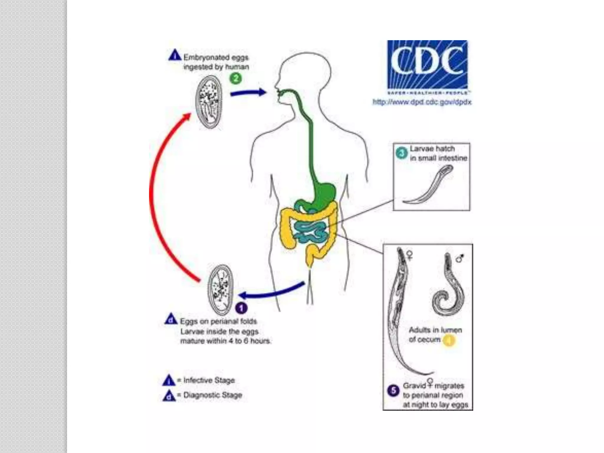
Enterobius vermicularis
Prepared by: Bishnu Prasad Timalsina
Common name: Pin worm, Seat worm,
thread worm
•Formerly, called oxyuris vermiculoris
History:
 The name Enterobius vermicularis means
tiny worm living in the intestine.
Greek: enteron-intestine, bios-life,
vermiculus -small worm.
 Term “oxyuris” means sharp tail, a feature
of the female worm, from which the name
pinworm is also derived.
 Leuckart(1865) first described the
complete life cycle of the parasite.
Geographical Distribution:
 worldwide in distribution(cosmopolitan)
 It is considered to be world’s most common
parasite, which affects the children.
Habitat:
Adult worms are found in the caecum,
appendix and adjacent portion of ascending
colon.
Morphology:
a. Adult worm:
 Short, white, fusiform worms with
pointed ends.
 Mouth is surrounded by cervical alae(3
no.)
 The oesophagus has a double –bulb
structure, a feature unique to this
worm.
b. Male worm:
 2.5 mm long and 0.1-0.2
mm thick.
 Posterior end is tightly
curved ventrally and
carries a copulatory spicule.
 Male survive for about 7-8
weeks.
c. Female worm:
 8-13 mm lond and 0.3-0.5 mm
thick.
 Posterior end is a thin pointed pin-
like tail.
 Single vagina present infront of the
middle third of the body which
leads to the paired uteri.
The uteri of gravid female is filled with
thousands of eggs.
Oviparous
Female survive for about 5-12 weeks.
d. Egg:
Egg is colourles and non bile stained
Floats in saturated salt solution
Planoconvex in shape
50-60 long,20-30 µm thick
Egg shell is double layer makes the egg stick
to each other and to clothing
Embryoated egg contain infective larva in
soil
Life cycle:
E. vermicularis is monoxenous, passing
its entire life cycle in the human host
Mode of tansmission:
Through contaminated hand, food ,
water,clothing and dust
Autoinfection: Due to scatching of
perianal area with fingers
:occurs most commonly in children
Retroinfection: The egg laid on the perianal
skin immediately hatch into the
infective stage larva and migrate through
the anus to develop into the adult worm in the
colon.
Pathogenicity and clinical features:
Enterobiasis:
• occurs mostly in children
•More common in females than in males
•About one- third of infections are symptomatic
•Intense irritation and pruritus of perianal and
perineal area(pruritis ani),when the worm
crawls out of the anus to lay eggs. This
leads to scratching of excoriation of the skin
around the anus .
•Irritation caused pruritis ani
•Nocturnal enuresis(disturb sleep)
•Chronic salpingitis
•Cervicitis,peritiontis
•UTI
•Responsible for appendicitis
Laboratory diagnosis:
A. Detection of egg:
 Examination of egg in feaces is not
useful in diagnosis because of present
of it in small proportion.
a.Under finge nails:
 Egg may be demostrated from the dirt
collected from the finger nails in
infected children.
Swab collected in early morning from
perianal folds are most often positive
b. NIH swab method
Named after national institute of health
By microscopic examination of
cellophane which is used for swabbing
by rolling over the perianal area
c. Scotch tape method:
By microscopic examine of scotch tape
(adhesive transparent cellophane tape)
which is used for collection of specimen
from anal margin.
B. Demostration of adult worm:
From stool
Occasionally from anus while children
are asleep
May be in appendix during appendectomy
Treatment:
Pyrantel pamoate(11mg/kg once,
maxm 1gram)
Albendazole(400mg once)
Mebendazole(100mg once) can be used
for single dose therapy while piperazine
has to be given daily for one week.
It is necessary to repeat the treatment
after 2 weeks to ensure elimination of
all worms
Prophylaxis
Maintainance of personal and community
hygiene such as frequent hand washing
finger nail cleaning and regular bathing
Frequent washing of night clothes and
bed linen
Awareness

Enterobius vermicularis

  • 1.
    Enterobius vermicularis Prepared by:Bishnu Prasad Timalsina
  • 2.
    Common name: Pinworm, Seat worm, thread worm •Formerly, called oxyuris vermiculoris
  • 3.
    History:  The nameEnterobius vermicularis means tiny worm living in the intestine. Greek: enteron-intestine, bios-life, vermiculus -small worm.  Term “oxyuris” means sharp tail, a feature of the female worm, from which the name pinworm is also derived.  Leuckart(1865) first described the complete life cycle of the parasite.
  • 4.
    Geographical Distribution:  worldwidein distribution(cosmopolitan)  It is considered to be world’s most common parasite, which affects the children. Habitat: Adult worms are found in the caecum, appendix and adjacent portion of ascending colon.
  • 5.
    Morphology: a. Adult worm: Short, white, fusiform worms with pointed ends.  Mouth is surrounded by cervical alae(3 no.)  The oesophagus has a double –bulb structure, a feature unique to this worm.
  • 6.
    b. Male worm: 2.5 mm long and 0.1-0.2 mm thick.  Posterior end is tightly curved ventrally and carries a copulatory spicule.  Male survive for about 7-8 weeks.
  • 7.
    c. Female worm: 8-13 mm lond and 0.3-0.5 mm thick.  Posterior end is a thin pointed pin- like tail.  Single vagina present infront of the middle third of the body which leads to the paired uteri.
  • 8.
    The uteri ofgravid female is filled with thousands of eggs. Oviparous Female survive for about 5-12 weeks.
  • 9.
    d. Egg: Egg iscolourles and non bile stained Floats in saturated salt solution Planoconvex in shape 50-60 long,20-30 µm thick Egg shell is double layer makes the egg stick to each other and to clothing Embryoated egg contain infective larva in soil
  • 10.
    Life cycle: E. vermicularisis monoxenous, passing its entire life cycle in the human host
  • 12.
    Mode of tansmission: Throughcontaminated hand, food , water,clothing and dust Autoinfection: Due to scatching of perianal area with fingers :occurs most commonly in children Retroinfection: The egg laid on the perianal skin immediately hatch into the infective stage larva and migrate through
  • 13.
    the anus todevelop into the adult worm in the colon. Pathogenicity and clinical features: Enterobiasis: • occurs mostly in children •More common in females than in males •About one- third of infections are symptomatic •Intense irritation and pruritus of perianal and perineal area(pruritis ani),when the worm
  • 14.
    crawls out ofthe anus to lay eggs. This leads to scratching of excoriation of the skin around the anus . •Irritation caused pruritis ani •Nocturnal enuresis(disturb sleep) •Chronic salpingitis •Cervicitis,peritiontis •UTI •Responsible for appendicitis
  • 15.
    Laboratory diagnosis: A. Detectionof egg:  Examination of egg in feaces is not useful in diagnosis because of present of it in small proportion. a.Under finge nails:  Egg may be demostrated from the dirt collected from the finger nails in infected children.
  • 16.
    Swab collected inearly morning from perianal folds are most often positive b. NIH swab method Named after national institute of health By microscopic examination of cellophane which is used for swabbing by rolling over the perianal area
  • 17.
    c. Scotch tapemethod: By microscopic examine of scotch tape (adhesive transparent cellophane tape) which is used for collection of specimen from anal margin.
  • 18.
    B. Demostration ofadult worm: From stool Occasionally from anus while children are asleep May be in appendix during appendectomy
  • 19.
    Treatment: Pyrantel pamoate(11mg/kg once, maxm1gram) Albendazole(400mg once) Mebendazole(100mg once) can be used for single dose therapy while piperazine has to be given daily for one week. It is necessary to repeat the treatment after 2 weeks to ensure elimination of all worms
  • 20.
    Prophylaxis Maintainance of personaland community hygiene such as frequent hand washing finger nail cleaning and regular bathing Frequent washing of night clothes and bed linen Awareness