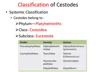
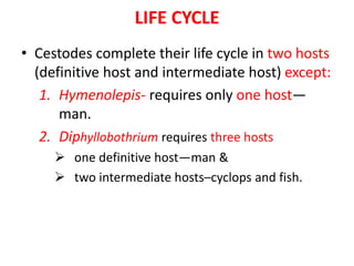
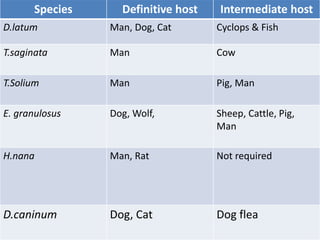
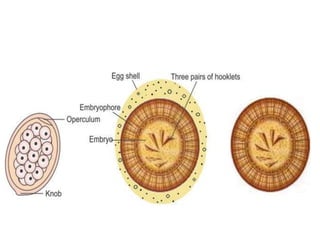
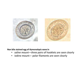
CESTODES
- Dr. ANKUR KUMAR
TOPICS for discussion-
• INTRODUCTION
• CLASSIFICATION
• MORPHOLOGY
• PSEUDOPHYLLIDEAN CESTODES
• CYCLOPHYLLIDEAN CESTODES
• SUMMARY
• LABORATORY DIAGNOSIS
• KATO KATZ TECHNIQUE
• Cestodes are helminthic parasites,
multicellular, bilaterally symmetrical
having three germ layers.
• The helminth of medical importance are
divided into three main groups-
1. Cestode
2. Trematode
3. Nematode
Introduction
CESTODE TREMATODE NEMATODES
SHAPE Tape-like; segmented Leaf-like; unsegmented Elongated, cylindrical;
unsegmented
SEXES Not separate i.e.
Monoecious,
hermaphrodite
Not separate , except
schistomes which are
diecious
Separate- Diecious
“HEAD” END Suckers, often with
hooks
Suckers, no hooks. No suckers, no hooks
ALIMENTARY
CANAL
absent Present but incomplete;
no anus
Present and complete
BODY CAVITY absent absent present
Life cycle Requires two hosts
(except Hymenolepis
and
Diphyllobothrium)
Requires three hosts
(except Schistosoma)
Requires one host
(except fi larial worms
and
Dracunculus)
Differences between cestodes, trematodes and nematodes
• Long, segmented and tape-like, hence called
tapeworms.
• They are flattened dorsoventrally.
• Size vary from few millimeters to several meters.
• Adult worms are found in intestinal canal of man
and animal.
• Excretory and nervous systems are present. The
excretory canals are built up of flame cells
(terminal cells) and canal cells.
• There is no circulatory system and no body cavity.
• Reproductive system is highly developed and
complete in each segment.
General characteristic of cestode
Classification of Cestodes
• Systemic Classification
• Cestodes belong to -
Phylum—Platyhelminths
Class- Cestoidea
Subclass- Eucestoda
Classification Based on the habitat of the Cestodes
• Intestinal Cestodes (Adult Worm Residing in
Human Intestine)
Diphyllobothrium species
Taenia solium and Taenia saginata causing
intestinal taeniasis
Hymenolepis species
Dipylidium species.
• Somatic/Tissue Cestodes (Larvae in Human
Muscles/Organs)
Taenia solium causing cysticercosis
Taenia multiceps
Echinococcus species
Spirometra species
Morphology of Cestodes
• Morphological forms:
1. Adult worm
2. Egg
3. Larva
There are three regions in an adult worm-
I. Head (scolex),
II. Neck
III. Strobila (a body or trunk) consisting of series of segments
(proglottides).
• Head is provided with suckers(slit-like or cup-like) and sometimes with
hooks which serve as organs of attachment
Head (scolex)
T. saginata T. solium
Adult worm
• Reproductive system is highly developed
and complete in each segment.
• According to maturity of reproductive
organs-
three types of segments of the strobila can be
recognized from the front backwards:
1. Immature segments
2. Mature segments
3. Gravid segments or fertilized segments
EGGS
• Pseudophyllidean cestodes:
 Eggs are ovoid, operculated,
surrounded by a single layer
called as egg shell (or
capsule), inside which the
embryo is present containing
hooklets (three pairs).
 Membrane lining the embryo
is ciliated. The eggs when laid
first in the feces, are not
embryonated.
 Maturation takes place later in
water.
• Cyclophyllidean cestodes:
 Eggs are round to oval,
covered by two layers—
 An outer egg shell (or
capsule) filled with yolk
material (thin, so might be
lost) and
 An inner thick radially striated
embryophore surrounding
the embryo.
 Eggs are embryonated from
the beginning, contains six
hooklets.
LARVA
• Pseudophyllidean
cestodes:
• Larva is solid without any
sac.
– They are:
Coracidium: First stage
larva of Diphyllobothrium
Procercoid: Second stage
larva of Diphyllobothrium
Plerocercoid: Third stage
larva of Diphyllobothrium
• Cyclophyllidean
Cestodes:
– Larvae contain bladder
like sacs.
– Cysticercus: Larval stage
of Taenia
– Hydatid cyst: larval stage
of Echinococcus
– Cysticercoid: Larval
stage of Hymenolepis
PSEUDOPHYLLIDEAN CESTODE CYCLOPHYLLIDEAN CESTODE
Head Bears 2 longitudinal slit- like grooves-
called bothria
Bears 4 cup-like muscular suckers
(acetabula). In T. solium and H.
nana, scolex has a beak like apical
protrusion called as rostellum
Uterus No branching; the convoluted uterine
tube assume the form of a rosette.
Branching may or may not be
present.
Uterine pore Present Absent
Common genital
pore
Ventral in the midline. Lateral
Eggs Operculated; give rise to ciliated
larvae.
Non operculated; do not give rise to
ciliated larvae.
Larval form Solid Contains bladder like sac
Difference b/n a Pseudophyllidean Cestode and a
Cyclophyllidean Cestode
LIFE CYCLE
• Cestodes complete their life cycle in two hosts
(definitive host and intermediate host) except:
1. Hymenolepis- requires only one host—
man.
2. Diphyllobothrium requires three hosts
 one definitive host—man &
 two intermediate hosts–cyclops and fish.
Species Definitive host Intermediate host
D.latum Man, Dog, Cat Cyclops & Fish
T.saginata Man Cow
T.Solium Man Pig, Man
E. granulosus Dog, Wolf, Sheep, Cattle, Pig,
Man
H.nana Man, Rat Not required
D.caninum Dog, Cat Dog flea
Laboratory diagnosis
• Direct wet mount (saline and iodine mount)
 Drops of saline and Lugol’s iodine are placed on two corners of a
slide.
 A small amount of feces is mixed by a stick to form a uniform
smooth suspension.
 Cover slip is placed on the mount and examined under low
power objective (10X); followed by high power objective (40X).
 Some times proglottids of Taenia also found in stool
examination.
• For Cysticercosis
 Radiodiagnosis— CT scan and MRI
 Antibody detection in serum or CSF
• For Echinococcus granulosus
 Hydatid fluid microscopy- brood capsules and protoscolices
 Antibody detection – IHA, LAT
 Antigen detection- ELISA
 Imaging methods—X-ray, USG, CT scan, MRI
 Skin test (Casoni test)
Concentration Techniques
1. Sedimentation techniques:
– Eggs settle down at the bottom following
centrifugation-
Formalin-ether concentration technique
2. Floatation techniques:
– The eggs float at the surface due to specific gravity
gradient
Saturated salt (sodium chloride) solution technique
Zinc sulphate floatation concentration technique
Formol-ether sedimentation technique (schematic diagram)
zinc sulfate flotation concentration technique (schematic) diagram;
Kato katz technique
Used for counting of eggs in stool.
• Place a plastic template with hole of size 6mm on top of clean glass
slide.
• Place a small amount of the faecal sample on a newspaper and
press a piece of nylon screen on top.
• With a spatula, scrape the faecal material through the nylon screen
so that only the debris remains.
• Transfer a small amount of the sieved faecal material into the
hole of the template & carefully fill the hole
• Lift off the template, now slide contain sieved fecal sample
• Place one piece of the cellophane (which has been soaked
overnight in methylene blue glycerol solution) over the faecal
sample.
• Place a clean slide over the top and press it evenly
downwards to spread the faeces in a circle.
• Remove the slide by gently sliding it sideways to avoid
separating the cellophane strip.
• Place the slide with the cellophane upwards for 30–60
minutes
• Place the slide under a microscope and examine the
whole area for eggs of schistosomes & soil transmitted
helminthes.
HYDATID CYST
MicroscopyMacroscopic picture
Non bile stained egg of Hymenolepis nana in
• saline mount—three pairs of hooklets are seen clearly
• iodine mount— polar filaments are seen clearly
THANK
YOU

Cestodes

  • 1.
  • 2.
    TOPICS for discussion- •INTRODUCTION • CLASSIFICATION • MORPHOLOGY • PSEUDOPHYLLIDEAN CESTODES • CYCLOPHYLLIDEAN CESTODES • SUMMARY • LABORATORY DIAGNOSIS • KATO KATZ TECHNIQUE
  • 3.
    • Cestodes arehelminthic parasites, multicellular, bilaterally symmetrical having three germ layers. • The helminth of medical importance are divided into three main groups- 1. Cestode 2. Trematode 3. Nematode Introduction
  • 4.
    CESTODE TREMATODE NEMATODES SHAPETape-like; segmented Leaf-like; unsegmented Elongated, cylindrical; unsegmented SEXES Not separate i.e. Monoecious, hermaphrodite Not separate , except schistomes which are diecious Separate- Diecious “HEAD” END Suckers, often with hooks Suckers, no hooks. No suckers, no hooks ALIMENTARY CANAL absent Present but incomplete; no anus Present and complete BODY CAVITY absent absent present Life cycle Requires two hosts (except Hymenolepis and Diphyllobothrium) Requires three hosts (except Schistosoma) Requires one host (except fi larial worms and Dracunculus) Differences between cestodes, trematodes and nematodes
  • 5.
    • Long, segmentedand tape-like, hence called tapeworms. • They are flattened dorsoventrally. • Size vary from few millimeters to several meters. • Adult worms are found in intestinal canal of man and animal. • Excretory and nervous systems are present. The excretory canals are built up of flame cells (terminal cells) and canal cells. • There is no circulatory system and no body cavity. • Reproductive system is highly developed and complete in each segment. General characteristic of cestode
  • 6.
    Classification of Cestodes •Systemic Classification • Cestodes belong to - Phylum—Platyhelminths Class- Cestoidea Subclass- Eucestoda
  • 7.
    Classification Based onthe habitat of the Cestodes • Intestinal Cestodes (Adult Worm Residing in Human Intestine) Diphyllobothrium species Taenia solium and Taenia saginata causing intestinal taeniasis Hymenolepis species Dipylidium species. • Somatic/Tissue Cestodes (Larvae in Human Muscles/Organs) Taenia solium causing cysticercosis Taenia multiceps Echinococcus species Spirometra species
  • 8.
    Morphology of Cestodes •Morphological forms: 1. Adult worm 2. Egg 3. Larva There are three regions in an adult worm- I. Head (scolex), II. Neck III. Strobila (a body or trunk) consisting of series of segments (proglottides). • Head is provided with suckers(slit-like or cup-like) and sometimes with hooks which serve as organs of attachment
  • 9.
  • 10.
  • 11.
    • Reproductive systemis highly developed and complete in each segment. • According to maturity of reproductive organs- three types of segments of the strobila can be recognized from the front backwards: 1. Immature segments 2. Mature segments 3. Gravid segments or fertilized segments
  • 12.
    EGGS • Pseudophyllidean cestodes: Eggs are ovoid, operculated, surrounded by a single layer called as egg shell (or capsule), inside which the embryo is present containing hooklets (three pairs).  Membrane lining the embryo is ciliated. The eggs when laid first in the feces, are not embryonated.  Maturation takes place later in water. • Cyclophyllidean cestodes:  Eggs are round to oval, covered by two layers—  An outer egg shell (or capsule) filled with yolk material (thin, so might be lost) and  An inner thick radially striated embryophore surrounding the embryo.  Eggs are embryonated from the beginning, contains six hooklets.
  • 13.
    LARVA • Pseudophyllidean cestodes: • Larvais solid without any sac. – They are: Coracidium: First stage larva of Diphyllobothrium Procercoid: Second stage larva of Diphyllobothrium Plerocercoid: Third stage larva of Diphyllobothrium • Cyclophyllidean Cestodes: – Larvae contain bladder like sacs. – Cysticercus: Larval stage of Taenia – Hydatid cyst: larval stage of Echinococcus – Cysticercoid: Larval stage of Hymenolepis
  • 14.
    PSEUDOPHYLLIDEAN CESTODE CYCLOPHYLLIDEANCESTODE Head Bears 2 longitudinal slit- like grooves- called bothria Bears 4 cup-like muscular suckers (acetabula). In T. solium and H. nana, scolex has a beak like apical protrusion called as rostellum Uterus No branching; the convoluted uterine tube assume the form of a rosette. Branching may or may not be present. Uterine pore Present Absent Common genital pore Ventral in the midline. Lateral Eggs Operculated; give rise to ciliated larvae. Non operculated; do not give rise to ciliated larvae. Larval form Solid Contains bladder like sac Difference b/n a Pseudophyllidean Cestode and a Cyclophyllidean Cestode
  • 15.
    LIFE CYCLE • Cestodescomplete their life cycle in two hosts (definitive host and intermediate host) except: 1. Hymenolepis- requires only one host— man. 2. Diphyllobothrium requires three hosts  one definitive host—man &  two intermediate hosts–cyclops and fish.
  • 16.
    Species Definitive hostIntermediate host D.latum Man, Dog, Cat Cyclops & Fish T.saginata Man Cow T.Solium Man Pig, Man E. granulosus Dog, Wolf, Sheep, Cattle, Pig, Man H.nana Man, Rat Not required D.caninum Dog, Cat Dog flea
  • 17.
    Laboratory diagnosis • Directwet mount (saline and iodine mount)  Drops of saline and Lugol’s iodine are placed on two corners of a slide.  A small amount of feces is mixed by a stick to form a uniform smooth suspension.  Cover slip is placed on the mount and examined under low power objective (10X); followed by high power objective (40X).  Some times proglottids of Taenia also found in stool examination. • For Cysticercosis  Radiodiagnosis— CT scan and MRI  Antibody detection in serum or CSF • For Echinococcus granulosus  Hydatid fluid microscopy- brood capsules and protoscolices  Antibody detection – IHA, LAT  Antigen detection- ELISA  Imaging methods—X-ray, USG, CT scan, MRI  Skin test (Casoni test)
  • 18.
    Concentration Techniques 1. Sedimentationtechniques: – Eggs settle down at the bottom following centrifugation- Formalin-ether concentration technique 2. Floatation techniques: – The eggs float at the surface due to specific gravity gradient Saturated salt (sodium chloride) solution technique Zinc sulphate floatation concentration technique
  • 19.
    Formol-ether sedimentation technique(schematic diagram) zinc sulfate flotation concentration technique (schematic) diagram;
  • 20.
    Kato katz technique Usedfor counting of eggs in stool. • Place a plastic template with hole of size 6mm on top of clean glass slide. • Place a small amount of the faecal sample on a newspaper and press a piece of nylon screen on top. • With a spatula, scrape the faecal material through the nylon screen so that only the debris remains. • Transfer a small amount of the sieved faecal material into the hole of the template & carefully fill the hole • Lift off the template, now slide contain sieved fecal sample • Place one piece of the cellophane (which has been soaked overnight in methylene blue glycerol solution) over the faecal sample.
  • 21.
    • Place aclean slide over the top and press it evenly downwards to spread the faeces in a circle. • Remove the slide by gently sliding it sideways to avoid separating the cellophane strip. • Place the slide with the cellophane upwards for 30–60 minutes • Place the slide under a microscope and examine the whole area for eggs of schistosomes & soil transmitted helminthes.
  • 24.
  • 25.
    Non bile stainedegg of Hymenolepis nana in • saline mount—three pairs of hooklets are seen clearly • iodine mount— polar filaments are seen clearly
  • 26.