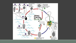
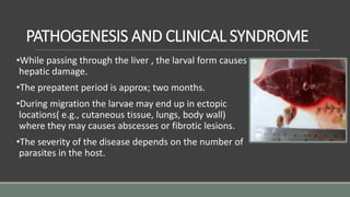
FASCIOLA HEPATICA
BAHIYA BASHEER
2nd year MBBS student
Fasciola hepatica
•Commonly called ‘sheep liver
fluke’.
•In sheep, the migratory phase of
the parasite produces severe
parenchymal disease of the liver
for which the parasite is called
sheep liver fluke and the disease is
known as liver rot.
HISTORY
•First trematode – 600 years ago
– Jehan de Briein -1379
•Linnaeus -1758
•Complete life cycle – Leuckart
and Thomas -1883
HABITAT
•Primarily a parasite of herbivores (particularly sheep
and cattle) causing zoonosis.
•Human beings are occasionally affected.
•Live for 9 years in the bile ducts of the liver of these
animals. Produce eggs that are carried by the bile
into the duodenum and then comes out with the
faeces.
MORPHOLOGY
ADULT
•Size : It is a large leaf-shaped fluke measuring
30×13 mm.
•Appearance : Brown and pale grey in colour with spiny
tegument.
•Suckers : There are two suckers, the oral(smaller) and
the ventral larger
•Intestine : Both the intestinal caeca show a number of
lateral compound branches
•Genital system : It is a hermaphrodite like other
trematodes.
•Longivity : Adult live for 5 years in sheep and 9-13
years in man. Adult lays approx. 20,000
eggs/day.
EGG
•Operculum :Operculated and unembryonated (contains a big
unsegmented ovum).
•Colour :Brownish yellow
•Size :130-150µm × 63-90µm, ovoid.
•Concentration: Eggs do not float in the saturated solution of
common salt.
•Egg can further develop only in water.
EPIDEMOLOGY
DISTRIBUTION
•Cosmopolitan disease found in all sheep-rearing and cattle-rearing
areas
•Russia, Europe.
•Latin American countries( Peru, Argentina)
•Egypt, Iran, Iraq, Northern Thailand , China, Japan and Autralia.
TRANSMISSION
•Transmission occurs by the ingestion of
metacerceriae encysted on aquatic
vegetation , such as watercress in
salads or contaminated water.
•In many areas of the world, animal
manure is used as primary fertiliser for
cultivation of watercress.
LIFE CYCLE
Fasciola hepatica requires two hosts to complete it life
cycle.
Definitive hosts: Herbivores like sheep, goat, cattle and
man. The adult worms lives in the biliary passage. These
animals serve also as reservoir hosts.
Intermediate hosts : Snails of the genus Lymnaea (e.g;
Ltruncalula) and Succinea in which larval development
occurs.
PATHOGENESIS AND CLINICAL SYNDROME
•While passing through the liver , the larval form causes
hepatic damage.
•The prepatent period is approx; two months.
•During migration the larvae may end up in ectopic
locations( e.g., cutaneous tissue, lungs, body wall)
where they may causes abscesses or fibrotic lesions.
•The severity of the disease depends on the number of
parasites in the host.
ACUTE INFECTION
•Migration through the liver parenchyma produces irritation
of the tissue and necrotic lesions in the liver.
•Acute disease is characterised by nausea, vomiting, pain,
chills and fever and marked eosinophilia are commonly
observed.
CHRONIC INFECTION
•As the worm take up residence in the bile ducts , numerous spines
covering the worm abrade the bile duct wall and leads to
inflammatory ,oedematous and fibrotic changes in the bile ducts.
•Some worms penetrate eroded areas in the duct and invade the liver
to produce necrotic foci which is called ‘LIVER ROT’, a condition more
frequently observed in sheep.
•In the chronic phase of severe infections, flukes are found both in
hepatic and extrahepatic ducts that cause mechanical obstruction
leading to cholangitis, cholecystits and gall stones.
DIAGNOSIS
CLINICAL DIAGNOSIS
•Diagnosis is based on an association of a sheep or cattle-
rearing and a history of consumption of home grown
vegetables or watercress.
•Together with some features of fever, eosinophilia, enlarged
liver and raised blood transaminase levels.
LABORATORY DIAGNOSIS
DEFINITIVE DIAGNOSIS
•It is established by finding large (150×80 µm) operculated eggs in faeces or
aspirates of duodenal fluid.
•F. hepatica eggs are morphologically similar to those of F. buski .
•Multiple stool samples may be required to find the trematode eggs.
•Eggs of hepatica may be passed in the stool following the ingestion of under-
cooked liver of infected sheep or cow. Stool should be examined for several days
after individuals have stopped eating liver to rule out spurious infections.
•Eggs are not seen in faeces in case of ectopic foci.
SEROLOGICAL TESTS
•Serological tests for antibodies by ELISA and
enzyme-linked immune-electro transfer blots using
F. hepatica excretion-secretion antigens have been
successfully used in areas of endemicity.
•These tests are useful when few eggs are found in
stool or when large populations are to be screened.
TREATMENT AND CONTROL
•In contrast of F. buski, F. hepatica responds poorly to
praziquantel. Treatment with BIOTHIONOL at 30 to 50 mg/kg
body weight on alternate days for 10 to 15 doses is
recommended.
•Alternatively , benzimidazole compound tricalbendazole 10
mg/kg as a single dose is recommended. Nitazoxanide is another
effective drug.
•Antibiotics are recommended to control secondary bacterial
infection. Prednisolone at a dose of 10-20 mg/day is used to
tackle toxaemia.
•People residing in areas frequented by sheep and cattle should
especially avoid ingestion of watercress and uncooked aquatic
vegetation.
THANK YOU

Fasciola hepatica

  • 1.
  • 2.
    Fasciola hepatica •Commonly called‘sheep liver fluke’. •In sheep, the migratory phase of the parasite produces severe parenchymal disease of the liver for which the parasite is called sheep liver fluke and the disease is known as liver rot.
  • 3.
    HISTORY •First trematode –600 years ago – Jehan de Briein -1379 •Linnaeus -1758 •Complete life cycle – Leuckart and Thomas -1883
  • 4.
    HABITAT •Primarily a parasiteof herbivores (particularly sheep and cattle) causing zoonosis. •Human beings are occasionally affected. •Live for 9 years in the bile ducts of the liver of these animals. Produce eggs that are carried by the bile into the duodenum and then comes out with the faeces.
  • 5.
    MORPHOLOGY ADULT •Size : Itis a large leaf-shaped fluke measuring 30×13 mm. •Appearance : Brown and pale grey in colour with spiny tegument. •Suckers : There are two suckers, the oral(smaller) and the ventral larger •Intestine : Both the intestinal caeca show a number of lateral compound branches •Genital system : It is a hermaphrodite like other trematodes. •Longivity : Adult live for 5 years in sheep and 9-13 years in man. Adult lays approx. 20,000 eggs/day.
  • 6.
    EGG •Operculum :Operculated andunembryonated (contains a big unsegmented ovum). •Colour :Brownish yellow •Size :130-150µm × 63-90µm, ovoid. •Concentration: Eggs do not float in the saturated solution of common salt. •Egg can further develop only in water.
  • 7.
    EPIDEMOLOGY DISTRIBUTION •Cosmopolitan disease foundin all sheep-rearing and cattle-rearing areas •Russia, Europe. •Latin American countries( Peru, Argentina) •Egypt, Iran, Iraq, Northern Thailand , China, Japan and Autralia.
  • 8.
    TRANSMISSION •Transmission occurs bythe ingestion of metacerceriae encysted on aquatic vegetation , such as watercress in salads or contaminated water. •In many areas of the world, animal manure is used as primary fertiliser for cultivation of watercress.
  • 9.
    LIFE CYCLE Fasciola hepaticarequires two hosts to complete it life cycle. Definitive hosts: Herbivores like sheep, goat, cattle and man. The adult worms lives in the biliary passage. These animals serve also as reservoir hosts. Intermediate hosts : Snails of the genus Lymnaea (e.g; Ltruncalula) and Succinea in which larval development occurs.
  • 11.
    PATHOGENESIS AND CLINICALSYNDROME •While passing through the liver , the larval form causes hepatic damage. •The prepatent period is approx; two months. •During migration the larvae may end up in ectopic locations( e.g., cutaneous tissue, lungs, body wall) where they may causes abscesses or fibrotic lesions. •The severity of the disease depends on the number of parasites in the host.
  • 12.
    ACUTE INFECTION •Migration throughthe liver parenchyma produces irritation of the tissue and necrotic lesions in the liver. •Acute disease is characterised by nausea, vomiting, pain, chills and fever and marked eosinophilia are commonly observed.
  • 13.
    CHRONIC INFECTION •As theworm take up residence in the bile ducts , numerous spines covering the worm abrade the bile duct wall and leads to inflammatory ,oedematous and fibrotic changes in the bile ducts. •Some worms penetrate eroded areas in the duct and invade the liver to produce necrotic foci which is called ‘LIVER ROT’, a condition more frequently observed in sheep. •In the chronic phase of severe infections, flukes are found both in hepatic and extrahepatic ducts that cause mechanical obstruction leading to cholangitis, cholecystits and gall stones.
  • 14.
    DIAGNOSIS CLINICAL DIAGNOSIS •Diagnosis isbased on an association of a sheep or cattle- rearing and a history of consumption of home grown vegetables or watercress. •Together with some features of fever, eosinophilia, enlarged liver and raised blood transaminase levels.
  • 15.
    LABORATORY DIAGNOSIS DEFINITIVE DIAGNOSIS •Itis established by finding large (150×80 µm) operculated eggs in faeces or aspirates of duodenal fluid. •F. hepatica eggs are morphologically similar to those of F. buski . •Multiple stool samples may be required to find the trematode eggs. •Eggs of hepatica may be passed in the stool following the ingestion of under- cooked liver of infected sheep or cow. Stool should be examined for several days after individuals have stopped eating liver to rule out spurious infections. •Eggs are not seen in faeces in case of ectopic foci.
  • 16.
    SEROLOGICAL TESTS •Serological testsfor antibodies by ELISA and enzyme-linked immune-electro transfer blots using F. hepatica excretion-secretion antigens have been successfully used in areas of endemicity. •These tests are useful when few eggs are found in stool or when large populations are to be screened.
  • 17.
    TREATMENT AND CONTROL •Incontrast of F. buski, F. hepatica responds poorly to praziquantel. Treatment with BIOTHIONOL at 30 to 50 mg/kg body weight on alternate days for 10 to 15 doses is recommended. •Alternatively , benzimidazole compound tricalbendazole 10 mg/kg as a single dose is recommended. Nitazoxanide is another effective drug. •Antibiotics are recommended to control secondary bacterial infection. Prednisolone at a dose of 10-20 mg/day is used to tackle toxaemia. •People residing in areas frequented by sheep and cattle should especially avoid ingestion of watercress and uncooked aquatic vegetation.
  • 18.