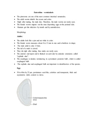
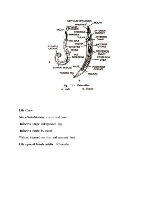
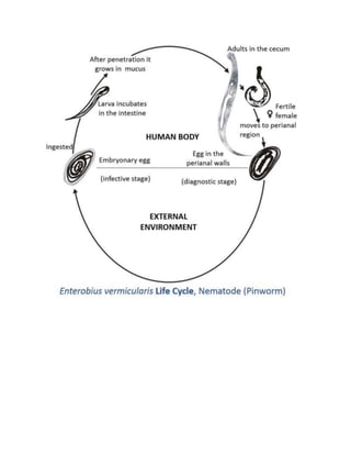
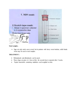
Enterobius vermicularis
 The pinworms are one of the most common intestinal nematodes.
 The adult worms inhabit the cecum and colon.
 Right after mating, the male dies. Therefore, the male worms are rarely seen.
 The female worms migrate out the anus depositing eggs on the perianal skin.
 Humans get this infection by mouth and by autoinfection.
Morphology
Adults:
 The adults look like a pin and are white in color.
 The female worm measures about 8 to 13 mm in size and is fusiform in shape.
 The male adult is only 2-5mm.
 The tail of a male is curved.
 They die right after mating, thus males are rarely seen.
 The anterior end tapers and is flanked on each side by cuticular extensions called
“cephalic alae”.
 The esophagus is slender, terminating in a prominent posterior bulb , which is called
esophageal bulb.
 The cephalic alae and esophageal bulb are important in identification of the species.
Egg:
 50 to 60m by 25 µm, persimmon seed-like, colorless and transparent, thick and
asymmetric shell, content is a larva.
Life Cycle
Site of inhabitation: cecum and colon
Infective stage: embryonated egg
Infective route: by mouth
Without intermediate host and reservoir host
Life span of female adults: 1-2 months
Pathogenesis
 The eggs hatch in the large intestine.
 Worms mature in 2-4 weeks and live for 2 months. Continuous reinfection is common.
 approximately one-third of infections are asymptomatic.
 The most common presentation is irritation and pruritus ani. sometimes itching is severe,
and secondary bacterial infection occurs.
 Occasionally, necrosis of the mucosal surface produces pain when nerve endings are
exposed.
 Worms often occur in the appendix and may be associated with appendicitis, but
causation has not been proved.
 Rarely, worms may migrate to ectopic sites, mostly within the female genitourinary tract
Laboratory Diagnosis
Cellophane tape test.
 E. vermicularis females lay their eggs on the perineum during the night.
 Touching the perianal skin with the sticky side of the tape will pick up the eggs; the tape
is affixed to a microscope slide and examined.
 Eggs are oval, approximately 55x25 µm in size, and flattened on one side, and they
contain a larva.
 Specimens should be collected prior to bathing or using the toilet.
 Four to six consecutive negative pinworm tape preparations are required to rule
out infection.
NIH swab
• Eggs are deposited in large number on the perianal and perineal skin at night can be
demonstrated by scraping this area by NIH swab in the morning before taking bath.
• Spread over glass slide and examined microscopically.
• This procedure should be repeated on three successive days
Stool samples.
 Eggs are only rarely seen in stool, but in patients with heavy worm burdens, adult female
worm may be seen in stool samples.
TREATMENT :
 Mebandazole and albendazole can be usesd .
 These drugs are given in 1 dose at first, the second dose is repeated after 2 weeks.
 Topical insecticide containing malathion can be applied on skin.

Enterobius vermicularis

  • 1.
    Enterobius vermicularis  Thepinworms are one of the most common intestinal nematodes.  The adult worms inhabit the cecum and colon.  Right after mating, the male dies. Therefore, the male worms are rarely seen.  The female worms migrate out the anus depositing eggs on the perianal skin.  Humans get this infection by mouth and by autoinfection. Morphology Adults:  The adults look like a pin and are white in color.  The female worm measures about 8 to 13 mm in size and is fusiform in shape.  The male adult is only 2-5mm.  The tail of a male is curved.  They die right after mating, thus males are rarely seen.  The anterior end tapers and is flanked on each side by cuticular extensions called “cephalic alae”.  The esophagus is slender, terminating in a prominent posterior bulb , which is called esophageal bulb.  The cephalic alae and esophageal bulb are important in identification of the species. Egg:  50 to 60m by 25 µm, persimmon seed-like, colorless and transparent, thick and asymmetric shell, content is a larva.
  • 2.
    Life Cycle Site ofinhabitation: cecum and colon Infective stage: embryonated egg Infective route: by mouth Without intermediate host and reservoir host Life span of female adults: 1-2 months
  • 4.
    Pathogenesis  The eggshatch in the large intestine.  Worms mature in 2-4 weeks and live for 2 months. Continuous reinfection is common.  approximately one-third of infections are asymptomatic.  The most common presentation is irritation and pruritus ani. sometimes itching is severe, and secondary bacterial infection occurs.  Occasionally, necrosis of the mucosal surface produces pain when nerve endings are exposed.  Worms often occur in the appendix and may be associated with appendicitis, but causation has not been proved.  Rarely, worms may migrate to ectopic sites, mostly within the female genitourinary tract Laboratory Diagnosis Cellophane tape test.  E. vermicularis females lay their eggs on the perineum during the night.  Touching the perianal skin with the sticky side of the tape will pick up the eggs; the tape is affixed to a microscope slide and examined.  Eggs are oval, approximately 55x25 µm in size, and flattened on one side, and they contain a larva.  Specimens should be collected prior to bathing or using the toilet.  Four to six consecutive negative pinworm tape preparations are required to rule out infection. NIH swab • Eggs are deposited in large number on the perianal and perineal skin at night can be demonstrated by scraping this area by NIH swab in the morning before taking bath. • Spread over glass slide and examined microscopically. • This procedure should be repeated on three successive days
  • 5.
    Stool samples.  Eggsare only rarely seen in stool, but in patients with heavy worm burdens, adult female worm may be seen in stool samples. TREATMENT :  Mebandazole and albendazole can be usesd .  These drugs are given in 1 dose at first, the second dose is repeated after 2 weeks.  Topical insecticide containing malathion can be applied on skin.